Wędrowska Ewelina, Pernak Mikołaj, Wędrowski Mateusz, Pankowska Violetta, Waśniowski Paweł, Piskorska Elżbieta, Zukow Walery. Impact of anti-inflammatory corticosteroids on changes in selected cytoimmunological parameters in selected interstitial lung diseases. Journal of Education, Health and Sport. 2019;9(2):334-348. eISNN 2391-8306. DOI
http://dx.doi.org/10.5281/zenodo.2576214
http://ojs.ukw.edu.pl/index.php/johs/article/view/6621
https://pbn.nauka.gov.pl/sedno-webapp/works/905450
The journal has had 7 points in Ministry of Science and Higher Education parametric evaluation. Part b item 1223 (26/01/2017). 1223 Journal of Education, Health and Sport eissn 2391-8306 7
© The Authors 2019;
This article is published with open access at Licensee Open Journal Systems of Kazimierz Wielki University in Bydgoszcz, Poland
Open Access. This article is distributed under the terms of the Creative Commons Attribution Noncommercial License which permits any noncommercial use, distribution, and reproduction in any medium, provided the original author (s) and source are credited. This is an open access article licensed under the terms of the Creative Commons Attribution Non commercial license Share alike.
(http://creativecommons.org/licenses/by-nc-sa/4.0/) which permits unrestricted, non commercial use, distribution and reproduction in any medium, provided the work is properly cited.
The authors declare that there is no conflict of interests regarding the publication of this paper.
Received: 01.02.2019. Revised: 01.02.2019. Accepted: 24.02.2019.
Impact of anti-inflammatory corticosteroids on changes in
selected cytoimmunological parameters in selected interstitial lung
diseases
Ewelina Wędrowska1, Mikołaj Pernak2, Mateusz Wędrowski3,4, Violetta Pankowska4,
Paweł Waśniowski4,5, Elżbieta Piskorska6, Walery Zukow7
1Department of Gene Therapy, Faculty of Medicine, Nicolaus Copernicus University
Collegium Medicum, Bydgoszcz, Poland
2Department of Plastic, Reconstructive and Aesthetic Surgery, Nicolaus Copernicus
University Collegium Medicum, Bydgoszcz, Poland
3Department of Positron Emission Tomography and Molecular Diagnostic, Nicolaus
Copernicus University, Collegium Medicum, Bydgoszcz, Poland.
4Nuclear Medicine Department, Oncology Centre, Bydgoszcz, Poland, 5Department of Inorganic and Analytical Chemistry, Nicolaus Copernicus University
Collegium Medicum, Bydgoszcz, Poland
6Department of Pathobiochemistry and Clinical Chemistry, Nicolaus Copernicus University
Collegium Medicum, Bydgoszcz, Poland
7Department of Spatial Management and Tourism, Faculty of Earth Sciences, Nicolaus
Copernicus University in Torun, Torun
Corresponding author:
Ewelina Wędrowska
ORCID ID: 0000-0003-4478-6749
Summary
Introduction. Glucocorticoids (GKS) hormones with strong immunosuppressive and anti-inflammatory activity are used, inter alia, in interstitial lung diseases (ILD). GKS inhibit excessive activity of inflammatory genes in the airways to induce apoptosis of immune cells, such as alveolar lymphocytes (AL).
Objective. Assessment of cytoimmunological changes including apoptosis occurring in alveolar lymphocytes in patients with selected ILD after treatment with systemic corticosteroids.
Methods. The material of the bronchoalveolar lavage (BAL) derived from patients with sarcoidosis (PS, n=66), idiopathic pulmonary fibrosis (IPF, n=27) and non-specific interstitial pneumonia (NSIP, n = 25; adequate number of patients treated with systemic GKS were 23, 8 and 6) were analyzed in cytoimmunological tests: a) percentage and the total values of BAL inflammatory cell populations; b) AL subset typing; c) CD4/CD8 index calculation; d) AL cell cycle analysis (DNA staining with propidium iodide, PI); in techniques mentioned in b) to d) items flow cytometry was used.
Results. In all patient groups, treatment with GKS resulted in a decrease in the total cell number, e.g. for PS (untreated: 310 ± 80x103/ml; treated: 188 ± 43x103/ml, median ± SEM,
p<0.05) and lymphocytes (untreated: 113 ± 71x103/ml; treated: 43 ± 25x103/ml median ±
SEM, p<0.05). There has also been significantly lower percentage of eosinophils in all groups in GKS-treated subgroups, e.g. for IPF (untreated: 4,6 ± 3,0%, treated: 0,3 ± 0,5%), for NSIP (untreated: 1,5 ± 0,7% and treated: with 0,4 ± 0,2%; median ± SEM, p<0.05 for both). In contrast, AL apoptosis rate was significantly higher in treated patients, e.g. for PS (untreated: 0,6 ± 0,5%; treated: 4,0 ± 2,5%), NSIP (untreated: 3,4 ± 1,8%; treated: 13,1 ± 5,1%; median ± SEM, p<0.05 for both). In all GKS-treated groups corticosteroid therapy caused lower CD4/CD8 index, but only on the level of statistical tendency (e.g. for PS untreated: 4,7 ± 0,5%, treated: with 2,9 ± 0,9%) for IPF (untreated: 1,2 ± 0,5%, treated: with 1,1 ± 0,5%; median ± SEM for both).
Conclusions. Systemic inflammatory glucocorticoid therapy (GKS) in all included groups of patients results in a decrease in the total number of alveolar lymphocytes, which is most likely related to the significant increase apoptosis rate of these cells. GKS medication in a similar extent caused probably both the death of helper (Th) and cytotoxic lymphocytes (Tc), since the decline in the value of CD4/CD8 index in treated patients compared with untreated ones was insignificant. A characteristic BAL cytological change in all tested ILD after GKS administration was a remarkable decrease in eosinophil percentage.
Key words: Apoptosis, BAL, corticosteroids, interstitial lung disease
Introduction
Sarcoidosis is a disease characterized by unknown etiology, the onset of the disease is chronic or subacute, fibrosis is rare, and inflammatory cells are lymphocytes and monocytes. In this disease, the population of CD4 + cells predominates. In IPF, the etiology is also not known, the beginning is of the type as in the case of sarcoidosis (the onset is chronic, when in PS - it is sometimes severe, as the so-called Loefgren syndrome). In addition, it differs from the previous disease entity, the presumed predominance of CD8 + lymphocytes, and the inflammatory cells are mainly eosinophils and neutrophils. Fibers are always present. However, fibrosis is common in NSIP, although in the form of so-called cell phones it appears late [5]. The treatment used in interstitial lung diseases is, among others corticotherapy.
Glucocorticoids are hormones that are important regulators of the body's homeostasis [6,7,8,9]. Due to their multi-faceted effect, they have found application in the treatment of inflammatory diseases, among others in interstitial lung diseases, acting in the genomic and non-genomic mechanism. Steroids penetrating the cell connect with their receptors (GR, glucocorticoid receptor), which causes the detachment of chaperone proteins, then the GKS-GR complex travels to the cell nucleus where dimerization of the receptor takes place. This results in the exposure of domains rich in zinc fingers, called DNA-binding domains. In this manner, the GKS-GR complex is combined with the GRE (glucocorticoid response element) within the promoters of the target genes. We differentiate GRE positive, which enhances gene activation and GRE negative, which by combining with the target gene inhibit its expression [10].
The most important effect of steroid therapy is the silencing of numerous pro-inflammatory genes activated in the course of long-lasting inflammation [11]. According to the literature, one of the most significant mechanisms of triggering T lymphocyte apoptosis by GKS is the induction of Bim protein expression, along with the parallel inhibition of gene expression for the Bcl-xL anti-apoptotic protein [12]. Another hypothesis suggests the effect of GKS on the release of cathepsins by disintegrating lysosomes
Purpose of work
The aim of the study was to determine cytoimmunological changes, with particular emphasis on the process of apoptosis in follicular lymphocytes from the lower respiratory tract in patients treated systemically with glucocorticosteroids in sarcoidosis, idiopathic pulmonary fibrosis and nonspecific interstitial pneumonias.
Materials and methods Examined groups
The study included 66 patients with sarcoidosis (pulmonary sarcoidosis, PS, including untreated, n = 43 and treated, n = 23), 27 with idiopathic pulmonary fibrosis (IPF, including untreated, n = 19 and treated, n = 8) and 25 with non-specific interstitial pneumonia (NSIP, including untreated n = 19 and treated n = 6). BAL material came from ill and untreated patients whose the therapy lasted for 6 to 22 months. The research included the consent of the Bioethical Commission of Collegium Medicum UMK (628/2014) with subsequent additions.
Bronchoalveolar lavage
material was aspirated, filtered through sterile gauze, combined, mixed, collected in sterile vessels and transported to the laboratory on ice [14].
Cytological and immunological tests of BAL material
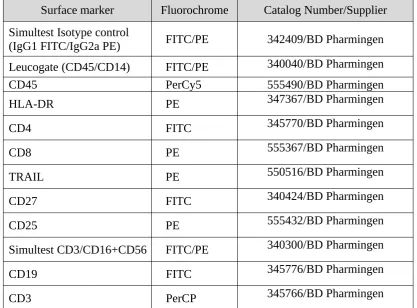
The principles of cytological and immunological analysis of BAL material have been previously reported [14,15]. The viability of BAL cells was determined with trypan blue, their total counts were calculated in a Bürker chamber; cytotoxic formulations (80g, 5min) were stained with the May-Grünwald-Giemsa method (MGG) and hematoxylin-eosin (HE); in the light microscope, the percentage formula of inflammation cells (inflow) BAL cells was calculated. The follicular lymphocyte (AL) subpopulations were typed by direct immunofluorescence technique. Briefly, the BAL material was centrifuged and rinsed in a PBS solution (300g, 8min), an amount of 1-5 x 10⁵ BAL cells was incubated with saturating amounts of mouse monoclonal antibodies (BD Pharmingen ™). A set of anti-CD45 FITC / CD14 PE antibodies ("leucogate", used to define the field of lymphocytes in flow cytometry) was used as a positive control. To determine the expression of the appropriate cytokine receptors on Th (CD4) and Tc (CD8) cells, a set of antibodies against human HLA-DR, TRAIL, CD27, CD25, CD16 + 56, CD19, CD3, CD4 and CD8 antigens were used, conjugated with fluorescent dyes FITC, PE and PE-Cy5. Cells expressing the test antigen emitted light in the standard FL1 fluorescence channels (green light), FL2 (bright red) and FL3 (dark red), respectively. As a negative control, isotype-compatible mouse antibodies with working antibodies used in the work were used. Details, including the type of antibody-conjugated fluorochromes, are given in Table 1. Incubation of BAL cells with antibodies (30 min. No light) was discontinued with PBS solution with 0.1% sodium azide, cells were rinsed and suspended in 0.3ml PBS with 1 % formaldehyde additive [16]
Table 1. Monoclonal antibodies used in flow cytometry analysis.
Surface marker Fluorochrome Catalog Number/Supplier
Simultest Isotype control
(IgG1 FITC/IgG2a PE) FITC/PE 342409/BD Pharmingen Leucogate (CD45/CD14) FITC/PE 340040/BD Pharmingen
CD45 PerCy5 555490/BD Pharmingen
HLA-DR PE 347367/BD Pharmingen
CD4 FITC 345770/BD Pharmingen
CD8 PE 555367/BD Pharmingen
TRAIL PE 550516/BD Pharmingen
CD27 FITC 340424/BD Pharmingen
CD25 PE 555432/BD Pharmingen
Simultest CD3/CD16+CD56 FITC/PE 340300/BD Pharmingen
CD19 FITC 345776/BD Pharmingen
CD3 PerCP 345766/BD Pharmingen
FITC - fluorescein isothiocyanate, PE - phycoerythrin, PerCy5 - phycoerythrin-cyanine 5, PerCP - peridinin chlorophyll protein
Cell cycle analysis in flow cytometry with staining of DNA with propidium iodide
The cell cycle was analyzed by flow cytometry with the staining of DNA with propidium iodide (PI). In this study, the ability of PI to stochiometric DNA binding is used, making it possible to assess cells in the late stage of apoptosis. In the histogram presentation of the surface area of pulses in the FL2 band (and therefore the specific for the excited PI light), typical cell cycle phases are distinguished: G0 / G1 peak, S phase and G2 / M peak. In order to fully penetrate the interior of the cell by PI, the cell suspension is incubated with surface-active substances (e.g., Nonidet detergent). Briefly, the BAL material was centrifuged and rinsed first in a PBS solution (400g, 5min), an amount of 1 x 10 6 BAL cells, and then incubated and under the same conditions 2 times centrifuged in NSS solution (Nonidet, propidium iodide, citrate buffer). 250μl of RNase solution was added to the cells and incubated for 15 min, then 0.5ml of PBS was added and tested in a flow cytometer. Caluza ®Flow Analysis Software was used to analyze and visualize the results.
Statistical methods
Results
Cytological results of BAL material
[image:6.595.137.463.198.393.2]Figure 1 presents the percentage recovery of fluid BAL material from patients with selected disease entities: PS, IPF, NSIP before and after glucocorticoid therapy (GKS). There was observed a statistically significant, with a significance level of p <0.05, percentage decrease in recovery after systemic treatment with GKS in the group of patients with IPF (untreated: 50.5 ± 1.8%; treated: 45.0 ± 2.9%; median ± SEM). In the remaining groups, the percentage of BAL fluid recovery after steroid treatment was reduced, however, it was not statistically significant.
Fig. 1. BAL fluid recovery %, the results are presented as median ± SEM values, p<0,05 was considered significant.
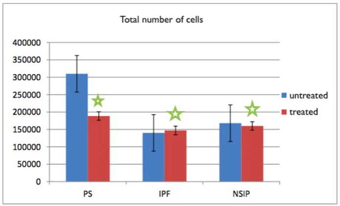
Interestingly, in sarcoidosis and nonspecific interstitial pneumonia a statistically significant decrease in the total number of cells was observed after treatment with GKS, PS (untreated 310 ± 80x103 cells/ml, treated: 188 ± 43x103 cells/ml, median ± SEM), for NSIP
(untreated: 168 ± 62x103 cells/ml, treated: 160 ± 32x103 cells/ml, median ± SEM). However,
in the case of IPF we observe a slight increase (untreated: 140 ± 38x103 cells/ml, treated: 147
Fig. 2. Total number of BAL cells, the results are presented as median ± SEM values, p<0,05 was considered significant.
[image:7.595.69.515.447.699.2]Table 2 provides us with the cytological results of the BAL material considering the selected cell populations in all the studied groups. In the BAL fluid of patients undergoing corticotherapy in all three diseases, a statistically significant decrease in eosinophils is noticeable. In PS (untreated: 1.1 ± 0.3%, treated: 0.2 ± 0.3%, median ± SEM), in IPF (untreated: 4.6 ± 3.0%; treated: 0.3 ± 0.5%, median ± SEM), in NSIP (untreated: 1.5 ± 0.7%, treated: 0.4 ± 0.2%, median ± SEM).
Table. 2. Cytoimmunological results of BAL material [%]
Macrophage s %
Lymphocyte s %
Neutrophils
% Eosinophils %
PS
unreated 56,0±2,8 (12,7-87,4) 41,7±2,9 (11,0-85,5) 1,1±0,5 (0,0-11,6) 1,1±0,3 (0,0-7,0)
treated 63,2±4,1(25,6-94,7) 30,5±4,0 (2,8-74,8) 2,1±1,4 (0,0-29,0) 0,2±0,3* (0,0-6,9)
IPF
unreated 60,1±4,7 (7,0-89,8) 14,0±3,6 (2,5-59,0) 15,0±2,9 (0,0-40,1) 4,6±3,0 (0,0-50,0)
treated 62,0±5,7 (44,0-90,5) 17,3±6,2 (3,0-58,0) 6,7±5,5 (2,1-53,0) 0,3±0,5* (0,0-3,9)
NSI P
unreated 68,9±3,9 (33,1-85,2) 18,2±2,9 (4,6-45,0) 5,9±3,8 (0,9-57,3) 1,5±0,7 (0,0-12,2)
treated 78,1±4,8(64,9-95,4) 13,9±4,2 (3,5-28,6) 1,8±2,8(0,7-18,4) 0,4±0,2* (0,0-1,5)
In BAL fluid, the number of lymphocytes in all examined groups descents at a statistically significant level after the use of GKS preparations. For sarcoidosis (untreated: 112 ± 71x103 cells/ml, median ± SEM; treated: 43 ± 25x103 cells/ml), for idiopathic pulmonary
fibrosis (untreated: 32 ± 7x10³ cells/ml; treated: 18 ± 11x103 cells / ml; median ± SEM), for
non-specific interstitial pneumonia (untreated: 38 ± 30x103 cells / ml, treated: 20 ± 8x10³
[image:8.595.128.471.157.352.2]cells/ml, median ± SEM).
Fig. 3. Total number of alveolar lymphocytes, the results are presented as median ± SEM values, p<0,05 was considered significant.
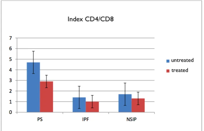
[image:8.595.124.474.500.725.2]Figure 3 displays the so-called the CD4/CD8 index, namely the ratio of CD4 + cells to CD8 +. There was visible, at the level of statistical tendency (p <0.1), decrease in all groups examined after treatment with glucocorticosteroids. For sarcoidosis (not treated: 4.7 ± 0.5%, treated: 2.9 ± 0.9%, median ± SEM), for idiopathic pulmonary fibrosis (untreated: 1.2 ± 0.5%; treated: 1, 02 ± 0.5%, median ± SEM), for non-specific interstitial pneumonia (untreated: 1.7 ± 0.7%, treated: 1.2 ± 0.6%, median ± SEM).
In the study of BAL cell apoptosis in the sub-G1 phase of the cell cycle, attention is paid to the higher percentage of apoptotic T lymphocytes in all examined groups after treatment with glucocorticosteroids (p <0.05) for PS (untreated: 0.6 ± 0.5%; 4.0 ± 2.5%), IPF (untreated: 5.0 ± 8.5%, median ± SEM, treated: 5.9 ± 1.3%), NSIP (untreated: 3.4 ± 1.8 %; treated: 13.1 ± 5.1%, median ± SEM, p <0.05.
Fig. 5. Alveolar lymphocytes (AL) apoptosis [%]; the results are presented as median ± SEM values, p<0,05 was considered significant.
Discussion
In studies carried out on experimental models, it was demonstrated that the Fas ligand transcription factor, specifically the NF-κB complex, is inhibited by GKS preparations. The beneficial effect of corticosteroids in sarcoidosis and in NSIP probably results from the inhibition of the NF-κB nuclear factor. In T lymphocytes, it becomes active due to ligation of the TCR receptor with antigen. It works anti-apoptotic because it enhances the expression of the Bcl-2 and Bcl-xL genes. Due to its activity, it influences the intrinsic pathway of apoptosis. The inhibition of its action by, for example, GKS preparations results in activation of the above-mentioned Bim protein, expression of the gene that suppresses the p73 cell cycle dependent on the p53 protein [18]. NF-κB is naturally blocked by an Iκβ inhibitor, whereas phosphorylation of this inhibitor by IKK kinase leads to the activation of the NFκB factor, which leads to the inhibition of a programmed cell death. In addition, the p22-FLIP factor degradation product activates IKK kinase owing to which it can perform its function [19].
lymphocyte [21]. Considering proteins from the Bcl-2 family, it is worth mentioning interleukin 7, which acts as a factor in the survival of follicular lymphocytes. Through JAK kinases and STAT-5 family transcription factors, it activates the expression of Bcl-2 factor [22]. Unfortunately, this cytokine promising to inhibit the inflammatory reaction seems to be insensitive to the use of preparations from the GKS group [17].
A noteworthy protein is tumor necrosis factor α (TNFα). T-lymphocytes expressing the TNFR2 receptor use TNFα as a survival signal. The TNFR2 receptor at the death domain has a structural TRAF motif that attaches the TRAF1 and 2 adapter proteins. The MAPK kinase, JNK kinase pathway is activated and, most importantly, the activation of the IKK inhibitor that triggers NFκB factors, the role of which has been described above. This mechanism is worth noting due to the fact that in one of the studies, researchers showed that in people suffering from sarcoidosis after using corticotherapy TNFα level decreased according to the above-mentioned mechanism of anti-inflammatory action of GKS preparations - by inhibition of proinflammatory cytokines as well as TNF [ 23].
GKS preparations also limit the expression of other inflammatory cytokines reactions, such as IL-1, IL-2, INFγ. It is these cytokines that are secreted by Th1 lymphocytes and, as is known, sarcoidosis is a disease in which the Th1 cell subpopulation predominates, while the steroids acting on the GILZ leucine zipper (glucocorticoid-induced leucine zipper) additionally inhibit gene expression for T-bet (a factor protein transcriptional genes promoting Th1 subpopulations [4] The above hypotheses quoted from the literature may be supported by the results obtained in the present study on the increase in alveolar cell (AL) apoptosis, especially in sarcoidosis and NSIP, thus in diseases with a key role of Th1 cells. this was statistically significant.
According to literature data, the problem of apoptotic effector T cells is not completely clear. It is not certain whether the apoptosis of these cells is determined by the mechanism of cell death induced by activation of AICD (activation of the induced cell death) or death by NID (neglect induced death as a special example of cell death in the mitochondrial pathway). The literature suggests that the interaction between Bim / Bcl-2 proteins may have a special role in the process of apoptosis. Patients with significantly low apoptosis of follicular lymphocytes showed high expression of BCL-2 protein. Conversely, significantly more frequent apoptosis was observed in groups of patients with a decreased percentage of BCL-2 + follicular lymphocytes [22]. Therefore, further research should focus on analyzing proteins, transcription factors that bind to particular signaling pathways, in order to be able to assess which one (extrinsic, internal, and possibly pseudoreceptor) is more often activated in the context of the death of follicular lymphocytes.
distinguished [25]. Perhaps that is why the results from previous work [26], that is statistically significant decrease in the CD4 / CD8 index in patients treated with sarcoidosis, were not confirmed. Well, in this study, this observation could not be unambiguously confirmed despite the fact that the group treated with GKS preparations had a marked decrease in CD4 / CD8 values, compared to untreated patients (more cases are necessary).
The conducted research also shows that the percentage and activity of follicular lymphocytes decreases in all three diseases in patients treated with glucocorticosteroids, which would indicate a beneficial effect of GKS preparations, and this is in accordance with the literature [27, 28]. An interesting fact from the point of view of this work are the results indicating an insignificant increase in the percentage of neutrophils, which is in conflict with literature reports that signal the reduction of neutrophil chemotaxis, eg by inhibiting the expression of IL-8 [29]. Nevertheless, this issue requires further research on a more representative group of patients.
For unknown reasons, lymphocytes at people suffering from IPF treated with GKS are unlikely to be resistant - the conclusion is due to the lack of statistically significant differences between the groups - despite the fact that the initial (in untreated patients) AL apoptosis is the highest of all three analyzed units disease. This corresponds to the increasingly common belief that GKS preparations do not benefit in the treatment of IPF [23] but they cause many side effects [30]. Despite this, doctors use GKS in default of other therapeutic options. In the light of the results of this work, this is not a rational position, there are no interest changes (in treated versus untreated) BAL neutrophils, currently considered important fibroblast inducing cells. The case is still unclear and may be the subject of further research, however, because another cell responsible for the deterioration of vital functions of IPF patients, probably the progression of pulmonary fibrosis, eosinophilia, seems to be very sensitive, according to these results, to the therapeutic effect of GKS. The results are confirmed in Meagher's work [31]. Finally, it should be noted that corticotherapy is not indicated if fibrosis has already occurred [32]. This would amplify the view present in the literature data about their inhibitory effect, first of all, differentiation into the Th1 subpopulation. The steroid preparations - as already mentioned - activate the GILZ protein (glucocorticoid-induced leucine zipper) which suppresses the expression of the T-beta transcription factor involved in the maturation of Th1 lymphocytes. It is believed that the predominance of Th2 (and perhaps also Th17) over Th1 is responsible for lung fibrosis and this is the case with IPF [33]. However, the current findings regarding modifications in the T cell subpopulations, and in particular the Th1 / Th2 / Th17 polarization disorder are not definitive. The next step that could be done is to study the markers of individual subpopulations of helper lymphocytes and to evaluate their expression.
According to the literature, little is known about the effect of neutrophils on lymphocyte apoptosis, and these cells are noteworthy because they are a source of oxygen free radicals and cathepsins, and recently discovered that such enzymes are involved in apoptosis of cells by inducing caspase-dependent death [35, 36], an interesting observation is the fact that in this study after steroid therapy only sarcoidosis had a non-significant increase in the percentage of neutrophils, and in the NSIP we noted a decrease, although the immune response in both cases shifted towards the polarization of the cell phenotype Th1.
Earlier, it was reported that steroid-induced apoptosis was primarily damaged by CD3 + lymphocytes [37]. However, in the light of this work, there is no unambiguous data confirming this hypothesis. Submission of two observations: 1) decrease in the number and percentage of lymphocytes in BAL material, 2) no statistically significant differences between subgroups of treated and untreated patients in the subpopulation of lymphocytes would suggest that apoptosis under the influence of GKS preparations in more or less the same range all three main BAL cell lymphocyte populations, namely T, B and NK cells [17].
In summary, in the light of the results we have received, we can come to three basic conclusions. In all diseases studied: sarcoidosis, idiopathic pulmonary fibrosis and non-specific interstitial pneumonia after the use of steroids, there is a significant decrease in the number of follicular lymphocytes (AL) and an increase in apoptosis of these cells. Therefore, the study of AL apoptosis can be used to assess the activity of the disease process, predict remission of the disease, namely the absence of symptoms, and determine the sensitivity of the inflammatory process to treatment with GKS. In addition, it is assumed that the main point of regulation of apoptosis are CD4 + accessory lymphocytes, because the value of the CD4 / CD8 index was reduced in all groups after taking steroid preparations (however unprecedentedly, which we emphasize once more, but was explained, too little of the examined cases).
IPF, because of the other two diseases, postulated by the literature response to glucocorticoids is a disorder that should be paid more attention. In the light of this research, the frequency of apoptosis increased after treatment with GKS. However, due to the conflict between the reports and the research carried out for the purpose of this study, further research into the determination of inflammatory mediators should be considered, among others interleukin 2, interferon γ, as well as BCL-2 protein, which overexpresses, as suggested by Herold and co-authors, has a protective function against apoptosis induced by GKS. Studies on transcription factors for individual lymphocyte subpopulations should not be omitted [17]. In the future, new therapeutic approaches, potentially protecting alveolar cells against apoptosis, may be considered, as the use of glucocorticosteroids is unfavorable in diseases of the occurring fibrosis, the more so as it has already been mentioned there probably is an unknown mechanism by which the Th2 subpopulation, which predominates in IPF escapes from the programmed death of a cell induced by steroids. Perhaps using gene therapy techniques and making genetic modifications of transcription factors inducing or inhibiting the programmed cell death process, we will be able to influence their expression in the course of interstitial diseases and presumably change the molecular mechanism of the action of steroid drugs.
Conclusions
2. The use of glucocorticosteroids causes a decrease in the percentage of eosinophils and lymphocytes significantly for all groups of patients, and an increase in the percentage of neutrophils for sarcoidosis.
3. The probable mechanism associated with a decrease in the number and percentage of follicular lymphocytes after GKS administration is a significant increase in the frequency of apoptosis, which was found in all examined groups. The frequency of BAL lymphocyte apoptosis significantly correlates negatively with BAL lymphocytosis.
4. In all examined groups, GKS treatment reduced the CD4 / CD8 lymphocyte index value, but that was not of significant nature. Presumably, GKS drugs in a similar range caused the death of accessory lymphocytes as well as cytotoxic lymphocytes
References
1. Wallis A, Spinks K: The diagnosis and management of interstitial lung diseases, BMJ 2015; 350: h2072
2. Gołecki M., Jankowska R.: Choroby śródmiąższowe płuc współczesne poglądy na immunopatogenezę, Adv Clin Exp Med., 2005; 171-174.
3. Wskazówki Polskiego Towarzystwa Chorób Płuc dotyczące metod pozyskiwania, opracowywania oraz oceny płynu z płukania oskrzelowo-pęcherzykowego (BAL), Pneumonologia i Alergologia Polska 2011; 79(2):75–89
4. Raghu G., Collard H.R., Egan J.J., Martinez F.J., Behr J., Brown K.K., et al., An official ATS/ERS/JRS/ALAT statement: idiopathic pulmonary fibrosis: evidence-based guidelines for diagnosis and management, Am J Respir Crit Care Med. 2011: 788–824. 5. Veeraraghavan S., Latsi P.I., Wells A.U., Pantelidis P., Nicholson A.G., Colby T.V., et
al., BAL findings in idiopathic nonspecific interstitial pneumonia and usual interstitial pneumonia, Eur Respir J., 2003; 22(2):239-244.
6. Baumann S., Dostert A., Novac N., Bauer A., Schmid W., Fas S.C., et al., Glucocorticoids inhibit activation-induced cell death (AICD) via direct DNA- dependent repression of the CD95 ligand gene by a glucocorticoid receptor dimer, Blood, 2005;106(2): 617-625.
7. Pałka J., Mazurek-Wądołkowska E., Molekularny mechanizm przeciwzapalnego działania glikokortykosteroidów, Farmaceutyczny Przegląd Naukowy, nr 4, 2008; 17-20. 8. Barnes P.J.: Anti-inflammatory actions of glucocorticoids: molecular mechanisms. Clin.
Sci (Lond). 1998; 94(6): 557-572.
9. Grzanka A., Genetyka astmy i molekularne podstawy działania leków stosowanych w astmie, [W:] Droszcz W. Astma, PZWL 2007: 33-93.
10. Bartholome B., Spies C.M. et al., Membrane glukocorticoid receptors (mGKR) are expressed in normal peripheral blood mononuclear cells and upregulated following in vitro stimulation and in patients with rheumatoid arthritis, FASEB J, 2004, 18:70-80. 11. Barnes P.J., How corticosteroids control inflammation, Quintiles Prize Lecture 2005, Br J
Pharmacol 2006; 148: 245-254.
12. Rocha-Viegas L., Vicent G.P., Baranao J.L., Beato M., Pecci A., Glucocorticoids repress bcl-X expression in lymphoid cells by recruiting STAT5B to the P4 promoter, J Biol Chem., 2006; 281(45): 33959-33970.
13. Pirożyński M.: Bronchofiberoskopia. Alfa Medica Press, Bielsko-Biała, 1999.
15. Kopiński P., Przybylski G., Balicka-Ślusarczyk B. i wsp.: Apoptoza limfocytów pęcherzykowych w sarkoidozie i w grupie kontrolnej występuje częściej u palaczy papierosów niż u osób niepalących. Przegl. Lek. 2006, 63, 841.
16. Kopiński P., Szczeklik J., Lackowska B. et al.: Flow cytometric characteristics of alveolar lymphocytes (AL) obtained by bronchoalveolar lavage (BAL) in the control group – proposal of normal value range of AL subsets in nonsmokers. Central Eur. J. Immunol. 2004; 29: 63.
17. Herold M.J., McPherson K.G., Reichardt H.M., Glucocorticoids in T cell apoptosis and function, Cell Mol Life Sci. 2006; 63(1): 60-72.
18. Arnold R., Brenner D., Becker M., Frey C.R., Krammer P.H.: How T lymphocytes switch between life and death. Eur. J. Immunol., 2006; 36:1654-1658.
19. Krammer P.H., Arnold R., Lavrik I.N.: Life and death in peripheral T cells, Nat. Rev. Immunol., 2007; 7: 532-542.
20. Rocha-Viegas L., Vicent G.P., Baranao J.L., Beato M., Pecci A., Glucocorticoids repress bcl-X expression in lymphoid cells by recruiting STAT5B to the P4 promoter, J Biol Chem., 2006; 281(45): 33959-70.
21. Haring J.S., Corbin G.A., Harty J.T., Dynamic regulation of IFN-γ signaling in antigenspecific CD8+ T cells responding to infection, J. Immunol., 2005; 174: 6791-6802.
22. Przybylski G., Wielikdzień J., Kopiński P., Mechanizmy zaprogramowanej śmierci efektorowych limfocytów T, Postępy Hig Med Dosw (online), 2013; 67: 1374-1390. 23. Barnes P.J., How corticosteroids control inflammation, Quintiles Prize Lecture 2005, Br J
Pharmacol 2006; 148: 245-254.
24. Ziegenhagen M.W., Rothe M.E., Schlaak M., Muller-Quernheim J., Bronchoalveolar and serological parameters reflecting the severity of sarcoidosis, Eur Respir J., 2003; 21(3): 407-413.
25. Kopiński P., Balicka-Ślusarczyk B., Dyczek A., Szpechciński A., Przybylski G., Jarzemska A., et al. Enhanced expression of Fas Ligand (FasL) in the lower air-ways of patients with fibrotic interstitial lung diseases (ILDs), Folia Histochem Cytobiol. 2011; 49(4): 636-645.
26. Kopiński P., Apoptoza limfocytów pęcherzykowych w wybranych śródmiąższowych chorobach płuc, Wydawnictwo Naukowe Uniwersytetu Mikołaja Kopernika Collegium Medicum im. Ludwika Rydygiera, 2012.
27. Kondoh Y., Taniguchi H., Yokoi T., Nishiyama O., Ohishi T., Kato T., et al. Cyclophosphamide and low-dose prednisolone in idiopathic pulmonary fibrosis and fibrosing nonspecific interstitial pneumonia., Eur. Respir. J., 2005; 25(3): 528-33.
28. Drent M., Mansour K., Linssen C., Bronchoalveolar lavage in sarcoidosis, Semin Respir Crit. Care. Med., 2007; 28(5): 486-495.
29. Sah, B.P., Goyal, S. & Iannuzzi, M.C., Novel Pharmacotherapy of Sarcoidosis, Pharmacology and Therapeutics, 2015.
30. De Lauretis A., Veeraraghavan S., Renzoni E., Review series: Aspects of interstitial lung disease: connective tissue disease-associated interstitial lung disease: how does it differ from IPF? How should the clinical approach differ? Chron Respir Dis. 2011; 8(1)s:53-82. 31. Meagher L.C., Cousin J.M., Seckl J.R., Haslett C. Opposing effects of glucocorticoids on
32. Wells A.U., Hirani N. et al, Interstitial lung disease guideline: the British Thoracic Society of Australia and New Zealand and the Irish Thoracic Society. Thorax, 2008; 63(5): 1-58.
33. Cannarile L., Fallarino F., Agostini M., Cuzzocrea S., Mazzon E., Vacca C., et al., Increased GILZ expression in transgenic mice up-regulates Th-2 lymphokines, Blood, 2006; 107(3): 1039-1047.
34. Nagai S., Handa T., Ito Y., Takeuchi M., Izumi T., Bronchoalveolar lavage in idiopathic interstitial lung diseases., Semin Respir Crit Care Med. 2007; 28(5): 496- 503.
35. DrogaMazovec G., Bojic L., Petelin A., Ivanova S., et al: Cysteine cathepsins trigger caspase-dependent cell death through cleavage of bid and antiapoptotic bcl-2 homologue. J. Biol. Chem., 2008, 283, s. 19140-150
36. McIlwain D. R., Berger T., Mak T. W.: Caspase Functions in Cell Death and Disease. Cold Spring. Harb. Perspect. Biol., 2013, 1, s. 1-28.

![Fig. 5. Alveolar lymphocytes (AL) apoptosis [%]; the results are presented as median ± SEM values, p<0,05 was considered significant.](https://thumb-us.123doks.com/thumbv2/123dok_us/8221202.266042/9.595.118.480.159.401/alveolar-lymphocytes-apoptosis-results-presented-median-considered-significant.webp)
