Introduction
Because of some problems in about fifty per-cent of the current cancer treatments, the alter-native methods are being actively considered
[1]. The interest in using bacteria as an anti-can-cer therapeutic agent dates back to the end of the 19th century [2,3]. It has been shown that some bacterial strains like: Salmonella choler-aesuis, Vibrio cholera, Listeria monocyto-genes, and Escherichia coli replicate effective-Original Research
Medical Journal of the Islamic Republic of Iran.Vol. 23, No. 4, February, 2010. pp. 207-217
Cell death induction by Streptococcus pyogenes in four types of
malignant cell lines
Zahra Eslami-Nejad, PhD.1, S. Noureddin Nematollahi-Mahani, PhD.2, Fereshteh Saffari, MS.3
Hamid Mollaii, MS.4, S. Ali Mohammad Arabzadeh, PhD.5
Abstract
Background:The interest in using bacteria as anti- cancer therapeutic agents dates back to the end of the19th century. Some bacteria like Salmonella and Listeria replicate effective-ly inside malignant cell lines and suppress their growth. The bacterium Streptococcus pyo-genes has become medically famous as a flesh-eating pathogen since mid-1980s. It is the causative agent of a life threatening clinical condition called necrotizing fasciitis. S. pyo-genes usually produces a range of lytic enzymes that promote bacterial pathopyo-genesis. With these characters, could this bacteria. be employed as a curing agent for certain cancers? The aim of this study was to determine the influence of S. pyogenes on malignant cellular death (apoptosis or necrosis)- in an ex-vivo "experimental- interventional" study.
Methods: The cytotoxicity of fifteen internalized streptococcal strains( including 12 clinical isolates, 2 known M types [M1, M3] and standard strain), on four types of malignant cell lines- A549, BT-20, PC-3, L-929- were tested by Trypan blue exclusion, DNA fragmen-tation and WST-1 methods. The streptococcal protease, lipase, DNase and serum opacity factor (SOF) were tested concurrently. The standard strain of Streptococcus (Enterococcus) faecalis was employed as negative control. The results were analyzed by statistical Minitab software.
Results:The overall cytotoxicity rate of -internalized- S. pyogenes was 57% by trypan blue method and 50 % by DNA electrophoresis. False positive results occurred for the neg-ative control in WST-1; therefore this test did not present reasonable results. The correlation between production of SOF, lipase, DNase and cytotoxicity of S. pyogenes was not signifi-cant (p > 0.05). However, 67% of the protease positive strains induced cellular death in at least one type of - malignant cell line (p<0.05).
Conclusion: Our findings indicated that, some non-invasive S. pyogenes that cause be-nign infection like pharyngitis can induce cell death in various cancerous cell lines. It seemed that among bacterial products, the proteolytic enzymes- linked to the streptococcal pyrogenic exotoxin B (spe-B)- were more related to bacterial invasion.
Keywords: Streptococcus pyogenes, cancer treatment, malignant cell, apoptosis.
1.Corresponding author, Associate Professor of Microbiology, Department of Microbiology. Afzalipour Medical School, Kerman Univer-sity of Medical Sciences. Kerman, Iran. Tel: +98913 3404754 , +98341 2114414. Fax: +98341-2113378
E-mail: [email protected]
2. Associate Professor of Anatomy. Department of Anatomy. Afzalipour Medical School. Kerman University of Medical Sciences. 3. Medical technologist of Microbiology. Department of Microbiology. Afzalipour Medical School, Kerman University of Medical sciences.
Departments of Microbiology and Anatomy, Afzalipour Medical School, Kerman University of Medical Sciences. Kerman, Iran.
ly inside malignant tumors [3,4]. Recently it was found that the metabolites of some sea-wa-ter bacsea-wa-teria and gliding bacsea-wa-teria have strong ac-tivity against human breast adeno-carcinoma, colon cancer, cervical cancer and oral cancer cell lines. The 16S rDNA sequencing analysis of these bacteria are related to Bacillus-vallis-mortis and Cytophaga-Flavobacterium-Bacte-riodes respectively [5, 6]. The exact mecha-nisms of tumor suppression have not been fully understood [3]. Among pathogenic bacteria, the Streptococcus pyogenes (group A streptococci-GAS), is responsible for a wide range of human diseases [6]. Since mid-1980s S. pyogenes has become medically famous as a flesh-eating pathogen [8]. As it causes a life threatening clinical condition called necrotizing fasciitis [8]. Viable S. pyogenes have been found inside the epithelial, endothelial, neutrophil and some other cell lines. However, this bacterium is known as an extra-cellular microbial agent [7]. Among its toxin-like products, the streptococ-cal pyrogenic exotoxin B (Spe B), potentially enhances tissue damage [10,11]. Essentially, Spe B is a cysteine protease with a considerable role in streptococcal pathogenesis [8]. Other extra- cellular products that may promote cell invasion are lipase, DNase, SOF,... [10,12, 13,14]. However, these enzymes are not unique for streptococcal strains [14,15]. This study was conducted to determine the anti-cancer po-tency of S. pyogenes in an ex-vivo "experimen-tal- interventional" trial. The main purpose was to test the cytotoxicity of some - internalized-clinical isolates of S. pyogenes on some carci-noma cell lines. The enzymatic properties of isolates were also analyzed concurrently.
Methods
Twelve clinical isolates of S. pyogenes (10 isolates from pharyngitis cases, 1 from a syn-ovial culture and finally 1 from a blood culture) and 2 known M type strains ( M1 and M3) that were kindly provided by the Pasteur Institute(Tehran- Iran) as well as the standard
strain( ATCC 8668) were examined. The ente-rococcus faecalis standard strain (NCTC 8213) was employed as the negative control in all steps.
Initially, the stationary phase of bacterial growth was determined by standard growth-curve analysis [12]. Accordingly, a fresh sus-pension (Mc Farland No. 0.5) of the over-night (~18 hours) culture- in the Todd-Hewitt broth (Himedia-india) was prepared. The serial dilu-tion was then prepared and used for each cyto-toxicity or enzymatic test.
The Staphylococcus aureus (ATCC 25923) and Staphylococcus epidermidis (PTCC 1435) were used as the positive or negative control in some bacterial enzyme tests.
Bacterial biochemical tests
i) Serum Opacity Factor(-SOF), The overnight culture of bacterial sample in the Todd- Hewith broth was centrifuged, and then 0.1 ml of filtrat-ed supernatant (0.45 Pm, Millipore-USA) was added to 1 ml of sterile horse serum. After incu-bation (37oC) for 16-18 hrs, the opacity density
of the mixture was evaluated with naked eye [14]. The M3 type of S. pyogenes and the Staphylococcus epidermidis were included as the positive and negative controls respectively [16,17].
ii) Protease test, A few numbers of fresh bac-terial colonies were inoculated onto Litmus milk agar (BBL- UK) and incubated (37oC ) up
to 10 days [16]. Positive result of casein (pro-tein) hydrolysis was indicated by the formation of a clear zone around colonies. Staphylococ-cus aureus was included as the positive control [15,19].
iii) Lipase test, Bacterial strains were first plated onto Margarine containing (1%) Brucel-la agar (Difco-USA). After incubation (48 hrs at 37o C), the cultures were kept in sealed plates
for 2 weeks at room temperature [20]. The pos-itive result was assessed by formation of irides-cence zone and/or white precipitation around colonies [20]. S. aureus was included as the Cell death induction by Streptococcus..
positive control [15].
iv) DNase test, The bacterial strains were inoculated in DNase test media (Merck - Ger-many). After incubation (37o C for 24- 48hrs),
the hydrochloric acid -HCl (1%) was added to culture plates. Formation of a clear zone around bacterial colonies was indicative of the positive result [21]. The S. aureus was included as the
positive control [15]. Fig1. Bacterial internalization into A 549 indicated by arrows
Table 1. The results of bacterial biochemical tests
Cell lines
To perform the research [19,22], four differ-ent types carcinoma cell lines including A549 (human respiratory epithelial cell), BT-20 (hu-man breast epithelial cells), PC3 (hu(hu-man prostate epithelial cell) and L929 (mouse fi-broblast cells) purchased from cell bank of Pas-teur institute (Tehran-Iran) were examined. The
cell culture used in Dulbeccos Modified Eagles Medium-DMEM (sigma-USA) was supple-mented with 10% fetal bovine serum (FBS) in optimal condition (370 C, CO2 10% and hu-midity ~ 95%), and subcultures were done every 3 days by standard method [7,19, 23].
Bacterial internalization assay, the freshly prepared cell suspension was seeded in 24-well Cell death induction by Streptococcus...
Fig. 2. Demonstration of A: infected, a: infected cell line (A549) B: infected, b: infected cell line (BT-20) C: un-infected, c: infected cell line (PC3) D: un- un-infected, d: infected cells line (L929).
tissue culture plates (~5× 10⁴ /well) and
incu-bated in optimal condition for 24hrs [19,22]. Then the prepared monolayers were infected with approximately 5× 10⁷ bactria/well, to ob-tain a multiplicity of infection-m.i.o- of 1000 [19,22]. The mixture was incubated in DMEM + 20% FBS - without any antibiotics- for 2 hours (37° C, 5% CO2) [19, 24]. In order to eliminate unbounded bacteria, they were washed up 3 times and re- incubated in a peni-cillin containing fresh medium (3ȝg/ ml) for additional one hour [25, 26]. Finally, the infect-ed monolayers were thoroughly rinsinfect-ed with PBS and incubated in an off-antibiotic medium, for 24- 48 hrs [19,22]. The viability of the inter-nalized bacteria was randomly monitored by the physical cell lysis method in ice- water and then the lysate inoculated on a blood agar plate [27].
Cytotoxicity Assays
i) Trypan blue exclusion stain, tripsizing the cell lines, the prepared cell suspension was mixed with an equal volume (1:1) of trypan blue- 0.4% solution. After 1-2 minutes, the
number of total and stained cells were counted in the Neubauer chamber [22,28]. The calculat-ed percentage of staincalculat-ed cells revealcalculat-ed the per-centage of dead cells [28].
ii) Analysis of DNA fragmentation. For preparation of infected cells, the cell-lines were washed and trypsinized 24 hrs after incubation. DNA extraction was done by the manufacturer protocal (Qiagen- Germany). Electrophoresis of The extracted DNA was electrophoresed in the 2% agarose gel (Fermentas- Lithuania), and the gel product stained by ethidium bromide and visualized by UV in Gel-Doc [7,19, 22]. The non- infected cell line was treated in the same way as the negative control.
iii) WST-1 method. According to Patrick et al's proposed protocol [22], the prepared cell suspension was seeded in a 96-well micro-titer plate (~104/ well) and infected with S. pyogenes
at m.i.o of 1000. The infectivity of the bacteria in cell line was continued as described above. After the planned time of incubation, the WST-1 dye (Roche- Germany) was added to each well, and the optical density (OD ) of each well read with a kinetic micro-plate reader at a Fig. 3. AfD : formation of smear from degradation of DNA in apoptotic cells by 2% agarose gel electrophoresis.
wavelength of 450 nm twice. First immediately after adding the dye then 1 hr after incubation (37oC) [22]. Triple cell cultures were tested for
each cell line and repeated at least three times-for an individual isolate/strain- on different days.
iv)Assessment of the morphological changes. An inverted phase contrast microscope-equipped with a camera (Olympus IX71-Japan) was used to visualize any changes in the morphology and cytopathic effects (CPEs) of infected cell line. The assessment of CPE pro-vided a useful way for following up of cell lines infectivity.
The un-infected cell line (of each type) was treated as negative control in all of the cytotoxi-city tests.
Statistical analysis
Quantitative numbers were expressed as means. Qualitative data were categorized as ei-ther positive or negative. The Minitab software was employed for determination of Pearson correlation, Odd ratio, Chi- square and P- value.
The P- values of less than 0.05 were considered significant. For WST-1 the one-way ANOVA and the post-Hoc turkey methods were applied to the results.
Results
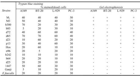
The results of bacterial biochemical tests are summarized in table 1. The rates of SOF+, Pro-tease+, Lipase+ and DNase+ among tested strains were 75%, 56%, 88% and 88% respec-tively.
Fig. 1 demonstrates the internalized bacteria in A549 cell line and Fig. 2 (Afd) shows the CPE of each type of the infected cell line. Fig. 3(AfD) is a part of the results of DNA elec-trophoresis for the dead cells. The results of en-zymatic biochemical tests are gathered in table 1. The data within table 2 is consisted of the re-sults of the cytotoxicity of all strains using Try-pan blue and electrophoresis. Although the quantities of three cytotoxicity methods are not exactly comparable, for ease of assessment, the percents of Trypan blue stain and WST-1 have been changed to positive or negative (compare Cell death induction by Streptococcus..
Table 2. Comparison of the results of cytotoxicity of all 15 strains of S. pyogenes + standard strain of S. faecalis on 4 types of malignant cell lines by 2 methods: Trypan blue, and gel electrophoresis.
with negative control ) in Table 3.
The correlation between Trypan blue stain and electrophoresis for 4 type's of cell lines: A549, BT-20, L-929 and PC-3- were 0.86, 0.63, 0.60 and 0.75(p<0.05). But the correla-tion between Trypan blue staining and WST-1 was not significant (p>0.5) for A549 and BT-20 and (p>0.05) for L-929, it was o.73 (p<0.05) for PC-3.
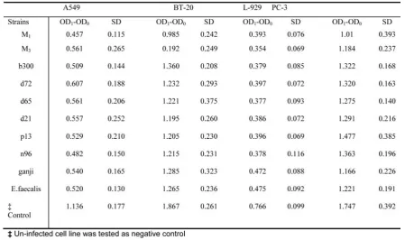
In this study the electrophoresis of DNA dis-played the basic method for evaluation of cyto-toxicity. Detailed data on the subject of optical densities (OD1-OD0) and standard deviations (SD) in WST-1 are included in table 4. Each strain was tested for three times in parallel.
Discussion
Bacteria are rich sources of natural products. Some have attracted the attention of the drug in-dustry as potent protective agents against natu-ral threats like infection and cancer [29].
Historically, Streptococcus pyogenes has opened a new window on cancer treatment since two German physicians W. Busch and F. Fehleisen -separately- found that certain types of cancers regressed following streptococcal erysipelas in hospitalized patients [3].
The bacterium Streptococcus pyogenes is not considered a significant intracellular pathogen like Listeria or Shigella however, it can effi-ciently enter a variety of mammalian cells [6,7]. The S. pyogenes releases a wide range of lytic
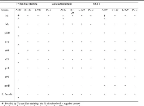
Table 3. Comparison of the results of cytotoxicity of 9 strains of S. pyogenes + standard strain of E. faecalis on 4 types of malignant cell lines by 3 methods: Trypan blue, electrophoresis and WST-1 assay.
enzymes /toxins e.g hemolysin and erythro-genic toxin [30]. Therefore the cellular death-as a consequence of streptococcal tissue infec-tions- could be the outcome of bacterial prod-uct's activity and/ or induction of apoptosis in the infected cell [7].
Among more than 80 M types of S. pyo-genes, the M3 has been recognized as the most invasive strain [31]. This type was employed as the positive control.
In the present study, the results of three cyto-toxicity methods were not equal. For the estab-lishment of cell death (apoptosis or necrosis), the electrophoresis of DNA was the method of choice [7, 22, 23], hense the results of two other methods were compared with this technique. Accordingly we got similar results from Trypan blue stain and DNA electrophoresis for 60 to 86% of the tests. Simplicity is the best known criterion of Trypan blue stain, but the technique might be accompanied by a high human error [32].
The cell viability was also determined by WST-1 reagent. The rate of positive result was 97% including for negative control (table 3). For one out of four cell lines (PC-3), the corre-lation of Trypan blue stain and WST-1 was sig-nificant (p <0.05). The colorimetric WST-1 as-say indicated the early cell-damage resulting from mitochondrial disorder [33, 34]. This as-say has been employed in a few similar experi-ences [22] however we do not recommend it for tests where two types of viable cells (bacteria and eukaryotic cells) are involved.
Isolates of S. pyogenes induced various level of cell death (Table 2). In Bennett study (Aus-tralia- 1999), those isolates that recovered from invasive cases of S. pyogenes diseases, invaded HEp-2 to a significantly lesser extend than those from superficial sites [32]. In other study it was shown that the invasion of epithelial cells may not be related to the invasive disease of S. pyogenes [36].
Moreover, each types of malignant cell lines, Cell death induction by Streptococcus..
Table 4. The mean of difference of optical densities (OD1-OD0) and standard deviations (SD) in WST-1. Every strain was tested three times on triple cultures of each cell line.
affected variably by streptococcal and even M3 invasions. The DNA electrophoresis, results indicated that the human prostate epithelial cell (PC3) resisted against M3 but this was not con-firmed by Trypan blue nor by WST-1assay (Table 2 and 3).
Experimental investigations have indicated that, invasion of S. pyogenes is under the influ-ence of streptococcal exoenzymes [10, 22, 37]. According to the results of the present study, the correlation between SOF production, lipase production, DNase production and cytotoxicity of S. pyogenes was not significant (p > 0.05, odd ratios: 0.37, 2.19 and 2.5 respectively) . However 67% of protease positive strains were cytotoxic, and the correlation of these charac-ters was 0.78 by Trypan blue stain ( p< 0.05 , odd ratio: 11.37). For S. pyogenes protease positive, is linked to the production of strepto-coccal pyrogenic exotoxin (SPE) [7,19 ,22, 23]. The Spe B, significantly mediates the enhance-ment of invasion [7,19,38], when compared with other types of SPEs (Spe A, and Spe C)
Cancer treatment is encountered by various significant problems [1]. Bacteria could unlock secrets that aid cancer treatment [29]. In this way, a number of familiar human pathogens like Salmonella typhimurium, E.coli, Clostridi-um novyi, Pseudomonas aeroginosa, Strepto-myces spp and their natural products have man-ifested excellent results [2,29,39,40].
The findings of present ex-vivo study showed that some S. pyogenes as a cusative agaent of benign infection like pharyngitis can effectively induce cell death in some carcinoma cell lines( within 24 hrs). It seemed that among streptococcal products, the proteolytic en-zymes related to Spe-B - were more associated with bacterial invasion.
Hopefully it will be possible to design re-combinant anti-cancer invasive bacteria by lig-anding to specific malignant cell receptors in the near future.
Acknowledgement
The budget for this study was provided by the research administration Center of Kerman Uni-versity of Medical Sciences. We are very grate-ful to Dr Nakhaii, Dr Najafi, Dr Bahrampour, for their superb cooperation, in this matter.
References
1. Weia M q, EllembK A o, Dunna P, Westa M J, Baic C X, Vogelsteind B. Facultative or obligate anaerobic bacteria have the potential for multimodality therapy of solid tumors. European Journal of Cancer 2007; 43(3): 490-496.
2. Avogadri F, Martinoli C, Petrovska L, Chiodoni C, Transidico P, Bronte V, et al. Cancer immunotherapy based on killing of Salmonella-infected tumor cells. Can-cer Res 2005; 65(9):3920-7.
3. Springer CJ, Lehouritis P, Marais R. Bacteria in cancer therapy. Microbiology today 2005; 56:113-115.
4. Medina E, Goldman O, Toppel AW, Chhatwal GS. Survival of Streptococcus pyogenes within host cells: A pathogenic mechanism for persistence and systemic in-vasion. JID 2003; 187: 597-603.
5. Jeong SY, Park SY, Kim YH, Kim M, Lee S J: Cyto-toxicity and apoptosis induction of Bacillus vallismortis BIT-33 metabolites on colon cancer carcinoma cells. Journal of Applied Microbiology, Volume 104, Number 3, March 2008 ; pp. 796-807(12).
6. Sangnoi Y, Srisukchayakul P, Arunpairojana V, Akkharawit Kanjana-Opas A: Diversity of marine glid-ing bacteria in Thailand and their cytotoxicity. Mirobial Biotechnology, Marine Biotechnology.
7. Marouni MJ, Sela S. Fate of Streptococcus pyo-genes and epithelial cells following internalization. J Med Microbiol 2004; 53(Pt 1):1-7.
8 . Levine GE, Manders SM. Life-threatening necro-tizing fasciitis. Clinics in Dermatology 2005; 23: 144-147.
9. Thulin P, Johansson L, Low DE, Gan BS, Kotb M, McGeer A, et al. Viable group A streptococci in macrophages during acute soft tissue infection. PLoS Medv 2006; 3(3): 53.
10. Saouda M, Wu W, Conran P, Boyle MD. Strepto-coccal pyrogenic exotoxin B enhances tissue damage ini-tiated by other Streptococcus pyogenes products. J Infect
Dis 2001; 184(6):723-31.
11. Lukomski S, Sreevatsan S, Amberg C, Reichardt W, Woischnik M, Podbielski A, et al. Inactivation of Streptococcus pyogenes extracellular cysteine protease significantly decreases mouse lethality of serotype M3 and M49 strains. J Clin Invest 1997; 99(11):2574-80.
12. Timmer AM, Kristian SA, Datta V, Jeng A, Gillen CM, Walker MJ, et al. Serum opacity factor promotes group A streptococcal epithelial cell invasion and viru-lence. Mol Microbiol 2006; 62(1):15-25.
13.Minkel JR. Role of Flesh-Eating Bacteria?s Toxin Identified. cited 2006 oct 16. Available from: http://www.scientificamerican.com/article.cfm?id=role -of-flesh-eating-bacteria.
14.Sumby P, Barbian KD, Gardner DJ, Whitney AR, Welty DM, Long RD. Extracellular deoxyribonuclease made by group A streptococcus assists pathogenesis by enhancing evasion of the innate immune response. PNAS 2005; 102(5): 1679-1684.
15. Brooks GF, Carroll KC, Butel JS, Morse SA. Jawetz, Melnick & Adelberg's Medical Microbiology. 24nd ed. New York: McGraw-Hill companies; 2007; pp. 203-211.
16.Johnson DR, Kaplan EL. Microtechnique for serum opacity factor characterization of group A strepto-cocci adaptable to the use of human sera. J Clin Microbi-ol 1988; 26(10):2025-30.
17.Tayeb ES, Nasr EM. Serum opacity factor of Staphylococcus epidermidis. Infect Immun 1977; 15(1):335-6.
18. McCarty M. Microbiology. In: Davis(ed) BD, edi-tor. Streptococci. Philadelphia: JB.Lipincott Compa-ny1990; pp.150.
19.Tsai PJ, Kuo CF, Lin KY, Lin YS, Lei HY, Chen FF, et al. Effect of group A streptococcal cysteine protease on invasion of epithelial cells. Infect Immun 1998; 66(4):1460-6.
20. Murray PR, Baron EJ, Jorgensen JH, Pfaller MA. Manual of clinical microbiology. 9nd ed. New York : St.Louis. The C.V. Mosby Company; 2007. pp.217-225.
21. Betty A. Forbes, Daniel F. Sahm and Alice S. Weissfeld. Baily and Scott's Diagnostic Microbiology. 12nd ed. New York : St.Louis. The C.V. Mosby Compa-ny 2007; pp. 171-172.
22. Tsai PJ, Lin YS, Kuo CF, Lei HY, Wu JJ. Group A Streptococcus induces apoptosis in human epithelial cells. Infect Immun 1999; 67(9):4334-9.
23. Kuo CF, Wu JJ, Tsai PJ, Kao FJ, Lei HY, Lin MT, et al. Streptococcal pyrogenic exotoxin B induces apop-tosis and reduces phagocytic activity in U937 cells. In-fect Immun 1999; 67(1):126-30.
24. Cue D, Cleary PP. High-frequency invasion of ep-ithelial cells by Streptococcus pyogenes can be activated by fibrinogen and peptides containing the sequence
RGD. Infect Immun 1998; 66(9):45-77.
25. Kaplan EL, Chhatwal GS, Rohde M. Reduced ability of penicillin to eradicate ingested group A Strepto-cocci from epithelial cells: clinical a pathogenetic impli-cations. Clinical infectious disease 2006; 43: 1398-1406. 26. Hamrick TS, Diaz AH, Havell EA. Influence of extracellular bactericidal agents on bacteria in macrophages. Infect Immun 2003; 71(2): 1016-1019.
27. Sela S, Neeman R, Keller N, Barzailai A. Relation-ship between asymptomatic carriage of Streptococcus pyogenes and the ability of the strains to adhere to and be internalized by cultured epithelial cells. J.Med.Microbiol 2000; 49:499-502.
28. Li PT, Lee YC, Elongovan N, Cho ST. Mouse 24p3 protein has an effect on L-929 cell viability. Int. J. Biol. Sci 2007; 3(2): 100-107.
29. Sakai J. Bacteria unlock secrets that may aid can-cer treatment. cited 2008 Feb 27. available from: http://www.news.wisc.edu/14816.
30. Talkington DF, Schwartz B, Black CM, Todd JK, Elliott J, Breiman RF, et al. Association of phenotypic and genotypic characteristics of invasive Streptococcus pyogenes isolates with clinical components of strepto-coccal toxic shock syndrome. Infect Immun 1993; 61(8):3369-74.
31.Cue D, Dombek Pe, Lam H, Cleary PP. Strepto-coccus pyogenes Serotype M1 encodes multiple path-ways for entry into human epithelial cells. Infect Immun 1998; 4593-4601.
32.Sarma KD, Ray D, Antony A. Improved sensitivity of trypan blue exclusion assay with Ni2+ or Co2+ salts. Cytotechnology 2000; 32(2): 93-95.
33. Strayer DS, Rubin E. Cell injury. In: Rubin R, Strayer DS, editors. Rubin Pathology. 5nd ed. Philadel-phia: Lippincott Williams & Wilkins; 2008; pp: 1-35.
34. Ngamwongsatit p, Banada PP, Panbangred W, Bhunia AK. WST-1 based cell cytotoxicity assay as a substitute for MTT- based assay for rapid detection of toxigene bacillus species using CHO cell line. Journal of microbiological methods. 2008; 73(3):211-5.
35. Bennett-Wood VR, Carapetis JR, Robins-Browne RM. Ability of clinical isolates of group A streptococci to adhere to and invade HEp-2 epithelial cells. J Med Mi-crobiol 1998; 47(10):899-906.
36. Molinari G, Chhatwal GS. Invasion and survival of Streptococcus pyogenes in eukaryotic cells correlates with the source of the clinical isolates. J Infect Dis 1998; 177(6):1600-7.
37. Kuo CF, Wu JJ, Lin KY, Tsai PJ, Lee SC, Jin YT, et al. Role of streptococcal pyrogenic exotoxin B in the mouse model of group A streptococcal infection. Infect Immun.1998; 66(8):3931-5.
38. Von Pawel-Rammingen U, Johansson BP, Bjorck L. IdeS, a novel streptococcal cysteine proteinase with Cell death induction by Streptococcus..
unique specificity for immunoglobulin G. Embo J. 2002; 21(7):1607-15.
39. Olle D. bacterial toxins for the treatment of cancer. [cited 2003 Dec 29]. Available from:http://www.suite101.com/article.cfm/new_can-cer_treatments/105463.
40.Brown CJ. Bacterial toxin kills most common form of brain cancer. CMAJ. 1999; 161(5): 481.