Jolanta Kaszuba-Zwoińska
1, Edyta Zdziłowska
2, Paulina Chorobik
3,
Zofia Słodowska-Hajduk
1, Kajetan Juszczak
1, Wiesław Zaraska
4, Piotr J. Thor
1Pulsing Electromagnetic Field and Death of Proliferating
Peripheral Blood Mononuclear Cells from Patients
with Acute Myelogenic Leukemia*
Pulsacyjne pole elektromagnetyczne a śmierć komórkowa
proliferujących leukocytów jednojądrzastych pochodzących
od pacjentów z ostrą białaczką mieloidalną
1 Department of Pathophysiology, Jagiellonian University – Medical College, Cracow, Poland 2 Clinic of Hematology, Jagiellonian University – Medical College, Cracow, Poland
3Department of Immunology, Jagiellonian University – Medical College, Cracow, Poland 4 Institute of Electron Technology, Cracow, Poland
Abstract
Background. A pulsing electromagnetic field (PEMF) influenced the viability of proliferating in vitro peripheral blood mononuclear cells isolated from Crohn’s disease patients by induction of cell death but did not cause any vital changes in the cells from healthy donors. Experiments with lymphoid cell line U937 have shown a protective effect of PEMF on puromycin-treated cells.
Objectives. The current study aimed to investigate the influence of PEMF on native proliferating leukocytes origi-nating from newly-diagnosed acute myelogenous leukemia (AML) patients.
Material and Methods. The effects of exposure to PEMF were studied in peripheral blood mononuclear cells from 8 patients with AML. Peripheral blood mononuclear cells (PBMCs) were stimulated with three doses of PEMF for 3h each with 24h intervals. After the last stimulation, the cells were double stained with Annexin V and 7-amino-actinomycin D (7-AAD) dye to estimate viability by flow cytometry analysis.
Results. Results indicated an increase of Annexin V-positive as well as double stained- and 7-AAD-positive cells after exposure to threefold PEMF stimulation.
Conclusions. A low-frequency pulsing electromagnetic field induces cell death in native proliferating cells isolated from AML patients. The increased vulnerability of proliferating PBMCs may be potentially applied in the therapy of AML (Adv Clin Exp Med 2011, 20, 6, 721–727).
Key words: pulsing electromagnetic field, acute myelogenous leukemia, apoptosis, necrosis.
Streszczenie
Wprowadzenie. Pulsacyjne pole elektromagnetyczne (PEMF) wpływa na żywotność proliferujących in vitro
jednojądrzastych leukocytów krwi obwodowej (PBMCs) izolowanych od pacjentów z chorobą Crohna przez wywołanie śmierci komórkowej, a nie powoduje zmian w żywotności komórek od zdrowych dawców. Eksperymenty prowadzo-ne na linii komórkowej U937 wykazały ochronny wpływ PEMF na komórki poddaprowadzo-ne działaniu puromycyny.
Cel pracy. Bieżące badania mają na celu przetestowanie wpływu PEMF na natywnie proliferujące leukocyty pochodzące od pacjentów z AML.
Materiał i metody. Działanie wywierane przez PEMF było badane na jednojądrzastych leukocytach krwi obwodowej uzyskanej od 8 nowo zdiagnozowanych pacjentów. PBMCs były poddawane 3-krotnie po 3 godz. jednorazowo działaniu PEMF w odstępach 24 godz. Po ostatniej stymulacji wybarwiono komórki aneksyną V i 7-amino-aktynomycyną D do analizy żywotności metodą cytometrii przepływowej.
Wyniki. Uzyskano wzrost liczby komórek pozytywnie barwiących się aneksyną V, 7-amino-aktynomycną D oraz obydwoma markerami po 3-krotnej ekspozycji PEMF.
Adv Clin Exp Med 2011, 20, 6, 721–727 ISSN 1230-025X
oRIgINAL PAPERS
© Copyright by Wroclaw Medical University
Previous studies have shown that a pulsing electromagnetic field (PEMF) reduces the vi-ability of inflammatory peripheral blood mono-nuclear cells (PBMCs) originating from patients with Crohn’s disease (CD) when stimulated with a mitogen. The effect on viability was doubled in Crohn’s patients [1]. Contrarily, lymphoid cell line U937 treated with PEMF and an apoptosis induc-tion agent, puromycin, was protected from cell death [2].
Acute myeloid leukemia (AML) and acute lymphoblastic leukemia (ALL), on the other hand, belong to a group of rapidly progressing cancers of the bone marrow and blood with non-inflammato-ry and multifactor genetic pathogenesis [3]. Acute myeloid leukemia is the most common form of adult acute leukemia, the product of several coex-isting genetic alterations and characteristic feature clonal expansion of hematopoietic stem and pro-genitor cells with blocked differentiation [4, 5].
The genetic changes encompass multiple chromosomal translocations leading to expres-sion of leukemogenic fuexpres-sion proteins. Analyses of leukemic blasts have revealed heterogeneity with abundant gene mutations and changes in expres-sion of microRNA. Multiple AML-associated le-sions target chromatin regulators like histone methyltransferases or histone acetyltransferases, including mixed-lineage leukemia 1 (MLL1] or CREB binding protein/p300 [6]. The pathogenetic mechanism responsible for the AML disorder phenotype plays a pivotal role in clinical efforts to therapeutically exploit the potential reversibility of epigenetic mechanisms and the leukemic state to develop efficient, targeted therapeutic strategies because, for the majority of advanced-age AML patients, the prognosis is poor, 40–50% long-term survival for younger patients [7, 8].
Quite a number of studies have indicated that exposure to extremely low frequency electromag-netic fields could cause DNA damage [9–24] and two studies [25, 26] have reported the effects of EMF fields on DNA repair mechanisms. Since the energy level associated with EMF exposure is not sufficient to cause direct breakage of chemical bonds within molecules, the effects are probably indirect and secondary to other induced biochem-ical changes in cells. one possibility is DNA dam-age by free radicals that are formed inside cells and disturb macromolecules like DNA, protein, and membrane lipids. Several reports have indicated
that EMF enhances free radical activity in cells [16–17, 27–30], particularly via the Fenton reac-tion [17, 31]. Free radicals and interacreac-tion with transitional metals (e.g. iron) [15–18, 24] have also been implicated as playing a role in the genotoxic effects observed after exposure to these fields. This data confirmed the genotoxic and cytotoxic influ-ence of PEMF on AML-originating leukocytes in the studies.
The present study was designed to evaluate the influence of low energy PEMF on natively prolif-erating leukocytes isolated from newly-diagnosed AML patients. The project is directed at consid-ering new procedures, alternatives to highly-toxic and poorly-tolerated standard chemotherapy, es-pecially in older patients.
Material and Methods
Patient Characteristics
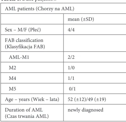
For this experiments, the authors obtained blood samples after a hematological classification procedure from newly diagnosed AML patients of the Department of Hematology of the Medi-cal College of Jagiellonian University (Cracow, Poland). Eight newly-diagnosed patients (4 men and 4 women, mean age: 53.4 ± 15.5) were tested (Table 1).
Wnioski. Pulsacyjne pole elekromagnetyczne wywołuje śmierć komórkową w natywnie proliferujących komórkach pochodzących od pacjentów z AML. Zwiększona podatność proliferujących leukocytów jednojądrzastych na działanie PEMF potencjalnie może być wykorzystana do leczenia AML (Adv Clin Exp Med 2011, 20, 6, 721–727).
Słowa kluczowe: pulsacyjne pole elektromagnetyczne, ostra białaczka mieloidalna, apoptoza, martwica.
Table 1. Patient characteristics
Tabela 1. Dane pacjentów AML patients (Chorzy na AML)
mean (±SD) Sex – M/F (Płeć) 4/4 FAB classification
(Klasyfikacja FAB)
AML-M1 2/2
M2 1/0
M4 1/1
M5 0/1
Age – years (Wiek – lata) 52 (±12)/49 (±19) Duration of AML
Isolation of Peripheral Blood
Mononuclear Cells (PBMCs)
PBMCs were isolated from remnant samples of the heparinized blood of 8 patients, taken for medical reasons by a standard Ficoll-Paque (Phar-macia, Sweden) density gradient procedure. The PBMCs were washed with an RPMI medium (gib-co, USA) and adjusted to 106 cells/ml in an RPMI
culture medium supplemented with L-glutamine + + gentamicin (0.2 M and 50 µg/ml) and 10% hu-man AB serum (both reagents from Sigma-Al-drich, germany) heat inactivated.
PBMC Cultures
PBMCs (0.2 ml aliquots, cell at three densi-ties – 1 × 10⁶ cells/ml, 05 × 106 cells/ml and 0.25 ×
106 cells/ml) were seeded in triplicate into 96-well
culture plates and incubated at 37oC in a
humi-fied atmosphere containing 5% Co2 for 24 h. The
24 h old cultures of PBMCs were then started on stimulation with a pulsed electromagnetic field for the first time.
Magnetic Stimulation
The generator produced a pulsating field of 50 Hz, 45(±5) mT inside the cell culture incubator. The rationale for choosing such a frequency of the PEMF stemmed from the following reasons: the frequency of magnetic stimulation is higher than the range which directly depolarizes autonomic fi-bers, and the heating effect is minimal. The 96-well plate with cells was placed in the generator pocket. The field was applied after the 24h culture three times for 3 hours per stimulation (3 ×) with 24 h intervals between stimulations. In the same way, control cultures unstimulated with PEMF were carried out in parallel.
Cell Death Evaluation
Annexin V-APC labeled (BD Biosciences, USA) was used to quantitatively determine the percentage of cells within the population that were undergoing apoptosis.
7-amino-actinomycin D (7-AAD, BD Biosci-ences, USA) as a standard flow cytometric viability probe was used to distinguish viable from non-vi-able cells. Annexin V-APC positive cells were ana-lyzed as apoptotic and both Annexin V-APC and 7-AAD were either in the end stage of apoptosis or undergoing necrosis analyzed as already dead.
For staining, U 937 cells were washed twice with cold PBS and resuspended in a 1 × binding
buffer (BD Biosciences, USA) at concentration 1 × 10⁶ cells/ml. Then, 100 μl of the solution was
trans-ferred to a 5 ml culture tube and 5 μl of Annexin V-APC and 5 μl of 7-AAD were added. The cells were gently vortexed and incubated in darkness for 15 min at RT. Prior to flow cytometric analy-sis, 400 μl of the 1 × binding buffer was added and the cells were analyzed on a FACS Calibur flow cytometer (Becton Dickinson, San Jose, CA) using Cell-Quest software.
Suggested controls to set up compensation and quadrants encompassed unstained cells, cells stained with Annexin V-APC alone (for FL-4 fluo-rescence) and cells stained with 7-AAD alone (de-tected in FL-3]. A minimum 10,000 events were collected on each sample.
Statistical Analysis
Data were expressed as mean and (±) standard deviation (SD) and compared using the Student’s t-test, with P < 0.05 considered as significantly dif-ferent.
Results
PBMCs isolated from the blood of AML pa-tients were cultivated at different ranges of culture densities – 1 × 10⁶ cells/ml, 0.5 × 10⁶ cells/ml and
0.25 × 10⁶ cells/ml,for 4 days. Starting from the
second day of culture, the cells were stimulated with low energy PEMF (50 Hz, 45 ± 5mT peak) three times for 3 h per a day in 24 hour intervals. After the last stimulation, the cells were stained with Annexin V and 7-AAD dye for flow cytom-etry analysis.
The percentages of apoptotic cells obtained after PEMF exposure were 34.83 (±14.38)%, 33.1 (±6.84)% and 57.51 (±13.28)% for density 1 × 10⁶
cells/ml, 0.5 × 10⁶ cells/ml and 0.25 × 10⁶ cells/ml,
respectively. PBMCs not stimulated with PEMF achieved the level of Annexin V-positive cells 20.12 (±7.46)%, 17.6 (±5.63)% and 35.11 (±13.73) % at their respective densities (Fig. 1).
PEMF induced the strongest apoptosis at 0.5 × 10⁶ cells/ml density of cultivated PBMCs.
As regards necrotic cells measured by 7-AAD binding, similar PEMF-induced effects were ob-tained as with apoptosis, but to a much smaller extent.
The most significant change in the amount of necrotic cells in the PBMC population was found at 1 × 10⁶ cells/ml density and equaled 6.08 (±1.27)%
Fig. 1. Apoptosis of AML-originated PBMCs cultivated in vitro at three different densities during a 4 day culture and simultaneously threefold stimulated with PEMF 3h per day with 24 h intervals; C – PBMCs not treated with PEMF; PEMF – PBMCs exposed to PEMF. Data is expressed as mean (±SD); statistical significance was determined by the Student t-test analysis as *P < 0.05
Ryc. 1. Apoptoza PBMCs pochodzących od pacjentów z AML hodowanych in vitro w 3 różnych gęstościach przez 4 dni i jednocześnie stymulowanych 3-krotnie PEMF przez 3 godz. w odstępach 24 godz.; C – PBMC niestymulow-ane PEMF; PEMF – PBMCs eksponowniestymulow-ane na PEMF. Dniestymulow-ane są wyrażone jako średnia (±SD); istotność statystyczną określono testem t-Studenta jako *P < 0,05
Fig. 2. Necrosis of PBMCs originating from AML patients, cultivated in vitro at three different densities during a 4 day culture and simultaneously threefold stimulated with PEMF 3 h per day with 24 h intervals; C – PBMCs not treated with PEMF; PEMF – PBMCs exposed to PEMF. Data is expressed as mean (± SD); statistical significance was determined by the Student t-test analysis as *P < 0.05
Ryc. 2. Martwica PBMCs pochodzących od pacjentów z AML hodowanych in vitro w 3 różnych gęstościach przez 4 dni i jednocześnie stymulowanych 3-krotnie PEMF przez 3 godz. w odstępach 24 godz.; C – PBMC niestymulowa-ne PEMF; PEMF – PBMCs eksponowaniestymulowa-ne na PEMF. Daniestymulowa-ne są wyrażoniestymulowa-ne jako średnia (± SD); istotność statystyczną określono testem t-Studenta jako *P < 0,05
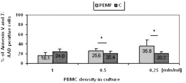
Late apoptotic and necrotic cells measured as a percentage of double stained cells – Annex-in V and 7-AAD positive, upon PEMF exposure, achieved levels 76% higher at the middle density of the culture than in the case of the (PEMF-unex-posed) control group (Fig. 3).
Discussion
Previously, the authors have described that PEMF caused apoptosis of the peripheral blood mononuclear cells (PBMC) originating from pa-tients with Crohn’s disease and the changed se-cretion of cytokines, but not triggered cell death induction in leukocytes from healthy donors.
Thus authors current investigations were aimed at researching the effect of 50 Hz, 45 (±5) mT PEMF interactions exerted on natively-pro-liferating leukocytes isolated from patients with acute myeloid leukemia.
AML is the result of the clonal transformation of hematopoietic precursors through the acquisi-tion of chromosomal rearrangements and mul-tiple gene mutations with the presence of more than 20% of undifferentiated cells [3, 4]. Leukemic blasts from patients with acute myeloid leukemia (AML) have revealed a marked heterogeneity with regard to the presence of acquired gene mutations and changes in gene and microRNA expression [8]. Chromatin-modulating mechanisms are also mediating the transforming activity of key driv-ers of leukemogenesis by aberrant recruitment of corepressors [5, 32]. Many of the identified genetic alterations not only represent independent prog-nosticators, but also may constitute targets for spe-cific therapeutic intervention like electromagnetic stimulation.
The main finding is that the damaging PEMF effect on the AML cell profile depends upon cell density, apoptosis is strongest at the middle but necrosis at the highest density of cultivated PB-MCs. Threefold stimulation of PBMCs with PEMF increased apoptotic cell numbers up to 87% at middle density and the number of necrotic cells at the highest density reached 83% compared to unexposed cells.
Differences in the extent of the PEMF-induced effects on PBMCs from AML patients may depend upon the heterogeneity of the isolated population of leukocytes from the blood (including blasts forms) as well as arrays of molecular changes in the genome responsible for pathomechanisms. [3–8, 32–34]. The results obtained strongly sup-port the hypothesis that PEMF activates the death pathways in proliferating cells. gluck et al. [40] confirmed our results on cell lines by showing that 50 Hz; 3,5 mT pulsed electromagnetic field with fourfold stimulation of the U937 line inhibited the proliferation of cells about 85% compared to their initial at a 1.5 × 10⁵ cells/ml concentration. The
parameters of the field and density of the culture were both similar conditions.
The death induction effect by PEMF exposure of the native proliferating PBMCs isolated from AML patients has been achieved for all densities of the cultured leukocytes but the strongest increase in the percentage of Annexin V and 7-AAD posi-tive cells was obtained at the middle density.
Considering the numerous side effects of con-ventional chemotherapy, the age of patients suf-fering from AML (the majority are elderly) and the complicated pathogenesis, electromagnetic exposure could have clinical implications as an al-ternative/additional, non toxic, noninvasive future prospective therapy of these proliferative diseases [7, 44].
Fig. 3. Percentage of the dead double-stained (late apoptotic and necrotic) PBMCs isolated from AML patients after a 4-day in vitro culture at different densities and threefold PEMF stimulation, 3 h per day with 24h intervals; C – PBMCs not stimulated with PEMF; PEMF – PBMCs stimulated with PEMF. Data is expressed as mean (±SD); statistical significance was determined by the Student t-test analysis as *P < 0.05
Ryc. 3. Liczba komórek apoptotycznych i martwiczych wyrażona jako procent podwójnie barwiących się komórek (aneksyną V i 7-AAD) po 4-dniowej hodowli in vitro prowadzonej przy różnych gęstościach początkowych i 3-krot-nej stymulacji PEMF po 3 godz. na dzień w odstępach 24 godz. C – PBMC niestymulowane PEMF; PEMF – PBMCs eksponowane na PEMF. Dane są wyrażone jako średnia (±SD); istotność statystyczną określono testem t-Studenta jako *P < 0,05
Acknowledgements
References
[1] Kaszuba-Zwoińska J, Ciećko-Michalska I, Madroszkiewicz D, Mach T, Słodowska- Hajduk Z, Rokita E, Zaraska W, Thor P: Magnetic field anti-inflammatory effects in Crohn’s disease depends upon viability and cytokine profile of the immune competent cells. J Physiol Pharmacol 2008, Mar 9[1], 177–8.
[2] Kaszuba-Zwoińska J, Wojcik K, Bereta M, Ziomber A, Pierzchalski P, Rokita E, Marcinkiewicz J, Zaraska W, Thor P: Pulsating electromagnetic field stimulation prevents cell death of puromycin treated U937 cell line. J Physiol Pharmacol 2010, Apr 61, 201–205.
[3] Breccia M, Alimena G: NF-k as a potential therapeutic target in myelodysplastic syndromes and acute myeloid leukemia. Expert opin Ther Targets 2010, 4, 1157–1176. Review.
[4] Vardiman JW, Thiele J, Arber DA et al.: The 2008 revision of the World Health organization (WHo) classifica-tion of myeloid neoplasm’s and acute leukemia: raclassifica-tionale and important changes. Blood 2009, 114, 937–951.
[5] Foran JM: New prognostic markers in acute myeloid leukemia: perspective from the clinic. Hematology Am Soc Hematol Educ Program 2010, 2010, 47–55.
[6] Peters AH, Schwaller J: Epigenetic mechanisms in acute myeloid leukemia. Prog Drug Res 2011, 67, 197–219.
[7] Pollyea DA, Kohrt HE, Medeiros BC: Acute myeloid leukaemia in the elderly: a review. Br J Haematol 2011, 152, 524–542.
[8] Marcucci G, Haferlach T, Döhner H: Molecular genetics of adult acute myeloid leukemia: prognostic and thera-peutic implications. J Clin oncol 2011, Feb 10, 29, 475–486.
[9] Ahuja YR, Vijayashree B, Saran R, Jayashri EL Manoranjani JK, Bhargava SC:In vitro effects of level, low-frequency electromagnetic fields on DNA damage in human leucocytes by comet assay. Indian J Biochem Biophys 1999, 36, 318–322.
[10] Delimaris J, Tsilimigaki S, Messini-Nicolaki N, Ziros E, Piperakis SM: Effects of pulsed electric fields on DNA of human lymphocytes. Cell Biol Toxicol 2006, 22, 409–415.
[11] Hong R, Zhang Y, Liu Y, Weng EQ: Effects of extremely low frequency electromagnetic fields on DNA of tes-ticular cells and sperm chromatin structure in mice. Zhonghua Lao Dong Wei Sheng Zhi Ye Bing Za Zhi 2005, 23, 414–417.
[12] Ivancsits S, Diem E, Pilger A, Rudiger HW, Jahn O: Induction of DNA strand breaks by intermittent exposure to extremely-lowfrequency electromagnetic fields in human diploid fibroblasts. Mutat Res 2002, 519, 1–13.
[13] Ivancsits S, Diem E, Jahn O, Rudiger HW: Age-related effects on induction of DNA strand breaks by intermittent exposure to electromagnetic fields. Mech Aging Dev 2003, 124, 847–850.
[14] Ivancsits S, Pilger A, Diem E, Jahn O, Rudiger HW: Cell typespecific genotoxic effects of intermittent extremely low-frequency electromagnetic fields. Mutat Res 2005, 583, 184–188.
[15] Jajte J, Zmyslony M, Palus J, Dziubaltowska E, Rajkowska E: Protective effect of melatonin against in vitro iron ions and 7mT 50Hz magnetic field-induced DNA damage in rat lymphocytes. Mutat Res 2001, 483, 57–64.
[16] Lai H, Singh NP: Melatonin, N-tert-butyl-alpha-phenylnitrone block 60-Hz magnetic field-induced DNA single and double strand breaks in rat brain cells. J Pineal Res 1997, 22, 152–162.
[17] Lai H, Singh NP: Magnetic-field-induced DNA strand breaks in brain cells of the rat. Environ Health Perspect 2004, 112, 687–694.
[18] Lourencini da Silva R, Albano F, Lopes dos Santos LR, Tavares AD Jr, Felzenszwalb I: The effect of electromag-netic field exposure on the formation of DNA lesions. Redox Rep 2000, 5, 299–301.
[19] Schmitz C, Keller E, Freuding T, Silny J, Korr H: 50-Hz magnetic field exposure influences DNA repair and mitochondrial DNA synthesis of distinct cell types in brain and kidney of adult mice. Acta Neuropathol (Berl) 2004, 107, 257–264.
[20] Svedenstal BM, Johanson KJ, Mild KH: DNA damage induced in brain cells of CBA mice exposed to magnetic fields. In Vivo 1999, 13, 551–552.
[21] Winker R, Ivancsits S, Pilger A, Adlkofer F, Rudiger HW: Chromosomal damage in human diploid fibroblasts by intermittent exposure to extremely low-frequency electromagnetic fields. Mutat Res 2005, 585, 43–49.
[22] Wolf FI, Torsello A, Tedesco B, Fasanella S, Boninsegna A, D’Ascenzo M, Grassi C, Azzena GB, Cittadini A: 50-Hz extremely low frequency electromagnetic fields enhance cell proliferation and DNA damage: possible involvement of a redox mechanism. Biochim Biophys Acta 2005, 1743, 120–129.
[23] Yokus B, Cakir DU, Akdag MZ, Sert C, Mete N: oxidative DNA damage in rats exposed to extremely low fre-quency electro magnetic fields. Free Radic Res 2005, 39, 317–323.
[24] Zmyslony M, Palus J, Jajte J, Dziubaltowska E, Rajkowska E: DNA damage in rat lymphocytes treated in vitro
with iron cations and exposed to 7 mT magnetic fields (static or 50 Hz). Mutat Res 2000, 453, 89–96.
[25] Chow K, Tung WL: Magnetic field exposure enhances DNA repair through the induction of DnaK/J synthesis. FEBS Lett 2000, 478, 133–136.
[26] Robison JG, Pendleton AR, Monson KO, Murray BK, O’Neill KL: Decreased DNA repair rates and protection from heat induced apoptosis mediated by electromagnetic field exposure. Bioelectromagnetics 2002, 23, 106–112.
[27] Lai H, Singh NP: Melatonin and a spin-trap compound block radiofrequency electromagnetic radiation- induced DNA strand breaks in rat brain cells. Bioelectromagnetics 1997, 18, 446–454.
[28] Lai H, Singh NP: Effects of microwaves and a temporally incoherent magnetic field on single and double DNA strand breaks in rat brain cells. Electromag Biol Med 2005, 24, 23–29.
[30] Simków M: Cell type specific redox status is responsible for diverse electromagnetic field effects. Curr Med Chem 2007, 14, 1141–1152.
[31] Phillips JL, Singh NP, Lai H: Electromagnetic fields and DNA damage. Pathophysiology 2009, 16, 2–3, 79–88.
[32] Hyde RK, Liu PP: The role of microRNAs in acute myeloid leukemia. F1000 Biol Rep 2010, 24, 2, 81.
[33] Ebert BL: genetic deletions in AML and MDS. Best Pract Res Clin Haematol 2010, 23, 4, 457–461.
[34] Marcucci G, Mrózek K, Radmacher MD, Garzon R, Bloomfield CD: The prognostic and functional role of microRNAs in acute myeloid leukemia. Blood 2011, 27, 117, 4, 1121–1129.
[35] Berg H: Problems of weak electromagnetic field effects in cell biology. Bioelectrochem Bioenerg 1999, 48, 2, 355–360.
[36] Narita K, Hanakawa K, Kasahara T, Hisamitsu T, Asano K: Induction of apoptotic cell death in human leukemic cell line, HL-60, by extremely low frequency magnetic fields: analysis of the possible electric mechanisms in vitro.
In Vivo 1997, 1 1, 4, 329–335.
[37] Wang X, Zhou A, Liu M, Yu H, Pang L, Zhu M, Wang L, Berg H: Effects of ELF capacitively coupled weak elec-tric fields on metabolism of 6B1 cells. Bioelectrochem Bioenerg 1999, 48, 2, 369–373.
[38] Aldinucci C, Garcia JB, Palmi M: The effect of strong static magnetic field on lymphocytes. Bioelectromagnetics 2003, 24, 109–117.
[39] Wetzel BJ, Nindl G, Vesper DN, Swez JA, Jasti AC, Johnson MT: Electromagnetic field effects: changes in pro-tein phosphorylation in the Jurkat E6.1 cell line. Biomed Sci Instrum 2001, 37, 203–208.
[40] Gluck B, Guntzschel V, Berg H: Inhibition of proliferation of human lymphoma cells U937 by a 50 Hz electro-magnetic field. Cell Mol Biol(Noisy-le-grand) 2001, 47, online Pub: oL115-7.
[41] Arafa HM, Abd-Allah AR, El-Mahdy MA, Ramadan LA, Hamada FM: Immunomodulatory effects of L-carnitine and q10 in mouse spleen exposed to low- frequency high-intensity magnetic field. Toxicology2003, 187, 171– 181.
[42] Johnson MT, Vanscoy-Cornett A, Vesper DN: Electromagnetic fields used clinically to improve bone healing also impact lymphocyte proliferation in vitro. Biomed Sci Instrum 2001, 37, 215–220.
[43] Onodera H, Jin Z, Chida S, Suzuki Y, Tago H, Itoyama Y: Effects of 10-T static magnetic field on human periph-eral blood immune cells. Radiat Res 2003, 159, 775–779.
[44] Johnson MT, Waite LR, Nindl G: Noninvasive treatment of inflammation using electromagnetic fields: current and emerging therapeutic potential. Biomed Sci Instrum 2004, 40, 469–474.
Address for correspondence:
Jolanta Kaszuba-Zwoińska Department of Pathophysiology
Jagiellonian University – Medical College Czysta Street 18
31-121 Cracow Poland
Tel.: +48 12 633 39 47
E-mail: [email protected] Conflict of interest: None declared Received: 7.04.2011