Mei Li*
1, A, C–FMinghai Wei
2, A, B, E, F, Zhenfu Jiang
2, B, Hong Wei
1, C, Weipeng Lu
1, C,
Wei Dou
1, C, Shen Lu
1, A, D–FBRAF Overexpression Induces Rampant
Glioma Proliferation Independent
of Phospho-EGFR Expression**
1 Laboratory Center, The Second Hospital of Dalian Medical University, Dalian, The People’s Republic of China 2 Neurosurgery Department, The Second Hospital of Dalian Medical University, Dalian,
The People’s Republic of China
A – research concept and design; B – collection and/or assembly of data; C – data analysis and interpretation;
D – writing the article; E – critical revision of the article; F – final approval of article; G – other
Abstract
Background. The prognosis for gliomas is still dismal when treated with traditional approaches. Molecular tar-geted therapy has become a trend in treating tumors, including gliomas. However, molecular characteristics vary among different kinds of tumors and ethnic groups.
Objectives. The aim of the research was to study the expression characteristics of key components in the EGFR pathway of gliomas in order to contribute new data for the molecular targeted treatment of gliomas.
Material and Methods. EGFR, KRAS, BRAF, PI3K, phospho-EGFR and Ki67 expression were detected with immunohistochemistry in 82 glioma specimens.
Results. The expression of EGFR was positively correlated with the patients’ age. There were no significant differ-ences in clinicopathological characteristics between gliomas with and without phospho-EGFR expression. EGFR overexpression was significantly correlated with phospho-EGFR expression. In gliomas with EGFR activation, over-expressions of EGFR, BRAF and PI3K were significantly correlated with proliferation. However, in gliomas without phospho-EGFR expression, only BRAF overexpression was significantly related to the proliferation of tumor cells.
Conclusions. BRAF overexpression could be an independent factor causing tumorigenesis in gliomas regardless of phospho-EGFR expression. The molecular characteristics can vary with the increasing age of glioma patients (Adv Clin Exp Med 2014, 23, 6, 893–899).
Key words: glioma, molecular targeted therapy, EGFR protein, BRAF protein, immunohistochemistry.
Adv Clin Exp Med 2014, 23, 6, 893–899 ISSN 1899–5276
ORIGINAL PAPERS
© Copyright by Wroclaw Medical University
Gliomas, which arise from glial cells, are the most common tumor of the central nervous sys-tem. They are characterized by rampant prolifer-ation and recurrence. Despite the availability of traditional treatment options such as surgical re-section, radiotherapy and traditional chemother-apy, the prognosis is often dismal. Recently, mo-lecular targeted drugs have become widely used in treating malignant tumors [1]. It has been prov-en that some drugs can significantly prolong sur-vival and improve the quality of life by specifical-ly blocking signal transduction in tumor cells to
inhibit their growth or suppress new blood vessel formation [2]. Additionally, unlike traditional che-motherapeutic drugs, molecular targeted drugs are more effective in treatment as they focus on spe-cific molecular changes, unrelated to the tumor’s tissue of origin. This makes it possible to treat gli-omas effectively by selecting molecular targeted drugs that address some specific molecular chang-es [3, 4]. Therefore, it has become necchang-essary to bet-ter understand the molecular characbet-teristics of gli-oma, in addition to their histopathological and morphological characteristics.
* Mei Li and Minghai Wei are co-first authors.
Aberrant activition of the EGFR signal trans-duction pathway plays a pivotal role in the tu-morigenesis of many malignant tumors, includ-ing gliomas [5]. Activated EGFR can initiate downstream signaling cascades, primarily in the KRAS/BRAF/ERK/MAPK pathway, and al-so in the PI3K/AKT pathway, thereby pushing cells into the process of proliferation [5, 6]. The EGFR family, which are receptor tyrosine kinas-es, is the key molecular node of signal transduc-tion, in which spontaneous activation is the most common event in malignant tumors. However, the initial agent of activation of the EGFR path-way varies among different tumors. It has been reported that Her-2 amplification or overexpres-sion is often found in breast cancer [7]. EGFR mu-tation in the intracellular domain (mainly in ex-on 19 and 21) is often seen in nex-on-small cell lung cancer (NSCLC), while in gliomas, EGFR mu-tation is mainly focused in the extracellular do-main (EGFRvIII) [8, 9]. Besides EGFR, any oth-er component of its downstream signaling that is activated abnormally can also cause cells to pro-liferate malignantly. KRAS, BRAF and PI3K mu-tations can activate the EGFR pathway, as found in colorectal carcinoma, thyroid carcinoma and melanoma, among others [10–12]. In prostate tu-mors, the PI3K signal pathway can be activated by PI3K overexpression or amplification, but not by its mutation [13].
Interestingly, even in the same kind of tumor, the cause of activation of the EGFR pathway var-ies among ethnic groups. The frequency of KRAS mutations was found to be about 30% and the fre-quency of EGFR mutation was less than 10% in NSCLC patients from Western countries [14, 15]; in contrast, the frequency of KRAS mutation was found to be less than 10%, while the frequency of EGFR mutation was 30–50% in NSCLC patients from East Asia [16, 17]. There is still uncertain-ty about the genetic alterations that occur in the genes of the EGFR signal transduction pathway in gliomas. The authors hope to contribute new data from glioma patients inChina.
It has been reported that EGFR overexpression, amplification and/or mutation (exon 2 to 7 dele-tion) are present in nearly half of all gliomas [18]. Although the abnormal activation of EGFR is an important factor in glioma tumorigenesis, there are other gliomas that proliferate without EGFR activation. Reports on the characteristics of gene alterations in gliomas without EGFR activation are few in number. EGFR activated in any way signals downstream by phosphorylation in the intracellu-lar domain of EGFR. Therefore, phospho-EGFR expression was used in this study as the marker for EGFR activation, and KRAS, BRAF, PI3K, Ki67
expression were detected simultaneously, in or-der to analyze the molecular characteristics of the EGFR signaling pathway in gliomas and better un-derstand its clinical significance.
Material and Methods
The Patients
and Tumor Specimens
In this study, 82 patients who were diagnosed with primary gliomas and treated with surgical re-section at the Second Hospital of Dalian Medical University between 2001 and 2008 were selected continuously. None had radiotherapy or chemo-therapy before surgery. Out of the total, 42 cases were female and 40 male, and they included 5 pa-tients under 18 years old (4 girls and 1 boy). The ages of the patients ranged from 3 to 75 years, with a mean ± SD age of 46.4 ± 17.5 years. There were 64 gliomas occurring in the telencephalon (35 in the right cerebral hemisphere and 31 in the left cerebral hemisphere, including 32 in the frontal lobe, 8 in the parietal lobe, 13 in the temporal lobe, 3 in the occipital lobe, 5 in the frontal and tempo-ral lobe, 1 in the frontal and parietal lobe, 2 in the parietal and occipital lobe), 6 gliomas in the cere-bellum, 1 glioma in the corpus callosum, 1 glioma in the pineal body, 1 glioma in the saddle ar-ea, 2 gliomas in the lateral ventricle, 1 glioma in the ventricle, 1 glioma in the brain stem and 5 gliomas in the spinal cord. After surgical removal the tumor specimens were incubated in 10% neu-tral-buffered formalin and paraffin embedded. All the tumor specimens were diagnosed and classi-fied by 2 licensed pathologists as grades I–IV ac-cording to the WHO classification system, includ-ing 4 grade I gliomas (all pilocytic astrocytomas), 28 grade II gliomas (consisting of 19 astrocytomas, 1 oligodendroglioma, 4 oligoastrocytomas and 4 ependymomas), 24 grade III gliomas (19 anaplas-tic astrocytomas, 4 anaplasanaplas-tic oligoastrocytomas and 1 anaplastic ependemoma), and 26 grade IV gliomas (24 glioblastomas and 2 medulloblasto-mas). All the specimens collected with the approval of the Ethics Committee of the Second Hospital of Dalian Medical University and with the informed consent of the patients before surgery.
Immunohistochemistry
Bioscience), phospho-EGFR (1 : 1600 dilution; clone 1H12; Cell Signaling Technology) and Ki67 (1 : 100 dilution; clone K-2; Life Technologies) were used as primary antibodies. Immunohistochemis-try was performed according to a previously de-scribed protocol [19]. Two cases failed to detect KRAS, and 10 cases failed to detect PI3K. As the negative control, phosphate buffered saline (PBS) was used to replace the primary antibody. The tu-mor cell was considered positive when there were brown granules in the membrane and cytoplasm, detecting EGFR and phospho-EGFR; when brown granules were only in cytoplasm, detecting KRAS, BRAF and PI3K; and when they were in the nu-cleus, detecting Ki67. The immunoreactivity of EGFR, KRAS, BRAF and PI3K was evaluated as negative when < 25% of the tumor cells showed positive; as weakly positive when 25–75% of the tu-mor cells showed positive; and as strongly positive when ≥ 75% of the tumor cells showed positive. For phospho-EGFR, the immunoreactivity was eval-uated as negative when < 25% of the tumor cells showed positive and positive when ≥ 25% of the tumor cells showed positive. For Ki67, the immu-noreactivity was evaluated as negative when < 1% of the tumor cells showed positive; weakly positive when 1–10% of the tumor cells showed positive; and strongly positive when ≥ 10% of the tumor cells showed positive. All the staining results were evaluated by two independent pathologists.
Statistical Analysis
The Pearson chi-square test and Fisher’s exact test were used to compare the differences in pos-itive expression frequencies between groups clas-sified according to their clinicopathological char-acteristics. Spearman’s rank-order correlation test was used to assess the relationships among differ-ent protein expression levels and the relationships of these proteins’ expression levels to the patients’ age and WHO classifications. All statistical analy-ses were performed with SPSS 15.0 software. Sig-nificance was set at the 0.05 level (2-tailed).
Results
EGFR, KRAS, BRAF, PI3K, Ki67
and Phospho-EGFR Expression
in Gliomas
Out of the total of 82 gliomas, positive ex-pression frequencies were 59.8% (49/82) for phos-pho-EGFR; 75.6% (62/82, including 38 weak pos-itives and 24 strong pospos-itives) for EGFR; 63.8% (51/80, including 27 weak positives and 24 strong
positives) for KRAS; 72.0% (59/82, including 18 weak positives and 41 strong positives) for BRAF; 68.1% (49/72, including 16 weak positives and 33 strong positives) for PI3K; 69.5% (57/82, includ-ing 41 weak positives and 16 strong positives) for Ki67 (Fig. 1).
The patients were divided into 3 groups accord-ing to their ages: children under 18 years, adults 18–50 years, and adults over 50 years. The ages of the glioma patients were positive correlated with the grades of the tumors (p = 0.003). The gliomas in children were mainly Grade II (4/5); those in the young adult patients (18–50 years) were main-ly Grades II (17/42) and III (16/42); while those in the older adult patients (over 50 years) were main-ly Grade IV (18/35).
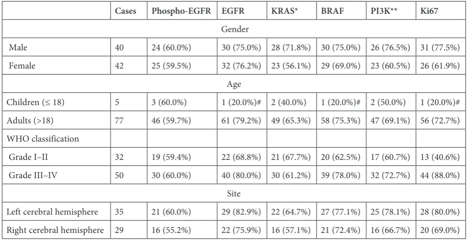
The relationship of all the proteins’ expres-sion to the histopathological characteristics is pre-sented in Table 1. The frequencies of EGFR, BRAF and Ki67 expression were all significantly high-er in adult glioma patients than in child patients (p < 0.05). The expression of EGFR was positive-ly correlated with age levels (p = 0.014, r = 0.270), while those of other proteins were not. The expres-sion of Ki67 was positively correlated with WHO classifications (p < 0.0005, r = 0.458), while those of other proteins were not. None of frequencies of the proteins’ expression differed significantly be-tween genders or bebe-tween tumor sites (left and right cerebral hemisphere).
Clinicopathological
Characteristics of Gliomas
with and Without
Phospho-EGFR Expression
Phospho-EGFR expression was detected as the marker of EGFR activation. All the gliomas were divided into 2 groups: those with phospho-EGFR expression (49 cases) and those without phos-pho-EGFR expression (33 cases). There were no significant differences in the ages and genders of patients or in tumor sites between the two groups (p > 0.05).
Molecular Characteristics
of Gliomas with and Without
Phospho-EGFR Expression
Fig. 1. Protein expression detected by immunohistochemistry, A1 – phospho-EGFR expression was negative; A2 – phospho-EGFR expression was positive. B, C D, E and F were EGFR, KRAS, BRAF, PI3K and Ki67 expression, respec-tively. 1 was weakly positive and 2 was strongly positive (×200)
Table 1. The relationships between clinicopathological characteristics and the expression of phospho-EGFR, EGFR, KRAS, BRAF, PI3K and Ki67
Cases Phospho-EGFR EGFR KRAS* BRAF PI3K** Ki67
Gender
Male 40 24 (60.0%) 30 (75.0%) 28 (71.8%) 30 (75.0%) 26 (76.5%) 31 (77.5%) Female 42 25 (59.5%) 32 (76.2%) 23 (56.1%) 29 (69.0%) 23 (60.5%) 26 (61.9%)
Age
Children (≤ 18) 5 3 (60.0%) 1 (20.0%)# 2 (40.0%) 1 (20.0%)# 2 (50.0%) 1 (20.0%)# Adults (>18) 77 46 (59.7%) 61 (79.2%) 49 (65.3%) 58 (75.3%) 47 (69.1%) 56 (72.7%) WHO classification
Grade I–II 32 19 (59.4%) 22 (68.8%) 21 (67.7%) 20 (62.5%) 17 (60.7%) 13 (40.6%) Grade III–IV 50 30 (60.0%) 40 (80.0%) 30 (61.2%) 39 (78.0%) 32 (72.7%) 44 (88.0%)
Site
Left cerebral hemisphere 35 21 (60.0%) 29 (82.9%) 22 (64.7%) 27 (77.1%) 25 (78.1%) 28 (80.0%) Right cerebral hemisphere 29 16 (55.2%) 22 (75.9%) 16 (57.1%) 21 (72.4%) 16 (66.7%) 20 (69.0%)
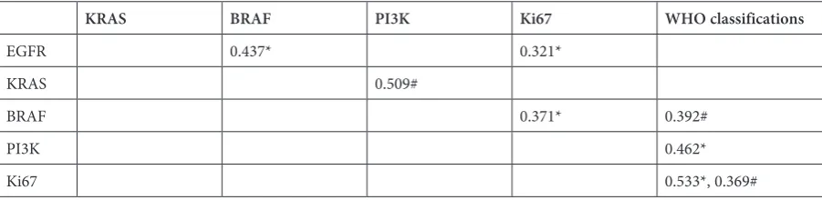
expression, EGFR expression was found to be posi-tively correlated with BRAF expression (p = 0.002). Expression of EGFR and BRAF were both positive-ly correlated with Ki67 expression (p = 0.024 and p = 0.009, respectively); and PI3K expression was positively correlated with tumor grade (p = 0.001) (Table 2). In gliomas without phospho-EGFR ex-pression, KRAS expression was positively correlat-ed with PI3K expression (p = 0.007); and BRAF expression was positively correlated with tumor grade (p = 0.024). However, expression of EGFR, KRAS, BRAF and PI3K were not significantly cor-related with either Ki67 expression or with tumor grade (Table 2).
Discussion
EGFR is a transmembrane receptor tyrosine kinase, and phosphorylation of EGFR will cause EGFR pathway activation. Aberrant EGFR is the key component activating the EGFR signal trans-duction pathway in many malignant tumors [5, 6, 20]. Gene mutation (including mutation in the ex-tracellular and inex-tracellular domain), amplification and overexpression are common ways EGFR is ac-tivated abnormally in gliomas [18, 21]. Therefore, the authors of this study selected monoclonal an-ti-EGFR, which can bind the extracellular domain of EGFR whether it is activated (in any way) or in-activated, to detect EGFR expression, and the anti-phospho-EGFR to indicate the state of EGFR acti-vation. No differences were found between gliomas with and without phospho-EGFR expression in re-lation to clinicopathological characteristics such as the patients’ gender and age or the tumor grade. KRAS, BRAF, PI3K and Ki67 expression did not vary with these characteristics. But it was found that the expression frequency of EGFR was sig-nificantly higher in gliomas with phospho-EGFR expression than in those without its expression.
Moreover, frequency was higher as the patients’ age increased, and it was significantly higher in adult patients as compared to children. It is thought that the distribution of the 4 grades of glioma varies with the patient’s age. Pilocytic astrocytoma (Grade I) and pleomorphic xanthoastrocytoma (Grade II) are commonly found in children, while glioblasto-ma mutiforme (Grade IV) is most often found in patients between 45 to 70 years [22]. In the pres-ent study it was also found that gliomas often had higher grades in older patients. This suggests that aging could be related to a more malignant his-tological type of glioma, which means the tumor cells have a stronger proliferation ability. It is not strange that aging is also related to EGFR overex-pression in gliomas (p = 0.014, r = 0.270). Howev-er, phospho-EGFR expression didn’t increase with age. This indicates that in gliomas from adult pa-tients, EGFR overexpression could not be the rea-son for abnormal EGFR activation.
Gliomas are different from most other malig-nant tumors. Metastasis is not common, but ram-pant proliferation is a main feature of gliomas and is closely related to the prognosis. The higher the grade of the glioma, the stronger its ability to pro-liferate, and the poorer the prognosis of the pa-tient [23, 24]. Ki67 is the most common molecu-lar marker indicating the ability to proliferate in gliomas [25]. The percentage of Ki67 expression in glioma cells reflects the potential ability to pro-liferate and is related to prognosis. Although the frequency of Ki67 expression overlaps in different histological types of gliomas, it is mainly 5–10% in Grade III gliomas and < 5% in Grade II glio-mas [30, 31]. Therefore, in the present study 1% and 10% were selected as the cut-off values, in or-der to clearly indicate the proliferation ability of gliomas. Ki67 expression has usually been shown to be significantly related to the grade of the gli-oma, and this was supported by the results of the present study. In gliomas with phospho-EGFR
Table 2. Relationships among EGFR, KRAS, BRAF, PI3K and Ki67 expressions and WHO classifications in gliomas with and without phospho-EGFR expression
KRAS BRAF PI3K Ki67 WHO classifications
EGFR 0.437* 0.321*
KRAS 0.509#
BRAF 0.371* 0.392#
PI3K 0.462*
Ki67 0.533*, 0.369#
Spearman’s rank-order correlation was used to separately analyze the relationship between protein expression levels and WHO classifications in gliomas with and without phospho-EGFR expression.
expression, EGFR expression was found to be sig-nificantly related to BRAF expression. At the same time, EGFR and BRAF expression were signifi-cantly correlated with separate Ki67 expression in gliomas, and PI3K expression was significant-ly correlated with tumor grade. This suggests that either EGFR, BRAF or PI3K overexpression could activate the EGFR signal pathway independently.
There could be multi-activating components par-ticipating in the rampant proliferation in gliomas with EGFR activation. In this situation, the use of combined modality therapy with EGFR, BRAF and PI3K inhibitors should be considered.
The PI3K/AKT signal pathway is often thought to be peripherally downstream of EGFR/ /KRAS. This pathway also regulates cell prolifer-ation, differentiation and anti-apoptosis [6, 26]. When the EGFR/KRAS/BRAF/ERK pathway is blocked, the PI3K/AKT pathway would be activat-ed, and this was often thought to be a mechanism of resistance to EGFR tyrosine kinase inhibitors in NSCLC [27]. Aberrant PI3K activation would also activate the PI3K/AKT pathway [28]. It has been reported that PI3K mutation and amplifica-tion have been found in some gliomas [29]. In this
study, KRAS expression was found to be signifi-cantly correlated with PI3K expression in gliomas without phospho-EGFR expression. This suggests that the PI3K/AKT pathway participates in the tu-morigenesis of gliomas without EGFR activation. Interestingly, it was BRAF expression, not that of PI3K expression, that was related to the grades of gliomas without phospho-EGFR expression. This suggested that BRAF overexpression could activate the BRAF signal pathway independently and then cause rampant proliferation in gliomas even with-out phospho-EGFR expression.
In conclusion, BRAF overexpression could be an independent factor that causes tumorigenesis in gliomas either with or without phospho-EGFR ex-pression. EGFR overexpression could be a result of increasing age. This should be considered careful-ly with regard to glioma patients treated oncareful-ly with EGFR inhibitors.
A limitation of the current study was that just one method of immunohistochemistry and one monoclonal antibody against phospho-EGFR were used. Further study is required to provide further proof of the possibility of BRAF as a molecular tar-get in treating gliomas.
Acknowledgements. We thank David Blette for his critical reading of the manuscript.
References
[1] Lurje G, Lenz HJ: EGFR signaling and drug discovery. Oncology 2009, 77, 400–410.
[2] Chappell WH, Steelman LS, Long JM, Kempf RC, Abrams SL, Franklin RA, Basecke J, Stivala F, Donia M, Fagone P, Malaponte G, Mazzarino MC, Nicoletti F, Libra M, Maksimovic-Ivanic D, Mijatovic S, Montalto G, Cervello M, Laidler P, Milella M, Tafuri A, Bonati A, Evangelisti C, Cocco L, Martelli AM, McCubrey JA: Ras/ Raf/MEK/ERK and PI3K/PTEN/Akt/mTOR inhibitors: rationale and importance to inhibiting these pathways in human health. Oncotarget 2011, 2, 135–164.
[3] Pollo B: Pathological classification of brain tumors. Q J Nucl Med Mol Imaging 2012, 56, 103–111.
[4] Mao H, Lebrun DG, Yang J, Zhu VF, Li M: Deregulated signaling pathways in glioblastoma multiforme: molecu-lar mechanisms and therapeutic targets. Cancer Invest 2012, 30, 48–56.
[5] Ebi H, Corcoran RB, Singh A, Chen Z, Song Y, Lifshits E, Ryan DP, Meyerhardt JA, Benes C, Settleman J, Wong KK, Cantley LC, Engelman JA: Receptor tyrosine kinases exert dominant control over PI3K signaling in human KRAS mutant colorectal cancers. J Clin Invest 2011, 121, 4311–4321.
[6] Shimizu T, Tolcher AW, Papadopoulos KP, Beeram M, Rasco DW, Smith LS, Gunn S, Smetzer L, Mays TA, Kaiser B, Wick MJ, Alvarez C, Cavazos A, Mangold GL, Patnaik A: The clinical effect of the dual-targeting strat-egy involving PI3K/AKT/mTOR and RAS/MEK/ERK pathways in patients with advanced cancer. Clin Cancer Res 2012, 18, 2316–2325.
[7] Azam M, Qureshi A, Mansoor S: Comparison of estrogen receptors, progesterone receptors and HER-2/neu expression between primary and metastatic breast carcinoma. J Pak Med Assoc 2009, 59, 736–740.
[8] Lynch TJ, Bell DW, Sordella R, Gurubhagavatula S, Okimoto RA, Brannigan BW, Harris PL, Haserlat SM, Supko JG, Haluska FG, Louis DN, Christiani DC, Settleman J, Haber DA: Activating mutations in the epidermal growth factor receptor underlying responsiveness of non-small-cell lung cancer to gefitinib. N Engl J Med 2004, 350, 2129–2139.
[9] Fukai J, Nishio K, Itakura T, Koizumi F: Antitumor activity of cetuximab against malignant glioma cells overex-pressing EGFR deletion mutant variant III. Cancer Sci 2008, 99, 2062–2069.
[10] Davies MA, Stemke-Hale K, Lin E, Tellez C, Deng W, Gopal YN, Woodman SE, Calderone TC, Ju Z, Lazar AJ, Prieto VG, Aldape K, Mills GB, Gershenwald JE: Integrated Molecular and Clinical Analysis of AKT Activation in Metastatic Melanoma. Clin Cancer Res 2009, 15, 7538–7546.
[11] Velho S, Oliveira C, Ferreira A, Ferreira AC, Suriano G, Schwartz S, Jr., Duval A, Carneiro F, Machado JC, Hamelin R, Seruca R: The prevalence of PIK3CA mutations in gastric and colon cancer. Eur J Cancer 2005, 41, 1649–1654.
[13] Agell L, Hernandez S, Salido M, de Muga S, Juanpere N, Arumi-Uria M, Menendez S, Lorenzo M, Lorente JA, Serrano S, Lloreta J: PI3K signaling pathway is activated by PIK3CA mRNA overexpression and copy gain in pros-tate tumors, but PIK3CA, BRAF, KRAS and AKT1 mutations are infrequent events. Mod Pathol 2011, 24, 443–452.
[14] Wu CC, Hsu HY, Liu HP, Chang JW, Chen YT, Hsieh WY, Hsieh JJ, Hsieh MS, Chen YR, Huang SF: Reversed mutation rates of KRAS and EGFR genes in adenocarcinoma of the lung in Taiwan and their implications. Cancer 2008, 113, 3199–3208.
[15] Li M, Zhang Q, Liu L, Liu Z, Zhou L, Wang Z, Yue S, Xiong H, Feng L, Lu S: The different clinical significance of EGFR mutations in exon 19 and 21 in non-small cell lung cancer patients of China. Neoplasma 2011, 58, 74–81.
[16] Rosell R, Moran T, Queralt C, Porta R, Cardenal F, Camps C, Majem M, Lopez-Vivanco G, Isla D, Provencio M, Insa A, Massuti B, Gonzalez-Larriba JL, Paz-Ares L, Bover I, Garcia-Campelo R, Moreno MA, Catot S, Rolfo C, Reguart N, Palmero R, Sanchez JM, Bastus R, Mayo C, Bertran-Alamillo J, Molina MA, Sanchez JJ, Taron M:
Screening for epidermal growth factor receptor mutations in lung cancer. N Engl J Med 2009, 361, 958–967.
[17] Li M, Liu L, Liu Z, Yue S, Zhou L, Zhang Q, Cheng S, Li RW, Smith PN, Lu S: The status of KRAS mutations in patients with non-small cell lung cancers from mainland China. Oncol Rep 2009, 22, 1013–1020.
[18] Yoshimoto K, Dang J, Zhu S, Nathanson D, Huang T, Dumont R, Seligson DB, Yong WH, Xiong Z, Rao N, Winther H, Chakravarti A, Bigner DD, Mellinghoff IK, Horvath S, Cavenee WK, Cloughesy TF, Mischel PS: Development of a real-time RT-PCR assay for detecting EGFRvIII in glioblastoma samples. Clin Cancer Res 2008, 14, 488–493.
[19] Li M, Liu L, Wang Z, Wang L, Liu Z, Xu G, Lu S: Overexpression of hMSH2 and hMLH1 protein in certain gastric cancers and their surrounding mucosae. Oncol Rep 2008, 19, 401–406.
[20] Corcoran RB, Ebi H, Turke AB, Coffee EM, Nishino M, Cogdill AP, Brown RD, Della Pelle P, Dias-Santagata D, Hung KE, Flaherty KT, Piris A, Wargo JA, Settleman J, Mino-Kenudson M, Engelman JA: EGFR-mediated re-activation of MAPK signaling contributes to insensitivity of BRAF mutant colorectal cancers to RAF inhibition with vemurafenib. Cancer Discov 2012, 2, 227–235.
[21] Vivanco I, Robins HI, Rohle D, Campos C, Grommes C, Nghiemphu PL, Kubek S, Oldrini B, Chheda MG, Yannuzzi N, Tao H, Zhu S, Iwanami A, Kuga D, Dang J, Pedraza A, Brennan CW, Heguy A, Liau LM, Lieberman F, Yung WK, Gilbert MR, Reardon DA, Drappatz J, Wen PY, Lamborn KR, Chang SM, Prados MD, Fine HA, Horvath S, Wu N, Lassman AB, DeAngelis LM, Yong WH, Kuhn JG, Mischel PS, Mehta MP, Cloughesy TF, Mellinghoff IK: Differential sensitivity of glioma versus lung cancer-specific EGFR mutations to EGFR kinase inhibitors. Cancer Discov 2012, 2, 458–471.
[22] Yang TH, Kon M, Hung JH, Delisi C: Combinations of newly confirmed Glioma-Associated loci link regions on chromosomes 1 and 9 to increased disease risk. BMC Med Genomics 2011, 4, 63.
[23] Borovski T, Verhoeff JJ, ten Cate R, Cameron K, de Vries NA, van Tellingen O, Richel DJ, van Furth WR, Medema JP, Sprick MR: Tumor microvasculature supports proliferation and expansion of glioma-propagating cells. Int J Cancer 2009, 125, 1222–1230.
[24] Glassmann A, Reichmann K, Scheffler B, Glas M, Veit N, Probstmeier R: Pharmacological targeting of the constitutively activated MEK/MAPK-dependent signaling pathway in glioma cells inhibits cell proliferation and migration. Int J Oncol 2011, 39, 1567–1575.
[25] Paulus W: GFAP, Ki67 and IDH1: perhaps the golden triad of glioma immunohistochemistry. Acta Neuropathol 2009, 118, 603–604.
[26] Rodriguez EF, Scheithauer BW, Giannini C, Rynearson A, Cen L, Hoesley B, Gilmer-Flynn H, Sarkaria JN, Jenkins S, Long J, Rodriguez FJ: PI3K/AKT pathway alterations are associated with clinically aggressive and his-tologically anaplastic subsets of pilocytic astrocytoma. Acta Neuropathol 2011, 121, 407–420.
[27] Kim SM, Kim JS, Kim JH, Yun CO, Kim EM, Kim HK, Solca F, Choi SY, Cho BC: Acquired resistance to cetux-imab is mediated by increased PTEN instability and leads cross-resistance to gefitinib in HCC827 NSCLC cells. Cancer Lett 2010, 296, 150–159.
[28] Huang TT, Sarkaria SM, Cloughesy TF, Mischel PS: Targeted therapy for malignant glioma patients: lessons learned and the road ahead. Neurotherapeutics 2009, 6, 500–512.
[29] Fan QW, Cheng C, Knight ZA, Haas-Kogan D, Stokoe D, James CD, McCormick F, Shokat KM, Weiss WA:
EGFR signals to mTOR through PKC and independently of Akt in glioma. Sci Signal 2009, 2, 4.
[30] Kleihues P, Cavanee WK (eds): World Health Organization classification of tumours. Pathology and genetics – tumors of the nervous system. IARC Press, Lyon 2000, 9–41.
[31] Montine TJ, Vandersteenhoven JJ, Aguzzi A, Boyko OB, Dodge RK, Kerns BJ, Burger PC: Prognostic sig-nificance of ki-67 proliferation index in supratentorial fibrillary astrocytic neoplasms. Neurosurgery 1994, 34, 674–678, 678–679.
Address for correspondence:
Shen Lu
Laboratory Center, The Second Hospital of Dalian Medical University No. 467 Zhongshan Road
Shahekou District, Dalian 116027 Liaoning Province
The People’s Republic of China Tel.: +86 411 84 68 75 54 E-mail: [email protected]
Conflict of interest: None declared