INCREASE OF INTRACELLULAR BAFF IN B CELLS
OF SJÖGREN’S PATIENTS IS NOT AFFECTED BY DECREASE OF BAFFR
Jan Krejsek1, Martina Koláčková1, Irena Lindrová2, Radovan Slezák2, Ctirad Andrýs1
Charles University in Prague, Faculty of Medicine and University Hospital in Hradec Králové, Czech Republic: Department of Clinical Immunology and Allergology1; Charles University in Prague, Faculty of Medicine and University Hospital in Hradec Králové, Czech Republic: Department of Stomatology2
Summary: The presence of a broad spectrum of autoantibodies in Sjögren’s syndrome (SjS) patients is the result of abnormal B-cell regulation that can be at least partially explained by abnormal BAFF/BAFFR regulation. The objective of this study was to determine both membrane and intracellular expression of BAFF/BAFFR in monocytes and B-cells in peripheral blood of 19 primary Sjögren’s syndrome patients and 20 healthy controls using flow cytometry. We also measured sBAFF in serum. Compared to healthy controls, both surface and intracellular expression of BAFF was significantly increased in monocytes and B-cells of SjS patients. Also serum sBAFF level was elevated. Expression of BAFFR on B-cells of SjS patients was surprisingly decreased, but there was no clear increase or decrease within monocytes. Our results indicate that activated monocytes communicate with B-cells via BAFF and BAFFR, so that B-cells are stimulated, but BAFF is also produced to stimulate cells in autocrine way. The decrease of BAFFR expression in SjS patients suggests that there is the mechanism that attempts to take over in order to balance the high level of BAFF.
Keywords: Sjögren’s syndrome; BAFF; BAFFR; intracellular expression; sBAFF
ORIGINAL ARTICLES
Introduction
Sjögren’s syndrome (SjS) is recognized as autoimmune exocrinopathy that primarily affects middle aged females (10, 12). Pathogenesis of SjS is caused by the immune system abnormalities. The exocrine glands are inflamed in SjS patients. Focal lymphocytic infiltrates surrounding the tubular epithelium are the hallmark of this inflammation. B-cell activity is excessive, resulting in wide spectrum of autoantibodies production (19). The pathogenesis of SjS still remains elusive with previous emphasis on the patho-genetic role of T cells. Current data indicates the substantial contribution of B-cells in the immunopathogenesis of SjS (1). B-cells may constitute germinal centers in affected sal-ivary glands. B-cells can act as antigen presenting cells, in this way fuelling abnormal T cells response. B-cells produce not only antibodies, but also several cytokines. Numerous cytokines are indispensable for B-cell functions. Among them, the most important role is deserved to B-cell activating factor belonging to the TNFα family (BAFF). The cytokine BAFF plays a key role in B-cell differen-tiation, survival, and activation (16). BAFF is a ligand for three membrane receptors BCMA (B-cell maturation receptor), TACI (transmembrane activator and calcium modulator and cyclophilin ligand interactor), and BAFFR the latter receptor is being chiefly involved in BAFF
sig-naling (11). Cells of innate immune system, including monocytes, macrophages, neutrophils, and dendritic cells, are the main producers of BAFF. Most recently, other cells that produce BAFF have been identified. These cells are of nonhematopoietic origin and include some types of epithe-lial cells, osteoclast, and astrocytes in CNS (6). BAFFR is widely expressed on B-cells. As for non-B-cells, BAFFR is upregulated on activated T cells and constitutively ex-pressed on Treg subset of T cells (21). It was predicted that excessive production of BAFF shall break B-cell self-tol-erance and allow self-reactive B-cells to survive. BAFF transgenic mice produce autoantibodies, leading to salivary gland destruction, the feature reminiscent of SjS (9). The serum level of soluble sBAFF is elevated in SjS patients. In addition, there is increased expression of BAFF on blood mononuclear cells (1).
with overall effort that shows further roles of BAFF/BAFFR in the immunopathogenesis of SjS. We evaluated the expres-sion of BAFF/BAFFR in peripheral blood monocytes and B-cells of SjS patients using flow cytometry and we also measured sBAFF in serum.
Patients and Controls
19 patients with SjS participated in this study. All of them were diagnosed with primary SjS. All patients fulfilled the European-American consensus group criteria (AECC). Diagnosis of Sjögren’s syndrome was based on routine evaluation of patient’s symptoms and laboratory results (autoantibodies analysis, test of salivary flow rate, Schirm-er’s test, medical records, etc.) performed at the Departments of Dentistry, Immunology and Allergy, Rheumatology and Ophthalmology at the University Hospital in Hradec Králové, Czech Republic. The control group enrolled in this study used no medication and consisted of 21 sex and age-matched individuals from the same geographical area. All participants confirmed their participation in the study by a written consent. The study project was approved by the Ethics Committee of the University Hospital in Hradec Králové, Czech Republic.
Tab. 1: Demographic and clinical data.
Controls (20) Patients (19)
Men/Women (n) 1/19 1/18
Age (years) 54 55
Symptoms of xerophthalmia (n) 0 17
Symptoms of xerostomia (n) 0 19
Dysphagia 0 14
Joint pain (n) 0 16
Thyropathy (n) 0 3
Autoantibodies (n) 0 15
NSAIDs (n) 0 4
Cystosporin A (n) 0 3
Corticosteroids (n) 0 4
Antimalarics (n) 0 1
Materials and Methods
Peripheral venous blood was collected into Vacutainer lithium heparin tubes (BD, UK). Mononuclear cells were separated from Hank’s-diluted blood sample (1:1) layered over Histopaque-1077 (Sigma-Aldrich, CR). Separated cells were washed twice with PBS containing 2% FBS and 1 mM EDTA (Sigma-Aldrich, CR). Cell number was count-ed with hemocytometer and monoclonal antibodies were added accordingly. Monoclonal antibodies against human
BAFF FITC (clone 1D6) and BAFFR PE (clone 8A7) were purchased from eBioscience, (UK) while anti-human CD14 PerCP (clone MEM-15) and anti-CD19 APC (clone LT19) antibodies were bought from Exbio, CR. Following incuba-tion, cells were fixed with 1% paraformaldehyde for 15 min and washed. Prior to intracellular staining with antibodies (anti-BAFF, anti-BAFFR), cells were permeabilized with 0.5% saponin (5 min). Cells were washed again and meas-ured immediately with CellQuest software on FACSCalibur (BD, USA). At least a minimum of 30,000 cells was ac-quired. Instrument setting and compensation was regularly adjusted using Calibrite beads with FACSComp software (BD, USA).
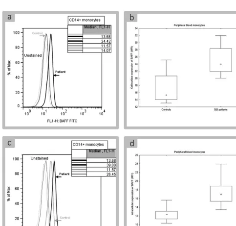
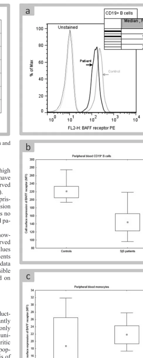
FlowJo software (TreeStar, USA) was used for analysis of data acquired by flow cytometry. Monocytes and B cells were distinguished on the basis of a presence of CD14 and CD19, respectively (Fig. 1). Expression of BAFF and BAFF receptor was characterized by median fluorescence intensity (MFI) that was further used for statistical analysis.
Histogram charts contain examples of flow cytometry data to show the differences between control group and group of patients. Every example that we selected was of value close to the median that described a measured param-eter in a given group.
Vacutainer tubes with a thrombin additive (BD, UK) were used to collect serum samples. These samples were stored at −70 °C. sBAFF was detected using anti-human sBAFF ELISA kit purchased from R&D Systems (USA). Sensitivity of the assay was 2.86 pg/mL. Concentration of sBAFF was measured on MRX microplate reader using Rev-elation software (Dynatech Laboratories, USA).
Statistical analysis
Expression of BAFF and BAFFR, and percentage of cell populations was compared between patients and controls. For this purpose, t-test or non-parametric tests (Man-Whitney test and Kolmogorov-Smirnov test) were used. Differences between clinical and demographic data were tested using χ2 test and Fisher exact test. Before all these comparisons, normality of data sets was tested with Shapiro-Wilks test. Homoscedasticity was determined using Levene’s test. All tests were performed at the 5% significance level.
Plots display median values, quartiles (box), and ranges of non-outlier values (whiskers). If not stated otherwise, any value in the manuscript represents median.
Results
Similar number of mononuclear cells was isolated from patients and controls (data not shown). When compared to control group, there was the prevalence of lymphocytes in isolated mononuclear cells of SjS patients. This differ-ence was statistically significant (83% in control group, 87% in SjS patients, p < 0.05). However, the percentage
of B cells did not significantly differ between both groups (1.27% in controls and 1.71% in patients). The percentage of monocytes was higher in control group (16.2%) than in SjS patients (11.5%, p < 0.05). When monocytes were con-sidered as 100% population, the percentage of monocyte subpopulations (CD14dim, CD14brigh) was not different between controls and patients.
Cell surface expression of BAFF was very low on healthy monocytes, making the increased expression in SjS patient distinctive from control group (Fig. 2A). The difference between both groups was statistically signifi-cant (Fig. 2B). BAFF was also localized intracellularly (Fig. 2C). Similarly to cell surface BAFF, intracellular
BAFF was significantly increased in monocytes of SjS pa-tients (Fig. 2C).
There was no difference in expression of BAFF between CD14dim and CD14bright monocytes, therefore, monocytes were analyzed as a unified population.
As in case of monocytes, cell surface and intracellular expression of BAFF was significantly increased in patients’ B cells (Fig. 3). However, no correlation between increased BAFF on monocytes and increased BAFF on B cells was found. There was also no correlation in intracellular BAFF (data not shown).
Serum concentration of sBAFF displayed a wide range of values in patients and significantly differed from the
Fig. 4: Comparison of sBAFF concentration between controls and SjS patients.
Fig. 5: Differences in cell surface expression of BAFFR. tration in control group (Fig. 4). Patients who expressed high
BAFF either on monocytes or B cells did not necessarily have high concentration of sBAFF; no correlation was observed between sBAFF and cell-bound BAFF (data not shown).
Expression of cell surface BAFFR on B cells was surpris-ingly decreased in SjS patients (Fig. 5A, B). High expression of BAFFR was also found intracellularly, but there was no difference in intracellular BAFFR between controls and pa-tients (data not shown).
Cell surface BAFFR was expressed on monocytes, how-ever, intracellular localization of BAFFR was not observed in this population of cells. Distribution of BAFFR values differed significantly between control group and patients (Fig. 5C). Nonetheless, due to the fact that patients’ data set completely overlapped with controls, it was not possible to conclude that expression of BAFFR was increased on monocytes of SjS patients.
Discussion
documented risk of B-NHL lymphoma development. There are consistent reports that monocytes are contributing to the elevated of BAFF in SjS patients (8). Serum level of BAFF in SjS patients correlates with BAFF mRNA expression (2). Peripheral blood monocytes of SjS patients produce signifi-cantly higher amount of sBAFF in vitro both spontaneously and after stimulation with interferon γ (22). We found sig-nificantly higher presence of BAFF both on the surface and in the intracellular compartment of SjS patients’ monocytes when compared to healthy controls. BAFF can contribute to B-cells physiology via its binding to the three membrane receptors of TNFα receptor family: BCMA, TACI, and BR3. BAFF interacts chiefly with BR3 which is for this reason designated as BAFFR. BAFFR is expressed on B-cells, activated T-cells, and regulatory T-cells. The expression of BAFFR is increasing as B-cells mature (21). We detected low-level expression of this receptor on monocytes of SjS patients, but no significant differences between patients and healthy control were found. In addition, no intracellular BAFFR in monocytes was identified in our study.
It is now firmly established that abnormal production of BAFF in SjS patients is linked to enhanced activity of type I interferon system (11). Increased expression of in-terferons I – regulated genes was described in the salivary glands, in which plasmocytoid dendritic cells were found as a principal source of interferon α. This can represent the link between innate and adaptive immunity in SjS (6). The interferons type I signature in CD14 monocytes along with higher BAFF mRNA expression identifies SjS patients with higher clinical activity of the disease (2). BAFF is a TNFα-like cytokine essential for the maturation and sur-vival of peripheral B-cells. It exists in a membrane bound and secreted form. Cell sources of BAFF include dendrit-ic cells, macrophages, neutrophils, activated T-cells and B-cells (4). We detected significantly enhanced membrane expression of BAFF on CD19 B-cells in blood of patients with SjS compared to healthy controls. The elevated pres-ence of BAFF was also found in cytoplasma of SjS patients B-cells. BAFFR expression on B-cells of our patients was decreased. This is in accord with other study with no dif-ference between naive and memory B-cells. The BAFFR down-regulation correlated with BAFF level and could be reproduced ex vivo by long-term exposure of B-cell to BAFF. The decrease of BAFFR expression on B-cells was greater in patients with extraglandular involvement than in patients with glandular involvement only (15). The decrease in the expression of BAFFR on B-cells could be explained by very likely internalization of BAFF-occupied BAFFR.
Serum level of sBAFF displayed a wide range of val-ues in our patients and was significantly higher compared to controls. Patients who showed high expression of BAFF either on B-cells or monocytes did not necessarily have high concentration of sBAFF.
We observed no correlation between sBAFF and cell bound BAFF. Conflicting results exists for BAFF quantifi-cation. Furthermore, concentration of sBAFF fluctuates due
to changes in inflammatory activity and extent of the disease, as well as the type of treatment (13, 17). In-house assays are used by some research groups. We used commercially available kit to detect sBAFF and our results are comparable with results of Quartuccio et al. (14) who reported upregu-lation of sBAFF in SjS patients and significant association of elevated sBAFF with development of B-cell lymphopro-liferative disease.
In conclusion, BAFF controls B-cell proliferation sur-vival, and maturation (18). BAFF is mainly produced by monocytes and excerts its function trough BAFFR. BAFFR signaling involves canonical and non-canonical NFκB path-ways (5) but there is evidence that BAFFR induces other pathways (7). Our results indicate that activated mono-cytes communicate with B-cells via BAFF and BAFFR, so that B-cells are stimulated, but BAFF is also produced to stimulate cells in autocrine way. The decrease of BAFFR expression in SjS patients suggest that there is a mechanisms that attempts to take over in order to balance the high lev-el of BAFF. Since the levlev-el of BAFF is still high in SjS patient’s B-cells, therapy targeting BAFF is likely to bring benefits to these patients.
Dedication
This paper is dedicated to celebrate 70th anniversary of foundation of Faculty of Medicine in Hradec Králové, Charles University in Prague, Czech Republic.
Acknowledgements
This work was supported by Charles University in Prague, Faculty of Medicine in Hradec Králové, Czech Re-public, project “PRVOUK” P37/10.
The authors declare no conflict of interest.
References
1. Bowman S, Barone F. Biologic treatments in Sjögren’s syndrome. Presse Med 2012; 41: e495–e509.
2. Brkic Z, Maria NI, van Helden-Meeuwsen CG et al. Prevalence of interferon type I signature in CD14 monocytes of patients with Sjögren’s syndrome and associa-tion with disease activity and BAFF gene expression. Ann Rheum Dis 2013; 72(5): 728–735.
3. Cornec D, Devauchelle-Pensec V, Tobón GJ, Pers JO, Jousse-Joulin S, Saraux A. B cells in Sjögren’s syndrome: from pathophysiology to diagnosis and treatment. J Autoimmun 2012; 39(3): 161–167.
4. Daridon C, Devauchelle V, Hutin P et al. Aberrant expression of BAFF by B lymphocytes infiltrating the salivary glands of patients with primary Sjögren’s syndrome. Arthritis Rheum 2007; 56(4): 1134–1144.
5. Gardam S, Brink R. Non-canonical NF-κB signaling initiated by BAFF influences B cell biology at multiple junctures. Front Immunol 2014; 4(509): eCollection 2014.
6. Gottenberg JE, Cagnard N, Lucchesi C et al. Activation of IFN pathways and plasmacytoid dendritic cell recruitment in target organs of primary Sjögren’s syn-drome. Proc Natl Acad Sci USA 2006; 103(8): 2770–2775.
7. Groom J, Mackay F. B cells flying solo. Immunol Cell Biol 2008; 86(1): 40–46. 8. Lavie F, Miceli-Richard C, Ittah M, Sellam J, Gottenberg JE, Mariette X. B-cell
9. Mackay F, Woodcock SA, Lawton P et al. Mice transgenic for BAFF develop lymphocytic disorders along with autoimmune manifestations. J Exp Med 1999; 190 (11): 1697–1710.
10. Moutsopoulos HM. Sjögren’s syndrome: autoimmune epithelitis. Clin Immunol Immunopathol 1994; 72(2): 162–165.
11. Nikolov NP, Illei GG. Pathogenesis of Sjögren’s syndrome. Curr Opin Rheumatol 2009; 21(5): 465–470.
12. Pers JO, Youinou P. Are the B cells cast with the leading part in the Sjögren’s syn-drome scenario? Oral Dis 2014; 20(6): 529–537; doi: 10.1111/odi.12153. 13. Pollard RPE, Abdulahad WH, Vissink A, et al. Serum levels of BAFF, but
not APRIL, are increased after rituximab treatment in patients with primary Sjögren’s syndrome: data from a placebo-controlled clinical trial. Ann Rheum Dis 2013; 72(1): 146–148.
14. Quartuccio L, Salvin S, Fabris M et al. BLyS upregulation in Sjögren’s syndrome as-sociated with lymphoproliferative disorders, higher ESSDAI score and B-cell clonal expansion in the salivary glands. Rheumatology (Oxford) 2013; 52(2): 276–281. 15. Sellam J, Miceli-Richard C, Gottenberg JE et al. Decreased B cell activating factor receptor expression on peripheral lymphocytes associated with increased disease activity in primary Sjögren’s syndrome and systemic lupus erythematosus. Ann Rheum Dis 2007; 66(6): 790–797.
16. Schenider P, MacKay F, Steiner V et al. BAFF, a novel ligand of the tumor necrosis factor family, stimulates B cell growth. J Exp Med 1999; 189(11): 1747–1756.
17. Varin MM, Le Pottier L, Youinou P, Saulep D, Mackay F, Pers JO. B-cell tolerance breakdown in Sjögren’s syndrome: focus on BAFF. Autoimmun Rev 2010; 9(9): 604–608.
18. Vincent FB, Sulep-Easton D, Figgett WA, Fairfax KA, Mackay F. The BAFF/ APRIL system: emerging functions beyond B cell biology and autoimmunity. Cytokine Growth Factor Rev 2013; 24(3): 203–215.
19. Vitali C, Bombardieri S, Jonsson R et al. Classification criteria for Sjögren’s syndrome. A revised version of the European criteria proposed by the American-European Consensus Group. Ann Rheum Dis 2002; 61(6): 554–558. 20. Voulgarelis M, Tzioufas AG. Current aspects of pathogenesis in Sjögren’s
syn-drome. Ther Adv Musculoskelet Dis 2010; 2(6): 325–334.
21. Ye Q, Wang L, Wells AD et al. BAFF binding to T cell-expressed BAFF-R costimulates T cell proliferation and alloresponses. Eur J Immunol 2004; 34(10): 2750–2759.
22. Yoshimoto K, Tanaka M, Kojima M et al. Regulatory mechanisms for the pro-duction of BAFF and IL-6 are impaired in monocytes of patients of primary Sjögren’s syndrome. Arthritis Res Ther 2011; 13(5): R170.
Received: 22/01/2015 Accepted in revised form: 01/04/2015
Corresponding author: