ISSN Print: 2151-1934
DOI: 10.4236/jct.2019.107043 Jul. 11, 2019 518 Journal of Cancer Therapy
Array Comparative Genomic Hybridization as a
Diagnostic Tool in Cancer
Panagiotis Apostolou, Ioannis Papasotiriou
Research & Development Department, Research Genetic Cancer Centre S.A., Florina, Greece
Abstract
The knowledge of the primary origin of tumor is essential in designing an ef-ficient cancer treatment algorithm. Useful diagnostic tools enable determina-tion of primary origin of the tumor; however the majority of them require tissue examination. Recent years, exploration of circulating tumor cells enabled scien-tists to study different parameters using the painless liquid biopsy. The present study aimed to identify whether aCGH might be used as a diagnostic tool in cancer detecting the primary origin of the tumor. Blood was extracted from healthy individuals and cancer samples and CTCs isolated. DNA extracted from the above samples and aCGH experiments followed. The samples were blinded analyzed and then unmasked to calculate specificity and sensitivity of the method. The sensitivity was 94%, the specificity 88%, while the positive pre-diction rate of the primary tumor was 72%. aCGH is a powerful tool in cancer diagnosis and treatment plan with high sensitivity and specificity rates. It can be performed from blood sample, which makes it an appropriate method for every patient, mainly for patients with unknown origin of the primary tumor.
Keywords
Cancer of Unknown Primary Origin, Array Comparative Genomic Hybridization, Cytogenetic, Cancer
1. Introduction
Cancer of unknown origin (CUP) referred to metastatic in which the primary tumor has not been identified. The primary tumor may not be detected or it may disappear after having created the metastasis [1]. CUP accounts for approx-imately 3% - 5% of all malignancies and the median age of diagnosis is 60 years old. The majority of CUP patients (80%) have unfavourable prognosis [2]. Me-thods to detect primary origin include liquid microscopy evaluation, immuno-How to cite this paper: Apostolou, P. and
Papasotiriou, I. (2019) Array Comparative Genomic Hybridization as a Diagnostic Tool in Cancer. Journal of Cancer Therapy, 10, 518-524.
https://doi.org/10.4236/jct.2019.107043
Received: May 13, 2019 Accepted: July 8, 2019 Published: July 11, 2019
Copyright © 2019 by author(s) and Scientific Research Publishing Inc. This work is licensed under the Creative Commons Attribution International License (CC BY 4.0).
DOI: 10.4236/jct.2019.107043 519 Journal of Cancer Therapy
histochemical assays, detection of specific tumor markers and cytogenetics [3]. Chromosomal abnormalities have implications in tumorigenesis since 1960, when the Philadelphia chromosome was linked to chronic myeloid leukemia [4]. The mechanism of triggering cancer is the fused bcr-abl gene, which leads to rapid division of cells [5]. The above rearrangement does not only create a hy-brid gene, but also dysregulate other genes. The abnormal expression of genes might contribute to proliferation or inability of repairing mutations [6]. Not on-ly rearrangements but also deletions and duplications are important in cancer. Several losses in tumor suppressor genes or gains of proto-oncogenes contribute to tumorigenesis. Several cancer types are associated with such abnormalities, like Wilm’s tumor [7] or melanoma [8]. Therefore, whole genome cytogenetic profile could be useful in cancer diagnosis.
Array comparative genomic hybridization (aCGH) is a specific molecular cy-togenetic method that combines CGH and DNA microarrays and enables whole molecular cytogenetic profiling. It is proved to help identify primary tumors, thus contributing to more efficient therapy protocols [9]. In the present study based on liquid biopsy and particularly on Circulating Tumor Cells (CTCs), aCGH technique was used to identify the origin of the tumor based on a blinded genomic DNA analysis. The technique is not only able to discriminate healthy from cancer samples but also to identify the origin of the tumor.
2. Materials and Methods
2.1. Samples
40 ml of blood was collected from 34 patients suffering from different types of cancer, while the same amount was collected from 9 healthy donors. Blood was placed in sterile 50 ml Falcon tubes (4440100, Orange Scientific, Braine-l’Alleud, Belgium) containing 7 ml of 0.02 M EDTA (E0511.0250, Duchefa Biochemie B.V., Haarlem, The Netherlands). Healthy individuals contained five male and four female samples while the patients’ samples included 16 males and 18 fe-males. Distribution of cancer type in patients group was as follows: breast (8), prostate (4), lung (6), colorectal (4), gastrointestinal (5), ovarian (4) and other cancers including haematological, hepatocellular, melanoma, pancreatic, esopha-geal, and urothelial. There were no data concerning the stage of cancer. The major-ity of samples were received from USA (28) and Philippines (5) while there were sent also from Malaysia (2), Germany (1), United Kingdom (1), Canada (1), Poland (2), Israel (2) and South Africa (1). Samples’ age was 61.48 ± 16.04 years old. The samples that were used were collected randomly among cancer and healthy samples. The study was accomplished during January 2018 to May 2019.
2.2. Blood Sample Preparation
col-DOI: 10.4236/jct.2019.107043 520 Journal of Cancer Therapy
lected after centrifugation and washed with phosphate-buffered saline (PBS) (P3813, Sigma-Aldrich). The cells were incubated in lysis buffer (154 mM NH4Cl (31107, Sigma-Aldrich), 10 mM KHCO3 (4854, Merck, Darmstadt, Germany), and 0.1 mM EDTA in deionized water) for 10 min to lyse the erythrocytes. Samples were then centrifuged as above and washed with PBS. Cells from the healthy donor were incubated at 4˚C for 30 min with CD45 magnetic beads (39-CD45-250, Gentaur, Kampenhout, Belgium), whereas those from patients with cancer were incubated with pan-cytokeratin beads (recognizing CK4, CK5, CK6, CK8, CK10, CK13 and CK18) (5c-81714, Gentaur) at 4˚C for 30 min. Following incubation, the samples were placed in a magnetic field to collect microbead-bound cells for pan-cytokeratin and negative selection was performed for CD45 cells, which were washed with PBS. Molecular analysis was performed on the isolated CD45-negative cells (non-cancerous) and the pan-cytokeratin-positive cells (cancerous).
2.3. Array CGH
Genomic DNA was isolated with QIAamp DNA Mini Kit (51306, Qiagen, Hil-den, Germany) from the above cells and then, aCGH protocol with Sureprint G3 human CGH 8 × 60 K platform (G4450A, Agilent, CA, USA) followed according to manufacturer’s instruction. The analysis was performed with Cytogenomics. For each abnormality, the genes that were involved on the appropriate locus were further literately studied to identify potential involvement in any type of cancer. Following gene study, the researcher suggested the type of cancer based only on experimental data. Finally, the diagnosis obtained from experimental data was unmasked and compared with that of physicians. In all reactions there were used reference male and female samples as control.
2.4. Statistical Analysis
The data categorized first in two groups, as cancer and healthy and the positive and negative predictive values (PPV-NPV respectively), sensitivity as well speci-ficity were calculated. A second analysis included only cancer samples and PPV was calculated based on specific type of cancer between blinded experimental data and medical form’s data.
2.5. Ethics Approval
This study was not a clinical trial and did not include any interventions in the patients. All procedures were conducted according to the standards of Safety, Bio-ethics and Validation. The study was reviewed and approved by the Bioethical Committee of the Research Genetic Cancer Centre Group. All patients/donors provided written consent for the use of their samples in the present study. The patients retained the right to withdraw their samples until the date when the sample was received at the laboratory and tested.
3. Results
DOI: 10.4236/jct.2019.107043 521 Journal of Cancer Therapy
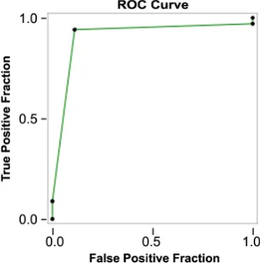
thirty-four while healthy were nine The aCGH results categorized samples based only on raw data and thirty-two cancer samples predicted as cancerous, while only one normal predicted as cancer. On the contrary, eight healthy samples predicted as normal and two cancer samples predicted as normal. Therefore sensitivity and specificity were calculated based on the above data. The analysis of aCGH data revealed sensitivity 94, 11% and specificity 88, 88% between healthy and cancer samples. Data are summarized in Table 1. Followed initially classification, cancer samples were further categorized according to their type. The performer predicted the type of cancer based once again on aCGH raw data, and then samples unmasked and the real type of cancer was compared. Among thirty-four cancer samples twenty-five were categorized correctly while in nine samples the type was not correctly predicted. The positive predictive value was calculated at 73.52% based on the above data. However it is noteworthy that the type of cancer predicted on the nine samples was similar with that one men-tioned in medical form. In Figure 1 are represented the above data.
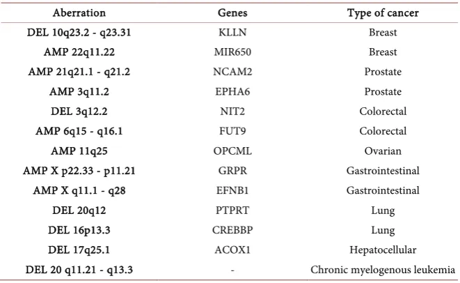
[image:4.595.209.540.393.464.2]As far as the types of abnormalities that were observed there was not specific pattern for each type of cancer. On Table 2 are summarized the most important and common abnormalities observed on specific types of cancer.
Table 1. Summarized results of patients as true positive and false positive. “Tested” refers
to the outcome from aCGH experiments, while “Real” represents the data from patients’ medical forms.
TESTED
REAL
POSITIVE (CANCER) NEGATIVE (HEALTHY) Total
POSITIVE (CANCER) 32 2 34
NEGATIVE (HEALTHY) 1 8 9
Total 33 10
Table 2. The most common aberrations observed in aCGH experiments. The middle
column referred to genes located on that locus and the final column represent the type of cancer correlated with each abnormality.
Aberration Genes Type of cancer
DEL 10q23.2 - q23.31 KLLN Breast
AMP 22q11.22 MIR650 Breast
AMP 21q21.1 - q21.2 NCAM2 Prostate
AMP 3q11.2 EPHA6 Prostate
DEL 3q12.2 NIT2 Colorectal
AMP 6q15 - q16.1 FUT9 Colorectal
AMP 11q25 OPCML Ovarian
AMP X p22.33 - p11.21 GRPR Gastrointestinal
AMP X q11.1 - q28 EFNB1 Gastrointestinal
DEL 20q12 PTPRT Lung
DEL 16p13.3 CREBBP Lung
DEL 17q25.1 ACOX1 Hepatocellular
[image:4.595.209.540.519.722.2]DOI: 10.4236/jct.2019.107043 522 Journal of Cancer Therapy
Figure 1. ROC curve analysis. The present figure represents the true positive rate
(sensi-tivity) together with the false positive rate (1-specificity). The results show a specificity of 88.88% and sensitivity 94.11%. The confidence interval was set to 95% both for sensitivity and specificity.
4. Discussion
The determination of the primary origin of the tumor, as it has been mentioned, requires the examination of tissue; therefore biopsy is essential. Several experi-mental data in CUP demonstrated are characterized by chromosomal instability
[10]. The existence of CTCs may be related with the metastatic ability of CUP as well as with other features, like sensitivity in therapy [11]. The identification of circulating tumor cells (CTCs), a population of cells derived from the primary tumor, gave a new impetus in biopsy, since all the examination require only a few milliliters of blood. CTCs arise from the primary tumor and throw through the blood stream, capable of creating new metastatic tumor [12]. The study of the above cells permitted scientists and physicians for timely and accurate re-sults.
Conventional cytogenetic techniques of karyotyping and FISH (fluorescence in situ hybridization) are widely used to detect abnormalities. Chromosome analysis through karyotyping performed with culture and analysis of lympho-cytes. Although scientists can observe the entire genome, the resolution is very limited. On the contrary, FISH has higher resolution than G-banding karyotyp-ing and there is no requirement for specific stage at the cell cycle. The main dis-advantage is that studied region is the one that is complementary to the probe
[13] [14].
chromo-DOI: 10.4236/jct.2019.107043 523 Journal of Cancer Therapy
somes. The main disadvantage of this method is the detection rate, since it can-not identify abnormalities less than 3 - 5 Mb [15]. The array CGH, which is a combination of microarrays and CGH enables detection of smaller abnormali-ties, depending each time on the probes that are used [16]. Genetic abnormali-ties have been associated with different diseases including cancer. On this field, genomic aberrations might contribute to tumorigenesis and have been con-nected with the progression of the disease. Array CGH is widely used for prenat-al and postnatprenat-al diagnosis of mentprenat-al retardation, development problems, conge-nital malformation syndromes [17], but it can also be applied in human genetic studies [18]. In neonates has improved determination of anomalies with un-known etiology, where G-banding results could not be obtained [19]. Array CGH has been used for tumor classification and prediction of progression and prognosis [20] [21].
According to our experimental data, aCGH as a technique has the potential to discriminate healthy and cancer samples and furthermore to identify the prima-ry origin of tumor with high sensitivity and specificity. Despite the fact that the size was not big enough, the data are encouraging and further experiments need to be performed in order to be used at clinical level.
Conflicts of Interest
The authors declare no conflicts of interest regarding the publication of this pa-per.
References
[1] Varadhachary, G.R. (2007) Carcinoma of Unknown Primary Origin. Gastrointes-tinal Cancer Research, 1, 229-235.
[2] Pavlidis, N. and Pentheroudakis, G. (2012) Cancer of Unknown Primary Site. The Lancet, 379, 1428-1435.https://doi.org/10.1016/S0140-6736(11)61178-1
[3] Varadhachary, G.R., Abbruzzese, J.L. and Lenzi, R. (2004) Diagnostic Strategies for Unknown Primary Cancer. Cancer, 100, 1776-1785.
https://doi.org/10.1002/cncr.20202
[4] Nowell, P.C. and Hungerford, D.A. (1960) Chromosome Studies on Normal and Leukemic Human Leukocytes. Journal of the National Cancer Institute, 25, 85-109. [5] Trask, B.J. (2002) Human Cytogenetics: 46 Chromosomes, 46 Years and Counting.
Nature Reviews Genetics, 3, 769-778.https://doi.org/10.1038/nrg905
[6] van Gent, D.C., Hoeijmakers, J.H. and Kanaar, R. (2001) Chromosomal Stability and the DNA Double-Stranded Break Connection. Nature Reviews Genetics, 2, 196-206.https://doi.org/10.1038/35056049
[7] Mitelman, F., Mertens, F. and Johansson, B. (2005) Prevalence Estimates of Recur-rent Balanced Cytogenetic Aberrations and Gene Fusions in Unselected Patients with Neoplastic Disorders. Genes Chromosomes Cancer, 43, 350-366.
https://doi.org/10.1002/gcc.20212
[8] Cairns, P., Polascik, T.J., Eby, Y., Tokino, K., Califano, J., Merlo, A., et al. (1995) Frequency of Homozygous Deletion at p16/CDKN2 in Primary Human Tumours.
DOI: 10.4236/jct.2019.107043 524 Journal of Cancer Therapy [9] Bertucci, F., Finetti, P., Guille, A., Adelaide, J., Garnier, S., Carbuccia, N., et al.
(2016) Comparative Genomic Analysis of Primary Tumors and Metastases in Breast Cancer. Oncotarget, 7, 27208-27219.https://doi.org/10.18632/oncotarget.8349 [10] Vikesa, J., Moller, A.K., Kaczkowski, B., Borup, R., Winther, O., Henao, R., et al.
(2015) Cancers of Unknown Primary Origin (CUP) Are Characterized by Chromo-somal Instability (CIN) Compared to Metastasis of Know Origin. BMC Cancer, 15,
151.https://doi.org/10.1186/s12885-015-1128-x
[11] Komine, K., Inoue, M., Otsuka, K., Fukuda, K., Nanjo, H. and Shibata, H. (2014) Utility of Measuring Circulating Tumor Cell Counts to Assess the Efficacy of Treatment for Carcinomas of Unknown Primary Origin. Anticancer Research, 34, 3165-3168.
[12] Williams, S.C. (2013) Circulating Tumor Cells. Proceedings of the National Acad-emy of Sciences of the United States of America, 110, 4861.
https://doi.org/10.1073/pnas.1304186110
[13] Manning, M. and Hudgins, L. (2010) Array-Based Technology and Recommenda-tions for Utilization in Medical Genetics Practice for Detection of Chromosomal Abnormalities. Genetics in Medicine, 12, 742-745.
https://doi.org/10.1097/GIM.0b013e3181f8baad
[14] Bridge, J.A. (2008) Advantages and Limitations of Cytogenetic, Molecular Cytoge-netic, and Molecular Diagnostic Testing in Mesenchymal Neoplasms. Journal of Orthopaedic Science, 13, 273-282.https://doi.org/10.1007/s00776-007-1215-1 [15] Kirchhoff, M., Gerdes, T., Maahr, J., Rose, H., Bentz, M., Dohner, H., et al. (1999)
Deletions below 10 Megabasepairs Are Detected in Comparative Genomic Hybridi-zation by Standard Reference Intervals. Genes Chromosomes Cancer, 25, 410-413. https://doi.org/10.1002/(SICI)1098-2264(199908)25:4<410::AID-GCC17>3.0.CO;2-J [16] Shaikh, T.H. (2007) Oligonucleotide Arrays for High-Resolution Analysis of Copy Number Alteration in Mental Retardation/Multiple Congenital Anomalies. Genetics in Medicine, 9, 617-625.https://doi.org/10.1097/GIM.0b013e318148bb81
[17] Kashork, C.D., Theisen, A. and Shaffer, L.G. (2008) Prenatal Diagnosis Using Array CGH. Methods in Molecular Biology, 444, 59-69.
https://doi.org/10.1007/978-1-59745-066-9_5
[18] Oostlander, A.E., Meijer, G.A. and Ylstra, B. (2004) Microarray-Based Comparative Genomic Hybridization and Its Applications in Human Genetics. Clinical Genetics, 66, 488-495.https://doi.org/10.1111/j.1399-0004.2004.00322.x
[19] Emy Dorfman, L., Leite, J.C., Giugliani, R. and Riegel, M. (2015) Microarray-Based Comparative Genomic Hybridization Analysis in Neonates with Congenital Ano-malies: Detection of Chromosomal Imbalances. The Journal of Pediatrics, 91, 59-67. https://doi.org/10.1016/j.jped.2014.05.007
[20] Jong, K., Marchiori, E., van der Vaart, A., Chin, S.F., Carvalho, B., Tijssen, M., et al.
(2007) Cross-Platform Array Comparative Genomic Hybridization Meta-Analysis Separates Hematopoietic and Mesenchymal from Epithelial Tumors. Oncogene, 26, 1499-1506.https://doi.org/10.1038/sj.onc.1209919