A functional MRI study of the influence of practice on component
processes of working memory
Susan M. Landau,
a,* Eric H. Schumacher,
aHugh Garavan,
bT. Jason Druzgal,
a,cand Mark D’Esposito
aaHenry H. Wheeler Brain Imaging Center, Helen Wills Neuroscience Institute, and Department of Psychology, University of California, Berkeley, CA 94720-1650, USA
bDepartment of Psychology, Trinity College, Dublin 2, Ireland
cDepartment of Neuroscience, University of Pennsylvania, Philadelphia, PA, USA
Received 28 July 2003; revised 7 January 2004; accepted 8 January 2004
Previous neuroimaging studies have shown that neural activity changes with task practice. The types of changes reported have been inconsistent, however, and the neural mechanisms involved remain unclear. In this study, we investigated the influence of practice on different component processes of working memory (WM) using a face WM task. Event-related functional magnetic resonance imaging (fMRI) methodology allowed us to examine signal changes from early to late in the scanning session within different task stages (i.e., encoding, delay, retrieval), as well as to determine the influence of different levels of WM load on neural activity. We found practice-related decreases in fMRI signal and effects of memory load occurring primarily during encoding. This suggests that practice improves encoding efficiency, especially at higher memory loads. The decreases in fMRI signal we observed were not accompanied by improved behavioral performance; in fact, error rate increased for high WM load trials, indicating that practice-related changes in activation may occur during a scanning session without behavioral evidence of learning. Our results suggest that practice influences particular component processes of WM differently, and that the efficiency of these processes may not be captured by performance measures alone.
D2004 Elsevier Inc. All rights reserved.
Keywords:Working memory; Neural mechanism; Performance measure
Introduction
The influence of practice on the efficient performance of a task is a fundamental aspect of human behavior. However, the cognitive and neural mechanisms mediating learning and practice are not well understood. Behavioral research suggests that performance of a novel task may initially require a great deal of executive control
(Anderson, 1982; Shiffrin and Schneider, 1984). With practice, however, better learning strategies may be implemented, and the processing required for successful task performance may become more efficient and automatic or proceduralized. In a variety of cognitive and motor tasks, this transition often results in improved task performance, which may be reflected by decreased reaction time and increased accuracy(Poldrack, 2000).
Research on the neural mechanisms underlying this shift from inexperienced to skilled task performance has produced inconsis tent results. Some of the inconsistency may result from the variety of tasks studied. Research on the neural effects of practice has included tasks ranging from motor learning(Karni et al., 1995; Petersen et al., 1998; Tracy et al., 2001)and passive visual perception(Gauthier et al., 1999; Rainer and Miller, 2000; van Turennout et al., 2000)to higher level ones like categorical and probabilistic learning(Poldrack et al., 1999; Seger et al., 2000), mirror reading(Kassubek et al., 2001; Poldrack et al., 1998), artificial grammar learning(Fletcher et al., 1999), and verb generation(Petersen et al., 1998; Raichle et al., 1994). Inconsis tency in the data may also be due to differences in the time course of practice related changes investigated (i.e., short term within session learning vs. long term task learning).
Reflecting these inconsistencies, wide variability has been reported both in the brain regions exhibiting practice related activation changes and in the patterns of activation in those regions. This variability can be characterized in two ways. First, the brain regions engaged by a task remain constant but the magnitude of the activation within these regions either increases
(Gauthier et al., 1999; Iacoboni et al., 1996; Karni et al., 1995)or decreases with practice of the task(Garavan et al., 2000; Jansma et al., 2001). This type of dynamic change in task related brain activity may reflect greater neural efficiency, more precise func tional circuitry(Garavan et al., 2000), or an expanded cortical representation of the task relevant information(Karni et al., 1995). Because both increases and decreases in activation with the development of task expertise have been reported, it is unclear how to interpret these changes with respect to neural efficiency.
Some researchers suggest that information processing efficien cy is associated with brain activation decreases. For example, * Corresponding author. Henry H. Wheeler Brain Imaging Center,
Helen Wills Neuroscience Institute, and Department of Psychology, University of California, 3210 Tolman Hall #1650, Berkeley, CA 94720-1650. Fax: +1-510-642-3004.
Jansma et al. (2001)conducted an fMRI study of practice on a verbal delayed recognition task, in which participants were given a set ofl etters to remember throughout a delay, and were asked to respond to a probe item based on whether or not it was in the initial set. They found that practice on this task resulted in activation decreases in working memory (WM) related areas (e.g., bilateral dorsal prefrontal cortex (PFC), precuneus, and right frontopolar area), presumably due to more efficient WM processing as task performance became automated. Similarly,Milham et al. (2003)
found decreases in anterior cingulate and dorsolateral PFC with practice on a variant of the Stroop task. Finally, decreases in neural activity with learning have been reported in primate electrophys iology experiments as well, supporting the hypothesis that neurons become increasingly efficient with stimulus familiarity and task exposure(Asaad et al., 1998).
In other studies, however, practice has produced increased activity with both short and long term training. For example,
Karni et al. (1995)found an expanded area of primary motor cortex active after several weeks of practice on a motor task.Olesen et al. (2004) found increased prefrontal and parietal activity after 5 weeks of training on a visuospatial working memory task. Simi larly, Iacoboni et al. (1996)reported increased activity in supple mentary motor area and PFC with short term practice on a motor association task. Increases in prefrontal, premotor, and basal ganglia regions have also been associated with motor sequence learning(Grafton et al., 1995), apparently reflecting plasticity within existing connections as the sequence becomes well learned. These studies suggest that improved neural efficiency and task performance may produce activationincreases. Taken together with studies reporting activation decreases with learning, it is clear that there is no simple relationship between practice and neuro physiological processes. Both increases and decreases in activity have been reported with task learning, suggesting that neural efficiency cannot be defined in terms of a monotonic change.
Other factors such as the time course ofl earning and behavioral changes in performance following practice further complicate the interpretation of how brain activation patterns reflect increases in neural efficiency. For example, in motor sequence learning,Karni et al. (1995)found short term decreases in activation in primary motor cortex followed by a long term increase in the extent of activation in the same region after 4 weeks of practice.Hund Georgiadis and von Cramon (1999)found that experienced pianists showed increases in primary motor cortex activation with practice, while non pianists showed decreases(Hund Georgiadis and von Cramon, 1999). Because both groups got faster with practice, it is unlikely that these effects are due to simple performance differ ences. A number of studies have addressed questions about the time course ofl earning by examining early and late phases of learning (e.g.,Tracy et al., 2001). These studies suggest that the time course ofl earning may be an important methodological consideration for examining neural activation associated with a task. Adding additional complexity to the issue, faster performance with practice may be confounded with neural efficiency because changes in neural activity may be due to changes in time spent on task rather than to more efficient processing(Poldrack, 2000).
A second source of variability in the effect of practice on brain activation may arise from a functional reorganization of task related brain regions. In contrast to the studies discussed above, several studies have found evidence for functional reorganization of brain activity with increased practice on a task(Petersen et al., 1998, 1999; Poldrack et al., 1998; Raichle et al., 1994; Sakai et al.,
1998; Shadmehr and Holcomb, 1997; Staines et al., 2002). These studies suggest that a shift in the location of active brain regions reflects a shift in the underlying processes required as task performance becomes skilled. A functional reorganization of activity with task practice may also reflect learning related changes in connectivity between regions over time(Buchel et al., 1999; Fletcher et al., 1999). Or, comparable to the short and long term effects mentioned above, some studies have suggested that reor ganization results from a transition from short term item specific learning to long term task learning(Fletcher et al., 1999; Poldrack and Gabrieli, 2001; Poldrack et al., 1998).
While learning is typically reflected in behavioral performance by greater accuracy or decreased reaction time with practice, some evidence exists showing discordance between behavioral and neural activation data. For example, on motor learning tasks, changes in activation have been observed even when reaction time does not decrease(Shadmehr and Holcomb, 1997; Staines et al., 2002), and on a working memory task, activation increased even when accuracy was at chance(Jaeggi et al., 2003). These findings call into question the assumption that behavioral data are coupled inseparably with neural activity when identifying learning related changes. Instead, the wide variability in practice related behavioral changes and activation patterns suggests that there is a complex relationship between the neural effects of practice and the cognitive processes engaged by a particular task.
To investigate the differential effect of practice on component WM processes, the current experiment examined how encoding, maintenance, and retrieval processes changed with practice on a visual WM task. We examined previously published data from an event related face WM task with varying levels of memory load
(Druzgal and D’Esposito, 2003). In that experiment, Druzgal and D’Esposito compared the influence of working memory load on activity on the temporal pattern and magnitude of activity in the fusiform face area (FFA) and prefrontal cortex (PFC). During encoding and delay periods, activation increased parametrically with memory load in both of these regions, but not in the fusiform object area (FOA). These findings suggest that both regions are sensitive to increasing demand for working memory processes.
Here, we investigate whether practice influences working memory processes differentially during repetitive performance of this face recognition task. The use of an event related fMRI design facilitates the ability to isolate brain activation during separate cognitive processes(Postle et al., 2000; Zarahn et al., 1997a). It also allows us to investigate different types of neural changes with practice (increases, decreases, or functional reorganization of activity) corresponding to these different processes.
We examined changes in activation from early to late in the scanning session in regions engaged by the task to determine the extent to which task related regions were influenced by practice and by memory load. We also examined mapwise activation changes to determine whether any regions (i.e., not necessarily task specific regions) could be identified based on a contrast of activity early versus late in the session and whether these regions were also influenced by memory load.
The day before scanning, participants were familiarized with the behavioral task. The initial learning period of a new task may engage psychological processes in addition to those specific to the task’s performance as participants may have to establish new performance strategies, consolidate the new task’s rules, and familiarize themselves with the task’s procedures. Initial learning periods may also contain a disproportionate number of errors and activation specific to error related processes could therefore con found a contrast between practiced and unpracticed activation patterns. Consequently, while these early learning processes are ofi nterest, they are not the focus of the present study, which instead focused on the effects of practice once a degree of stability on task performance had already been established.
Materials and methods
Participants
Ten right handed participants (age range 22 27) were recruited from the University of Pennsylvania Medical Center. All partic ipants gave written informed consent before participation in the study. Participants were screened against medical, neurological, and psychiatric illnesses, and also for use of prescription medications.
Behavioral task
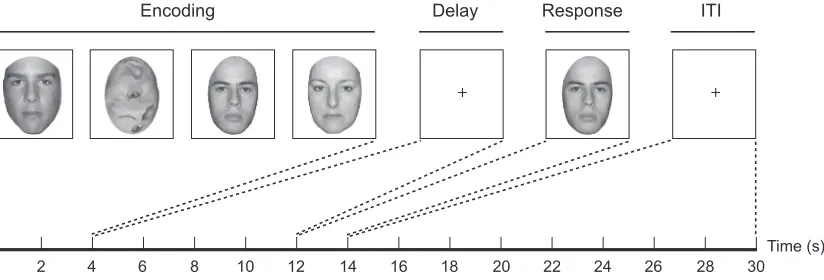
Each trial was composed of (1) a 4 s encoding period, (2) an 8 s delay period, (3) a 2 s retrieval period, and (4) a 16 s inter trial interval. At encoding, each participant saw four serially presented images that were a mixture of gray scale faces and gray scale scrambled faces. Each image was on screen for 1 s and participants had to remember all of the intact faces. Sets of encoding stimuli contained between one and four faces. The order off ace and scrambled face stimulus presentation was randomized so that participants did not know how many faces they would have to remember until the end of the encoding period. Faces were cropped to an ovoid shape so that peripheral face features (such as hair, ears, and neck) were not visible. During the delay period, participants were instructed to fixate on a crosshair at the center of the screen. At retrieval, a single gray scale face appeared and participants were
required to give a motor response indicating whether that face matched one of the faces presented at encoding. There were 12 trials per fMRI run, and 8 runs in the session per participant, for a total of 96 trials per participant. Participants practiced the behavioral task for 30 min on the day before the scanning session. For data analysis purposes, the first three runs in the session (36 trials) were defined asEarly and the last three runs (36 trials) were defined asLate. Trials with one or two faces at encoding were defined asLow Load trials, and trials with three or four faces at encoding were defined as High Loadtrials. Trials were balanced across early and late periods, low and high load conditions, and the number of match/non match motor responses(Fig. 1).
MRI technique
Imaging was carried out on a 1.5T SIGNA scanner (GE Medical Systems) equipped with a prototype fast gradient system for echo planar imaging. A standard radiofrequency (RF) head coil was used with foam padding to restrict head motion comfortably. High resolution sagittal and axial T1 weighted images were obtained in every participant. A gradient echo, echoplanar se quence (TR 2000 ms, TE 50 ms) was used to acquire data sensitive to the blood oxygen level dependent (BOLD) signal. Resolution was 3.75 3.75 mm in plane, and 5 mm between planes (21 axial slices were acquired). Twenty seconds of gradient and RF pulses preceded data acquisition to allow steady state tissue magnetization. Participants viewed a back lit projection screen from within the magnet bore through a mirror mounted on the head coil.
Data preparation
[image:3.595.90.502.539.679.2]Offline data processing was performed using the VoxBo analysis package (http://www.voxbo.org). Initial data preparation proceeded in the following steps: image reconstruction; sinc interpolation in time (to correct for the fMRI slice acquisition sequence); motion correction (six parameter, rigid body, least squares alignment); slice wise motion compensation (to remove spatially coherent signal changes via the application of a partial correlation method to each slice in time(Aguirre et al., 1998a; Zarahn et al., 1997b).
Derivation of the empirical hemodynamic response function (HRF)
The rationale for empirically deriving a HRF is described elsewhere(Aguirre et al., 1998b). An HRF was derived from primary sensorimotor cortex in each participant in the following manner. Before performing the WM task described above, each participant performed a task in which a central white fixation cross changed briefly (130 ms) to a flickering checkerboard every 20 s, cueing the participant to make a bilateral button press. Twenty such events occurred during the 400 s scan.
Statistical analysis
Since fMRI data are temporally auto correlated under the null hypothesis(Zarahn et al., 1997b), statistical analyses were con ducted within the framework of the modified general linear model (GLM) for serially correlated error terms(Worsley and Friston, 1995). A time domain representation of the expected 1/f power structure(Zarahn et al., 1997b)and a notch filter that removed frequencies above the Nyquist frequency and below 0.02 Hz (i.e., the portions of highest power in the noise spectrum) were placed in the convolution matrix(Worsley and Friston, 1995). Due to the event related nature of the behavioral paradigm, the data were not smoothed temporally. The data obtained from the HRF task were modeled by using a Fourier basis set off our sines and four cosines. A partialFtest was used to evaluate the significance of activity in sensorimotor cortical voxels, and an HRF estimate was extracted from the suprathreshold voxels by averaging their time series. This empirical estimate of the HRF was used in subsequent analyses for each participant.
The general linear model (GLM) describes fMRI signal change as a series of amplitude scaled and time shifted covariates or regressors. Each covariate modeled a series of a brief neural events convolved by the participant’s empirical HRF. Covariates were
used to model encoding, delay, and retrieval periods for both high and low levels of memory load (low: 1 or 2 faces, high: 3 or 4 faces) and for both early and late phases of practice in the scanning session (early: runs 1 3, late: runs 6 8). Thus, three trial periods with two load levels for each period and with two phases within the scanning session gave a total of 12 covariates ofi nterest. For each load and phase of the session, encoding modeledt 0 4 s of a trial; delay t 8 12 s; and retrievalt 12 16 s. Additional nuisance covariates were included to model an intercept, trial specific effects, and late encoding/early delay att 4 8 s(Fig. 1).
The nuisance late encoding/early delay covariate was included to avoid contamination of delay related activation by variance that was not captured by the encoding covariate(Zarahn et al., 1997a). Therefore, all delay related activity reported in this analysis arises from the delay covariate and not the nuisance late encoding/early delay covariate.
Our inferential statistics were derived with a multiple regression where the data for each participant were modeled by linear combinations of the covariates ofi nterest. For each participant, parameter estimates were obtained corresponding to the indepen dent variable that modeled each task period for a particular contrast. Specifically, we examined main effects of task and practice for each participant within each trial period. Mapwise and functional region ofi nterest analyses were conducted based on these contrasts (see below).
Mapwise group analyses
[image:4.595.148.460.462.708.2]To perform mapwise group analyses, a whole brain map oft values associated with a contrast ofi nterest (see below) was generated in each participant’s native anatomical space. Thet map for each participant was normalized to the Montreal Neuro logical Institute (MNI) reference brain template using algorithms from SPM96b (http://www.fil.ion.ucl.ac.uk/spm/distrib96.html) by applying a 12 parameter affine transformation with non linear
deformations routine. Normalizedtmaps were then smoothed using a Gaussian smoothing kernel (7.5 mm full width at half maximum). For each voxel, the group oftvalues (one derived from each of the participants) was tested for a significant difference from zero. The upper threshold (corrected) for significance [t(9) > 6.43] was adjusted for multiple comparisons given the smoothness of the map to correct to a mapwiseP < 0.05, two tailed. The lower threshold (uncorrected) for significance [t(9) > 4.30] was set to give a voxelwiseP< 0.0005, two tailed. A minimum cluster size of five contiguous voxels was used. The result was a whole brain map of voxels that showed the contrasts ofi nterest across participants. Ttests were run on the random effectstmaps rather than on the parameter estimate maps becausetmaps are scaled by the noise for each voxel, while parameter estimates are not. Scaling by the individual noise within each voxel can increases the power of the random effects analysis(Postle et al., 2000).
Two contrasts for the mapwise group analyses were conducted at the encoding, delay, and retrieval periods: (1) main effect of task and (2) main effect of practice. To approximately identify the Brodmann’s areas (BA) identified in these analyses, we converted the MNI coordinates to Talairach coordinates. As noted recently by
Brett et al. (2002), the MNI reference brain is not exactly the same size or shape as the brain shown in theTalairach and Tournoux (1988)atlas. Software for converting these coordinates to Talairach coordinates is available online (http://www.mrc cbu.cam.ac.uk/ Imaging/Common/mnispace.shtml). However, we should note that the algorithm provided does not always produce coordinates that correspond to those obtained via visual inspection using the Talairach and Tournoux atlas.
Functionally defined regions ofi nterest (ROI) analyses
Functionally defined ROI analyses were conducted as follows: regions showing a main effect of task and of practice from the mapwise analyses were separately further analyzed using a within subjects repeated measures ANOVA, conducted ata 0.05, with factors Practice (early, late) and Load (high, low). To carry out this analysis, local maxima within the regions showing a main effect of task at the corrected threshold were identified. For each local maximum, voxels that were contiguous with the local maximum and that also reached the uncorrected threshold for significance (P < 0.0005) were considered a functionally defined ROI. From each functionally defined ROI and for each participant, we examined practice and load effects by obtaining mean parameter estimates of all voxels in the region. This yielded four mean parameter estimate values for each participant in each ROI (low load early, low load late, high load early, high load late). We used these parameter estimates in the corresponding ANOVA.
Results
Behavioral data
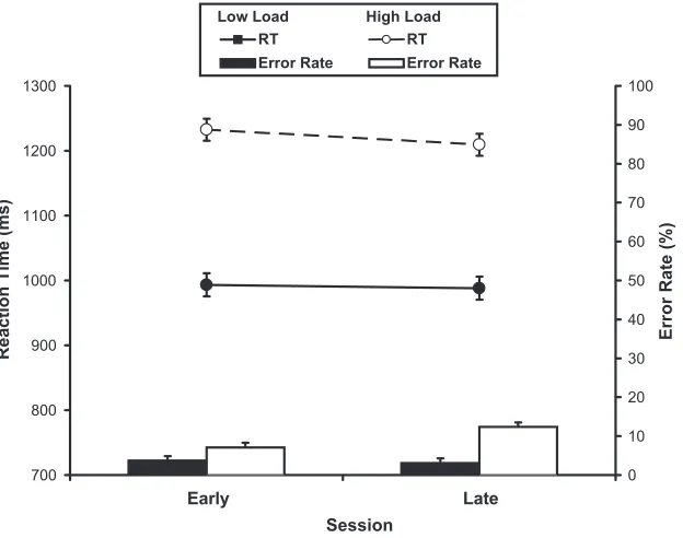
Fig. 2plots the mean reaction times (RTs) and error rates for the four conditions ofi nterest (i.e., high and low memory load and early and late in practice). The data for one participant were lost due to technical difficulties. Memory load had a significant effect on mean RTs, [F(1,8) 24.49, P < 0.01] and mean error rates [F(1,8) 46.39,P< 0.001]. Amount of practice had no significant
[image:5.595.95.496.409.708.2]effect on mean RTs [F(1,8) 0.218, P> 0.6] but did affect mean error rates reliably [F(1,8) 19.97, P< 0.01]. Finally, there was no significant interaction between the effects of memory load and amount of practice on mean RTs, [F(1,8) 0.645, P > 0.6], but these factors had significant interacting effects on mean error rates, [F(1,8) 5.44, P < 0.05]. A post hoc analysis showed that this interaction was driven by the significant increase in mean error rate from early to late for high memory load trials (7 12%) without a corresponding change for the low load trials (4% errors early compared with 3% errors late).
Imaging data mapwise analyses
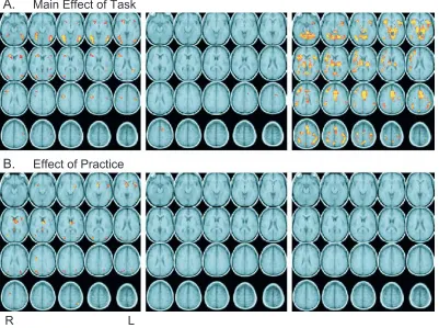
We identified regions showing a main effect of task and an effect of practice (Fig. 3 andTable 1) during each task period (encoding, delay, and retrieval). As shown inFig. 3a, there was a main effect of task across several frontal, temporal, parietal, and extrastriate regions in each of the task periods. The effects of practice within these regions are reported below in the ROI analyses.
The practice effect analysis revealed a network off rontal, parietal, temporal, and subcortical regions during encoding that were more active during early scans as compared to late scans in the session(Fig. 3b). No regions showed a significant practice effects during the delay and retrieval periods. All brain regions illustrated inFig. 3bshowed decreases in activation from early to late in the session. Furthermore, activity in all of these regions was significantly above baseline during the early scans. No region in any task period showed significant activation increases from early to late in the session.
Imaging data functionally defined ROI analyses
To determine whether regions showing a main effect of task were also influenced by practice and load, we carried out subse quent planned contrasts of the regions identified in the mapwise analyses reported above(Fig. 3a). For the main effects ROIs, during encoding, only three regions (in left middle temporal gyrus, left postcentral gyrus, and left insula) didnotalso show effects of practice. During delay and retrieval periods for the main effect ROIs, no regions showed practice effects. Several of these regions during delay and retrieval did, however, show practice by load interactions, with high load trials showing a disproportionate decrease from early to late compared with low load trials. The local maxima for these regions and the type of effect found in each region are listed in theTable 1.
We conducted further planned ANOVAs to examine the regions identified by the mapwise analyses of practice (regions shown in
Fig. 3b, left and middle panels) to determine whether they were also influenced by memory load. During encoding, we found load effects and practice by load interactions in several parietal, occip ital, and subcortical regions. We also found regions showing a practice effect only. Average signal in these regions decreased by 35% from early to late for high load trials and by 30% for low load trials. The local maxima of each region are listed in theTable 1, grouped by the type of effect (practice effect only, practice and load effects, practice by load interaction) found in each region.
The interaction between load and practice on mean error rate in the behavioral data raises the possibility that the changes in activation from early to late were a result of decreased accuracy, and not due to the effects of practice per se. To address this, we
[image:6.595.314.553.102.651.2]carried out the analysis for correct trials only. In other words, the regions we identified as showing practice effects for correct and incorrect trials(Fig. 3b)were re examined using data from correct Table 1
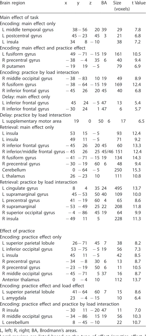
Local maxima of the statistical parametric maps for the main effect of task and effect of practice for all three task periods
Brain region x y z BA Size tValue
(voxels) Main effect of task
Encoding: main effect only
L middle temporal gyrus 38 56 20 39 29 7.8
L postcentral gyrus 45 23 45 3 21 6.8
8 4 3 a
l u s n i
L 10 38 7.2
Encoding: main effect and practice effect
L fusiform gyrus 49 71 15 19 161 10.5
R precentral gyrus 38 4 35 6 40 9.4
R putamen 19 19 5 79 6.9
Encoding: practice by load interaction
R middle occipital gyrus 38 83 10 19 49 8.9
R fusiform gyrus 38 64 15 19 169 12.4
R inferior frontal gyrus 45 26 20 45 40 6.8 Delay: main effect only
L inferior frontal gyrus 45 24 5 47 13 5.4
R inferior frontal gyrus 30 24 1 47 6 5.7
Delay: practice by load interaction
L supplementary motor area 19 0 50 6 17 6.5
Retrieval: main effect only
L insula 53 15 5 93 12.4
L insula 49 11 5 71 9.2
R inferior frontal gyrus 45 26 20 45 60 13.3 R inferior/middle frontal gyrus 45 26 25 45/46 151 12.4
R fusiform gyrus 41 71 15 19 134 14.3
R precentral gyrus 30 19 60 6 48 9.4
Cerebellum 0 64 5 250 15.3
L thalamus 26 23 10 111 10.8
Retrieval: practice by load interaction
L cingulate gyrus 8 4 35 24 495 13.7
L supramarginal gyrus 45 53 50 40 109 10.0
L precentral gyrus 41 19 60 4 65 8.6
R supramarginal 53 49 25 22 208 11.8
R superior occipital gyrus 4 86 45 19 64 9.9
R insula 49 11 5 228 11.3
Effect of practice
Encoding: practice effect only
L superior parietal lobule 26 71 45 7 38 8.2 L inferior occipital gyrus 53 75 5 19 56 7.3
L insula 45 11 5 42 8.5
R precentral gyrus 34 8 30 6 13 8.7
R precentral gyrus 23 19 50 6 11 10.5
R middle occipital gyrus 45 71 5 37 16 8.7
Anterior thalamus 0 4 10 112 13.7
Encoding: practice effect and load effect
L superior parietal lobule 41 64 60 7 15 8.6
L amygdala 23 4 15 10 6.4
Encoding: practice effect and practice by load interaction
R insula 30 11 20 47 11 7.0
R middle occipital gyrus 34 86 15 19 56 10.3
L cerebellum 8 45 10 22 10.7
L, left; R, right; BA, Brodmann’s areas.
trials only. We found no differences between data from correct trials only and data including all trials.
To distinguish between fatigue and practice as explanations for the activation decreases we observed, we carried out the following additional analysis: participants were divided into ‘‘higher error rate’’ and ‘‘lower error rate’’ groups based on a median split of early versus late accuracy scores. The higher error rate group (N 5) had an increase in error rate of 6.11% from early to late, and the lower error rate group (N 4) had an increase of only 2.78%.
If the decreases in activation we observed are due to increased participant fatigue, then the higher error rate group, which likely felt more fatigue, should show greater decreases in activation from early to late. This should be especially true for encoding, which showed the major effect of practice (seeFig. 3b). Therefore, we examined the percent signal change (based on mean parameter estimates) in the regions showing decreases during encoding (left panel inFig. 3b) for both higher error rate and lower error rate participant groups.
Although the groups differed in the overall amount of activa tion, in contrast to what one would expect iff atigue was the cause of the observed activation decreases, the decreases for these two groups were nearly identical (0.041 and 0.039 for the higher and lower error rate groups, respectively; [F(1,124) 0.166,P> 0.68] for the interaction).
Discussion
Our goal in the current experiment was to investigate whether practice influences WM processes differentially during repetitive performance of a face WM task. We investigated practice related activation changes from early to late in a scanning session across low (1 or 2 faces) and high (3 or 4 faces) memory loads for different task periods (encoding, delay, retrieval) of a face working memory (WM) task. We found effects of practice based on two types of analyses across a group of participants: (1) we identified regions in a mapwise analysis showing a main effect of task and further examined these regions for practice (changes in activation from early to late in the scanning session) and load (high load versus low load) effects, (2) we identified regions in a mapwise analysis showing a main effect of practice by comparing early versus late runs in the session and further analyzed these regions for load effects and practice by load interactions. In these analyses, practice was shown to produce activation decreases. We found no evidence for practice related increases in activation or for shifts in the locations of activated regions from early to late in the session. Importantly, the neural effects of practice we identified were independent of evidence ofl earning in the behavioral data. In other words, neural activity changes over time as the task is performed repetitively, but these changes are not dependent on behavioral changes typically associated with skill learning. Thus, our results challenge the idea that dynamic changes in activation are linked to faster or more accurate performance as has been commonly reported in experiments on cognitive and motor skill learning (e. g. Berns et al., 1997; Karni et al., 1998; Poldrack et al., 1998). Instead, the neural activity we observed changes over time, but is independent of task improvement, suggesting that there are important neural changes associated with learning that are not captured in the behavioral data.
These findings have several important implications for exam ining dynamic changes in neural activity as a task is performed
repetitively. Variability of neural activity across the time course of the scanning session (or over several sessions) is a common methodological ‘‘problem’’ for imaging. Unless an fMRI experi ment is specifically designed to examine skill learning, most studies are designed to capture a fixed neural profile associated with a particular cognitive process. To accomplish this, signal change values may be normalized to eliminate within session variance. While some of this variance is due to intrinsic scanner fluctuations, some are due to important learning related changes in neural activity. Thus, the process of discounting this ‘‘noise’’ is likely to also eliminate these experimentally relevant activation changes. Although several fMRI experiments have examined early and late phases ofl earning (e.g.,Muller et al., 2002; Sakai et al., 1998; Toni et al., 2001; Tracy et al., 2001), the present study employs a working memory task rather than a traditional motor or association learning paradigm, and does not involve improved performance associated with skill acquisition.
The influence of working memory load on task related activity has been reported elsewhere(Druzgal and D’Esposito, 2003). That study reported that activation increased parametrically with mem ory load in the prefrontal cortex (PFC) and the fusiform face area (FFA), but not in the fusiform object area (FOA), during encoding and delay periods. Those results suggest that the PFC and FFA are sensitive to increasing demand for WM resources. In the present study, we focus specifically on the influence of practice and the interaction between practice and memory load. To this end, the manipulation ofl oad was used to determine whether activation is changed differentially with practice as WM demands increase.
Practice effects
Of all the task related regions(Fig. 3a)we identified, we found regions showing additional effects of practice during the encoding period only. Interestingly, we found only three encoding related regions that did not show a practice effect, indicating that these regions are involved in encoding processes that are not influenced by task repetition. These regions are in left middle temporal gyrus, left postcentral gyrus, and left insula. Because activity in these regions does not change with practice, these regions are likely involved in encoding processes that are not sensitive to stimulus novelty or to changes in encoding efficiency. Because this practice related activation is primarily left lateralized, one candidate pro cess is subvocal verbalization (e.g., providing names or labeling) of faces during encoding.
The mapwise contrast of early versus late session changes in activation also indicates that the influence of practice is seen primarily during encoding. This finding is consistent with theories of memory that hypothesize that encoding is an active process requiring attention, whereas retrieval processes (e.g., motor re sponse) are more automatic(Naveh Benjamin et al., 2000). There is also evidence from divided attention studies that encoding is more strongly affected by dual task interference than retrieval
(Craik et al., 2000). Because ofi ts higher demands on attention, the active encoding process may more readily benefit from practice. This benefit may reflect improvements in general encod ing processes rather than in processes specific to particular stimuli. That is, practice related decreases are unlikely due to greater familiarity with the memoranda given that similar effects have been observed previously with highly familiar letter stimuli
Regions showing a practice effect without a corresponding main effect of task may reflect mediation of cognitive processes necessary for successful task performance during the initial phases of task learning only. All of these regions showed significant activity above baseline during the early period of the scanning session, indicating that these regions are related to task perfor mance. That these regions do not show significant task related activity across the session highlights a benefit of our practice related analysis. With this approach, we identified additional task related brain regions that traditional analyses fail to detect.
We observed activation decreases in several regions normally associated with motor processing.Murray et al. (2000) have proposed that a basal ganglia prefrontal network, including out puts to the thalamus, is critically involved in the formation of arbitrary visuomotor associations and abstract rules. Thus, the decreases we observed in the anterior thalamus, putamen, and right anterior frontal gyrus may result from subjects’ decreasing need to focus on task rules as the task became well practiced.
It is possible that repetition priming effects contributed to the decreases with practice we found in visual processing regions, as have been reported previously(Martin and van Turennout, 2002). It is unlikely, however, that repetition priming can fully explain the effects we found. First, if the practice related decreases were a result of repeated viewing of the same stimuli, asvan Turennout et al. (2000)found for objects during passive viewing andHenson et al. (2002)found for faces on an implicit task, we would expect to see decreases during the retrieval period as well as during encod ing, particularly in primary visual regions. Second, because the activation decreases occurred throughout the brain, it is likely that multiple systems are affected by practice, not just low level perceptual processing systems.
It is also possible that the null effects of practice during the delay and retrieval periods may reflect a lack of statistical power due to the small sample size and division of the functional data into early and late phases. However, the practice effects we observed during encoding (i.e., activation decreases) suggest that the statis tical power and sample size are adequate for detecting of effects of interest. While small (i.e., undetected) practice effects may have been present during the delay and retrieval periods, the encoding period clearly shows the greatest effects.
The changes in activation we observed in the absence of behavioral evidence ofl earning suggests that, in agreement with some motor learning studies(Shadmehr and Holcomb, 1997; Staines et al., 2002), neural changes with task experience may occur even without faster task performance. Although RT did not change across the session, error rates did increase from early to late on high load trials. This raises the possibility that the practice related changes we found were due to participant fatigue or decreased effort late in the session.
However, the analysis of the ‘‘higher error rate’’ and ‘‘lower error rate’’ groups (see Results) showed no difference between groups in percent signal change from early to late. Rather than implicating fatigue as the major cause of the observed activation decreases, these findings are consistent with our interpretation that the decreases we observed during encoding reflect changes in efficiency with task practice.
Several additional findings are also inconsistent with the observed decreases being related to fatigue rather than practice. First, participants’ reaction times were faster from early to late in the session (by an average of 5 ms for low load trials and 23 ms for high load trials). While these decreases were not significant, this
trend would not be expected if participants were experiencing greater fatigue and distraction during the late trials. Second, decreases in activation were observed for both low load and high load trials, while increased error rate occurred only during high load trials. Third, and most importantly, the main effect of task analysis showed that the extent of overall activity was greater during retrieval (2087 active voxels) than during encoding (626 active voxels). A generalized fatigue or attention effect would be expected to produce decreases in activity from early to late in both task periods, although we report decreases only during encoding.
Furthermore, while other fMRI studies have reported decreases in activation with practice on WM tasks(Garavan et al., 2000; Jansma et al., 2001; Milham et al., 2003), these studies did not isolate separate components of WM and therefore did not contain a ‘‘built in’’ control for general attention effects as in the current experiment. Finally, these experiments showed improvements in performance during the scanning session that accompanied activa tion decreases, suggesting that fatigue and attention effects cannot explain these decreases. Thus, the most relevant existing data, which are consistent with the data we present here, do not support the fatigue explanation.
Practice by load interactions
We conducted a further analysis in the functional ROIs to examine practice related changes across both high and low memory loads to investigate the nature of the effect of practice on specific WM processes. Regions showing effects of practice but not load may be involved in general learning processes: ones not specific to WM. Other regions, affected both by practice and memory load, may be more important for implementing WM specific encoding strategies, such as chunking, which may become optimized once the task has been well learned. Recent WM studies suggest that greater activation for increased memory load is due to additional recruitment of cognitive resources required for success ful maintenance in high WM load conditions(D’Esposito et al., 2000).
The pattern ofi nteraction between practice and memory load on activity within a particular brain region may indicate the nature of the WM processes instantiated there. For example, an interaction in which the load effect disappears with practice may indicate a process that is required when the task is novel but not when it is performed with expertise. Conversely, additive effects between practice and load may indicate a process that becomes more efficient with practice, but is required for the successful perfor mance of the task.
In our practice effect ROIs identified by the mapwise analyses, several regions (right insula, right middle occipital gyrus, and left cerebellum) showed practice by load interactions during encoding, with high load trials showing greater decreases from early to late than low load trials. In the main effect of task ROIs, we found practice by load interactions in several other regions. While we observed these interactions in all three task periods, only the interaction regions during encoding also showed effects of practice. These findings suggest that regions specialized for increasing efficiency with practice may also be influenced by load. Further more, it appears that practice interacts with load in a complex way that may not be apparent when examining the early versus late signal change alone.
indicates that the relationship between information being encoded and corresponding brain activation may change flexibly with task experience. Clearly, this result challenges any simple mapping between brain activation levels and information processing load. In revealing that this mapping is likely to be contingent on the participant’s familiarity with the task, it suggests that objective differences in task demands may be minimized by the efficiency of the encoding mechanisms.
Learning and neural efficiency
The finding that practice related decreases occurred predomi nantly during the encoding period, and not during the delay and retrieval periods, provides strong evidence that practice influences WM processes differentially. There are several theories of encod ing efficiency that offer insight into how subjects may have generated encoding strategies that were implemented more suc cessfully with practice. For example, chunking is a process that involves condensing information into discrete units to be held in WM. Greater knowledge and expertise of a particular domain allows more chunking ofi nformation related to that domain to occur(Gobet et al., 2001; Miller, 1956). In this experiment, faces can be considered visual stimuli for which subjects have special expertise(Gauthier et al., 1999). Subjects may use face expertise to organize individual facial features into chunks more efficiently as the task becomes familiar.
Related to the concept of chunking isEricsson and Kintsch’s (1995) model ofl ong term working memory, in which subjects develop skill in a particular domain by applying strategies for efficient encoding in long term memory. These strategies become refined with repeated exposure to the task and stimuli. Thus, in this experiment, early on in the session, subjects may carry out inefficient encoding procedures ofi ndividual features off aces early on in the session. With task practice, however, subjects may learn to use expert knowledge about faces to develop strategies for encoding relevant features that will allow them to successfully discriminate between target and distractor faces.
Finally, Glassman (1999)has applied the concept of proce dural knowledge to these models and has suggested that proce dural memory (based on task knowledge and skill) serves as a focusing mechanism that serves as a priming mechanism for efficient encoding and for facilitating chunking. He proposes that brain regions involved in this focusing mechanism must be highly multimodal to incorporate complex timing, perceptual, and mne monic systems. Thus, the regions we observed showing encod ing related decreases may be important early in the session for focusing knowledge about facial features, but less important late in the session when encoding strategies have already been established.
We found no brain regions showing activation increases with practice, and no new regions appearing late that were not active early in the session. This suggests that the change with practice was one ofi ncreased neural efficiency for processes (e.g., chunking) that remained constant across the task, and not a shift in strategy with increased practice. The present results of practice induced activation decreases complement two previous investigations of WM practice effects(Garavan et al., 2000; Jansma et al., 2001), both of which observed activation decreases with practice. Our interpretation is also consistent with results from a study that found practice related decreases in activity as participants learned to filter out task irrelevant responses on a version of the Stroop task.
Specifically, Milham et al. (2003) found different profiles of practice related decreases in dorsolateral PFC and anterior cingu late corresponding to decreasing need for attentional control.
In other neuroimaging studies investigating the relationship between neural efficiency and activation, results have been mixed.
Gray et al. (2003) reported a positive correlation between left lateral prefrontal activation and intelligence scores(Gray et al., 2003), suggesting that more prefrontal activity was associated with more efficient processing. Other studies have found that higher intelligence was associated with greater activation decreases with learning on a spatiomotor task(Haier et al., 1992b), and with less spatial dispersion of the source of activity(Jausovec and Jausovec, 2003). Other studies have reported correlations between better task performance and low activation levels(Haier et al., 1992a; Rypma et al., 2002), which is consistent with our findings that activation decreases as processing becomes more efficient.
The absence of behavioral evidence ofl earning raises the question of whether neural processingdidin fact become more efficient. It should be noted that response time measurements on trials such as these provide only indirect and uncertain informa tion about the efficiency of the preceding encoding processes. However, the neural efficiency hypothesis is supported by our findings with respect to memory load. While the behavioral data did not show evidence ofi mproved performance, it did show an effect ofl oad, with longer reaction times on high load trials than on low load trials. Correspondingly, brain activity was greater for high than low load trials in several regions showing practice effects and practice by load interactions. Consequently, that the more demanding task condition (high load) produced greater activation levels suggests that the ease of processing is inversely related to activation levels. The observation that the load effect dissipated with practice (i.e., activation on high and low load trials became indistinguishable with practice) suggests that pro cessing demands did decrease and that encoding processes became more efficient.
The suggestion that neural activation may change from early to late in a scanning session, even in the absence of behavioral evidence ofl earning, has implications for data analysis in any functional neuroimaging study involving repetition of a task or specific stimuli. The effects of an hour or two of practice with a task or specific stimuli typically are not considered in most experiments. Trials at the beginning of the scanning session, when the task and stimuli are unfamiliar, are averaged together with trials at the end of the session, when the task and stimuli are performed with greater ease and flexibility. By revealing dramatic changes from early to late in practice, this experiment shows that practice related variance may be of great interest even when the task does not require learning skills or associations. We suggest that the neural profile of most tasks is not stable but changes as a function of time and participant experience over the course of scanning. Future imaging experiments may benefit from viewing neural activation as a dynamic, rather than static, phenomenon. One way to address this issue is to modify standard fMRI data analysis procedures that eliminate within session signal variance in a way that accounts for changes with time and task expertise.
and stable way. Our data suggest, instead, that neural activation corresponding to repetitive task performance is both spatially and temporally dynamic.
Acknowledgments
This research was supported by a pre doctoral fellowship from the National Science Foundation and a grant from the National Institute of Health.
References
Aguirre, G.K., Zarahn, E., D’Esposito, M., 1998a. The inferential impact of global signal covariates in functional neuroimaging analyses. Neuro-Image 8, 302 306.
Aguirre, G.K., Zarahn, E., D’Esposito, M., 1998b. The variability of hu-man, BOLD hemodynamic responses. NeuroImage 8, 360 369. Anderson, J.R., 1982. Acquisition of cognitive skill. Psychol. Rev. 89,
369 406.
Asaad, W.F., Rainer, G., Miller, E.K., 1998. Neural activity in the primate prefrontal cortex during associative learning. Neuron 21, 1399 1407. Berns, G.S., Cohen, J.D., Mintun, M.A., 1997. Brain regions responsive to
novelty in the absence of awareness. Science 276, 1272 1275. Brett, M., Johnsrude, I.S., Owen, A.M., 2002. The problem off unctional
localization in the human brain. Nat. Rev., Neurosci. 3, 243 249. Buchel, C., Coull, J.T., Friston, K.J., 1999. The predictive value of changes
in effective connectivity for human learning. Science 283, 1538 1541. Craik, F.I., Naveh-Benjamin, M., Ishaik, G., Anderson, N.D., 2000.
Divid-ed attention during encoding and retrieval: differential control effects? J. Exp. Psychol. Learn Mem. Cogn. 26, 1744 1749.
D’Esposito, M., Postle, B.R., Rypma, B., 2000. Prefrontal cortical contri-butions to working memory: evidence from event-related fMRI studies. Exp. Brain Res. 133, 3 11.
Druzgal, T.J., D’Esposito, M., 2003. Dissecting contributions of prefrontal cortex and fusiform face area to face working memory. J. Cogn. Neuro-sci. 15, 771 784.
Ericsson, K.A., Kintsch, W., 1995. Long-term working memory. Psychol. Rev. 102, 211 245.
Fletcher, P., Buchel, C., Josephs, O., Friston, K., Dolan, R., 1999. Learn-ing-related neuronal responses in prefrontal cortex studied with func-tional neuroimaging. Cereb. Cortex 9, 168 178.
Garavan, H., Kelley, D., Rosen, A., Rao, S.M., Stein, E.A., 2000. Practice-related functional activation changes in a working memory task. Microsc. Res. Tech. 51, 54 63.
Gauthier, I., Tarr, M.J., Anderson, A.W., Skudlarski, P., Gore, J.C., 1999. Activation of the middle fusiform ‘face area’ increases with expertise in recognizing novel objects. Nat. Neurosci. 2, 568 573.
Glassman, R.B., 1999. A working memory ‘‘theory of relativity’’: elasticity in temporal, spatial, and modality dimensions conserves item capacity in radial maze, verbal tasks, and other cognition. Brain Res. Bull. 48, 475 489.
Gobet, F., Lane, P.C., Croker, S., Cheng, P.C., Jones, G., Oliver, I., Pine, J.M., 2001. Chunking mechanisms in human learning. Trends Cogn. Sci. 5, 236 243.
Grafton, S.T., Hazeltine, E., Ivry, R., 1995. Functional mapping of se-quence learning in normal humans. J. Cogn. Neurosci. 7, 497 510. Gray, J.R., Chabris, C.F., Braver, T.S., 2003. Neural mechanisms of general
fluid intelligence. Nat. Neurosci. 6, 316 322.
Haier, R.J., Siegel Jr., B.V., MacLachlan, A., Soderling, E., Lottenberg, S., Buchsbaum, M.S., 1992a. Regional glucose metabolic changes after learning a complex visuospatial/motor task: a positron emission tomo-graphic study. Brain Res. 570, 134 143.
Haier, R.J., Siegel Jr., B.V., Tang, C., Abel, L., Buchsbaum, M.S., 1992b.
Intelligence and changes in regional cerebral glucose metabolic rate following learning. Intelligence 16, 415 426.
Henson, R.N., Shallice, T., Gorno-Tempini, M.L., Dolan, R.J., 2002. Face repetition effects in implicit and explicit memory tests as measured by fMRI. Cereb. Cortex 12, 178 186.
Hund-Georgiadis, M., von Cramon, D.Y., 1999. Motor-learning-related changes in piano players and non-musicians revealed by functional magnetic-resonance signals. Exp. Brain Res. 125, 417 425.
Iacoboni, M., Woods, R.P., Mazziotta, J.C., 1996. Brain behavior relation-ships: evidence from practice effects in spatial stimulus response com-patibility. J. Neurophysiol. 76, 321 331.
Jaeggi, S.M., Seewer, R., Nirkko, A.C., Eckstein, D., Schroth, G., Groner, R., Gutbrod, K., 2003. Does excessive memory load attenuate activa-tion in the prefrontal cortex? Load-dependent processing in single and dual tasks: functional magnetic resonance imaging study. NeuroImage 19, 210 225.
Jansma, J.M., Ramsey, N.F., Slagter, H.A., Kahn, R.S., 2001. Functional anatomical correlates of controlled and automatic processing. J. Cogn. Neurosci. 13, 730 743.
Jausovec, N., Jausovec, K., 2003. Spatiotemporal brain activity related to intelligence: a low resolution brain electromagnetic tomography study. Brain Res. Cogn. Brain Res. 16, 267 272.
Karni, A., Meyer, G., Jezzard, P., Adams, M.M., Turner, R., Ungerleider, L.G., 1995. Functional MRI evidence for adult motor cortex plasticity during motor skill learning. Nature 377, 155 158.
Karni, A., Meyer, G., Rey-Hipolito, C., Jezzard, P., Adams, M.M., Turner, R., Ungerleider, L.G., 1998. The acquisition of skilled motor perfor-mance: fast and slow experience-driven changes in primary motor cor-tex. Proc. Natl. Acad. Sci. U. S. A. 95, 861 868.
Kassubek, J., Schmidtke, K., Kimmig, H., Lucking, C.H., Greenlee, M.W., 2001. Changes in cortical activation during mirror reading before and after training: an fMRI study of procedural learning. Brain Res. Cogn. Brain Res. 10, 207 217.
Martin, A., van Turennout, M., 2002. Searching for the neural correlates of object priming. In: Squire, L.R., Schacter, D.L. (Eds.), Neuropsychol-ogy of Memory, vol. 3. Guilford Press, New York, pp. 239 247. Milham, M.P., Banich, M.T., Claus, E.D., Cohen, N.J., 2003.
Practice-related effects demonstrate complementary roles of anterior cingulate and prefrontal cortices in attentional control small star, filled. Neuro-Image 18, 483 493.
Miller, G.A., 1956. The magical number seven, plus or minus two: some limits on our capacity for processing information. Psychol. Rev. 63, 81 97.
Muller, R.A., Kleinhans, N., Pierce, K., Kemmotsu, N., Courchesne, E., 2002. Functional MRI of motor sequence acquisition: effects ofl earning stage and performance. Brain Res. Cogn. Brain Res. 14, 277 293. Murray, E.A., Bussey, T.J., Wise, S.P., 2000. Role of prefrontal cortex in
a network for arbitrary visuomotor mapping. Exp. Brain Res. 133, 114 129.
Naveh-Benjamin, M., Craik, F.I., Gavrilescu, D., Anderson, N.D., 2000. Asymmetry between encoding and retrieval processes: evidence from divided attention and a calibration analysis. Mem. Cogn. 28, 965 976.
Olesen, P.J., Westerberg, H., Klingberg, T., 2004. Increased prefrontal and parietal activity after training of working memory. Nat. Neurosci. 7, 75 79.
Petersen, S.E., van Mier, H., Fiez, J.A., Raichle, M.E., 1998. The effects of practice on the functional anatomy of task performance. Proc. Natl. Acad. Sci. U. S. A. 95, 853 860.
Petersson, K.M., Elfgren, C., Ingvar, M., 1999. Dynamic changes in the functional anatomy of the human brain during recall of abstract designs related to practice. Neuropsychologia 37, 567 587.
Poldrack, R.A., 2000. Imaging brain plasticity: conceptual and methodo-logical issues A theoretical review. NeuroImage 12, 1 13.
Poldrack, R.A., Desmond, J.E., Glover, G.H., Gabrieli, J.D., 1998. The neural basis of visual skill learning: an fMRI study of mirror reading. Cereb. Cortex 8, 1 10.
Poldrack, R.A., Prabhakaran, V., Seger, C.A., Gabrieli, J.D., 1999. Striatal activation during acquisition of a cognitive skill. Neuropsychology 13, 564 574.
Postle, B.R., Zarahn, E., D’Esposito, M., 2000. Using event-related fMRI to assess delay-period activity during performance of spatial and nonspatial working memory tasks. Brain Res. Brain Res. Protoc. 5, 57 66.
Raichle, M.E., Fiez, J.A., Videen, T.O., MacLeod, A.M., Pardo, J.V., Fox, P.T., Petersen, S.E., 1994. Practice-related changes in human brain func-tional anatomy during nonmotor learning. Cereb. Cortex 4, 8 26. Rainer, G., Miller, E.K., 2000. Effects of visual experience on the
repre-sentation of objects in the prefrontal cortex. Neuron 27, 179 189. Rypma, B., Berger, J.S., D’Esposito, M., 2002. The influence of
working-memory demand and subject performance on prefrontal cortical activity. J. Cogn. Neurosci. 14, 721 731.
Sakai, K., Hikosaka, O., Miyauchi, S., Takino, R., Sasaki, Y., Putz, B., 1998. Transition of brain activation from frontal to parietal areas in visuomotor sequence learning. J. Neurosci. 18, 1827 1840.
Seger, C.A., Poldrack, R.A., Prabhakaran, V., Zhao, M., Glover, G.H., Gabrieli, J.D., 2000. Hemispheric asymmetries and individual differ-ences in visual concept learning as measured by functional MRI. Neuro-psychologia 38, 1316 1324.
Shadmehr, R., Holcomb, H.H., 1997. Neural correlates of motor memory consolidation. Science 277, 821 825.
Shiffrin, R.M., Schneider, W., 1984. Automatic and controlled processing revisited. Psychol. Rev. 91, 269 276.
Staines, W.R., Padilla, M., Knight, R.T., 2002. Frontal-parietal event-relat-ed potential changes associatevent-relat-ed with practising a novel visuomotor task. Brain Res. Cogn. Brain Res. 13, 195 202.
Talairach, J., Tournoux, P., 1988. Co-planar Stereotactic Atlas of the Hu-man Brain. Thieme, New York, NY.
Toni, I., Ramnani, N., Josephs, O., Ashburner, J., Passingham, R.E., 2001. Learning arbitrary visuomotor associations: temporal dynamic of brain activity. NeuroImage 14, 1048 1057.
Tracy, J.I., Faro, S.S., Mohammed, F., Pinus, A., Christensen, H., Burk-land, D., 2001. A comparison of ‘Early’ and ‘Late’ stage brain activa-tion during brief practice of a simple motor task. Brain Res. Cogn. Brain Res. 10, 303 316.
van Turennout, M., Ellmore, T., Martin, A., 2000. Long-lasting cortical plasticity in the object naming system. Nat. Neurosci. 3, 1329 1334. Worsley, K.J., Friston, K.J., 1995. Analysis off MRI time-series revisited
Again. NeuroImage 2, 173 181.
Zarahn, E., Aguirre, G., D’Esposito, M., 1997a. A trial-based experimental design for fMRI. NeuroImage 6, 122 138.