Original Article
JARID1B modulates breast cancer
cell apoptosis by regulating p53 expression
Jinghao Wang1*, Xiaosong Wu1*, Jingxin Wang2, Luchen Shan3
1Department of Pharmacy, The First Affiliated Hospital, Jinan University, Guangzhou, China; 2Department of
Pharmacy, Hongqi Hospital of Mudanjiang Medical University, Heilongjiang, China; 3Institute of New Drug
Research and Guangzhou Key Laboratory of Innovative Chemical Drug Research in Cardio-cerebrovascular Diseases, Jinan University College of Pharmacy, Guangzhou, China. *Equal contributors.
Received May 15, 2018; Accepted July 10, 2018; Epub September 1, 2018; Published September 15, 2018
Abstract: Jumonji AT-rich interactive domain 1B (JARID1B) has been implicated in breast cancer progression, but its role in apoptosis has not been explored. The present study was designed to investigate the effect of JARID1B on breast cancer cell apoptosis. Apoptosis was assessed by TUNEL, flow cytometry and caspase-3 activity. JARID1B and p53 expression were examined by Western blot. Cell viability was measured by an MTT assay. We found that JARID1B is overexpressed in the breast cancer cell line and in breast cancer tissues. Upregulated expression of JARID1B in breast cancer tissues correlates with poor patient prognosis. The apoptosis of breast cancer cells is significantly increased by RNA interference targeting JARID1B. Moreover, the expression of p53 is modulated by JARID1B; the silencing of JARID1B exhibits greatly increased p53 expression at the protein level. The inhibition of p53 by small interfering RNA (siRNA) reverses the JARID1B siRNA-induced increase of apoptosis. Our results col-lectively suggest that JARID1B plays a key role in breast cancer cell apoptosis, and it may partially achieve this role through p53.
Keywords: Breast cancer, JARID1B, p53, apoptosis
Introduction
Breast cancer is one of the most frequent carci-nomas and the second leading cause of can-cer-related mortality in women, with an esti-mated 1.5 million new cases per year [1, 2]. The pathological progression of breast cancer is multistage and complicated, consisting of oncogenesis, proliferation, apoptosis, invasion, and metastasis [3]. Apoptosis, or programmed cell death, is an important control mechanism
of normal cell physiology [4]. A deficiency in
apoptosis is one of the key features of cancer cells; restoring and activating apoptosis in can-cer cells is a major target of cancan-cer treatment [5-7]. Consequently, targeting the induction of apoptosis might be a good therapeutic strategy to combat breast cancer.
JARID1B specifically removes the trimethyl modification of H3K4, inhibiting gene transcrip -tion [8]. Previous studies have suggested that JARID1B plays a vital role in the development of breast cancer, and it is therefore considered to
be an important drug target protein [9-12]. Some studies have demonstrated that JARID1B is a critical player in the regulation of apoptosis in cancer progression [13, 14]. For instance, JARID1B knockdown results in G1 arrest and early apoptosis by suppressing Bcl-2 family members in head and neck squamous carci- noma cells [13]. The down-regulation of JARI- D1B expression inhibits cell proliferation, in- duces apoptosis and blocks the cell cycle in human acute lymphoblastic leukemia cells [14]. Although JARID1B expression has been studied in breast cancer, little is known about the func-tion and mechanism of JARID1B in breast can-cer cell apoptosis.
expression [16]. Thus, we speculated that JARID1B might regulate p53 in breast cancer cell apoptosis. We aimed to determine whether JARID1B inhibition could induce apoptosis in breast cancer cells, and if so, to elucidate the mechanisms involved.
Materials and methods
Patients and tissue samples
A total of 100 breast cancer tissue samples, along with matched adjacent normal tissues, were used in this study. All of the samples were obtained from patients who were diagnosed with stage IA to IIIA breast cancer and who underwent breast surgery at the Second
Affiliated Hospital of Harbin Medical University
between 2015 and 2017. The patients ranged in age from 21 to 73 years old, with a mean of 42 years. None of the patients received adju-vant chemotherapy, radiotherapy or immuno-therapy before surgery. Written informed con-sent was obtained from all of the patients who participated in this study, which was approved
by the Ethical Committee of Harbin Medical
University.
Immunohistochemistry
5-μm thick specimen sections were embedded in paraffin. The sections were deparaffinized
in xylene, rehydrated with graded alcohol, and endogenous peroxide activity was blocked with
3% H2O2 for 15 min. Tissue sections were incu-bated with JARID1B mouse polyclonal antibody (1:100 dilution, Abcam, USA) at 4°C overnight in a humid chamber. A secondary biotinylated antibody was used for 30 min at room tempera-ture. Antigen-antibody complexes were detect-ed using the streptavidin-peroxidase method
(15 min exposure) with diaminobenzidine (DAB)
as the chromogen substrate (Vectastain Elite ABC kit, Vector Laboratories, Burlingame, CA, United States). The peroxidase signal was
visu-alized by treatment with a DAB substrate-chro -mogen system for 8 min. Finally, the sections were stained lightly with hematoxylin, and PBS was used in place of the primary antibody as a negative control. Five views were examined per slide, and 100 cells were observed per view at
200× magnification. Positive reactions were defined as those showing brown immunostain -ing in the cell cytoplasm, nucleus, and mem-brane. The intensity of staining was determined
as: 0 = no staining; 1 = weak staining; 2 = mod-erate staining; and 3 = strong staining. Tumor cell area: 0 = positive staining in less than 5% of tumor cells; 1 = positive staining in 5-25% of tumor cells; 2 = positive staining in 26-50% of tumor cells; 3 = positive staining in 51-75% of tumor cells.
Cell culture and transfection of JARID1B siRNA The human breast carcinoma cell line MCF- 7 and the normal breast epithelial cell line (MCF-10A) were purchased from the Shanghai Institute of Cell Biology, Chinese Academy of Sciences (Shanghai, China). The cells were cul-tured in DMEM (Invitrogen, USA) supplemented with 10% fetal bovine serum (Gibco, USA) and antibiotics in a 5% CO2 incubator at 37°C. JARID1B siRNA and p53 siRNA were purchased
from Santa Cruz Biotechnology (Santa Cruz,
CA). siRNA negative control (NC) was
synthe-sized by Shanghai GenePharma Co., Ltd. The
siRNA NC sequence was: forward (5’-UUCUCC- GAACGUGUCACGUTT-3’) and reverse (5’-ACGU- GACACGUUCGGAGAATT-3’). MCF-7 cells (1×10⁵ per well) were starved in serum-free medium for 24 h before transfection with the X-treme GENE siRNA transfection reagent (Roche,
Penzberg, Germany) according to the manufac
-turer’s instructions. The final concentration of
JARID1B siRNA and p53 siRNA was 200 nM. Cell viability assay
Cells (2×10⁴ cells/well) were seeded in a 96- well culture plate. Cell viability was measured
by a 3-(4,5-dimethylthiazol-2-yl)-2,5-diphenyl
-tetrazolium bromide (MTT) assay according to
the manufacturer’s instructions. The absor-bance was measured at 490 nm. The cell viabil-ity was expressed as a percentage of relative viable cells to the control cells.
Terminal deoxynucleotidyl transferase dUTP nick end labeling (TUNEL)
Apoptosis of MCF-7 was detected with the in
apoptotic cells is presented as a percentage of
annexin V and 1 μL of 100 μg/ml PI working solution into each 100 μL cell, the suspension
cells were incubated at room temperature for 15 minutes. Stained cells were detected by
flow cytometry measuring the fluorescence
emission at 530 nm and 575 nm. Western blot analysis
Total protein samples were extracted from MCF-7 and MCF-10A cells for immunoblotting analysis.Protein samples (70 µg) were fraction-ated by SDS-PAGE (10% polyacrylamide gels) and transferred to nitrocellulose membranes. The membranes were blocked in 5% non-fat milk PBS for 2 h and then incubated at 4°C overnight with the following primary antibodies: JARID1B (1:100, Abcam, Cambridge, MA, USA), p53 (1:1000, Abcam, Cambridge, MA, USA),
and GAPDH (1:2000, ZSJZ-Bio, Beijing, China).
After washing, the membranes were incubated with a secondary antibody for 1 h. Images were captured on the Odyssey CLx Infrared Imaging System (LI-COR Biosciences, Lincoln, NE, USA).
Western blot bands were quantified using
Odyssey CLx v2.1 software. The data were
nor-malized to GAPDH as an internal control.
Caspase-3 activity assay
Caspase-3 activity in MCF-7 cells was deter-mined with colorimetric assay kits (Beyotime Institute of Biotechnology, China) according to the manufacturer’s instructions. MCF-7 cells were lysed in an ice-cold cell lysis buffer for 15 min, and then centrifuged at 20,000× g for 10
min at 4°C. 30 μl of the supernatant was incu -Figure 1. The expression of JARID1B was upregulated in breast cancer
tis-sues, and increased JARID1B expression correlates with poor patient prog-nosis. A. Representative images of JARID1B immunohistochemical staining in normal breast tissue and breast cancers. 200×. B. The numbers of JA-RID1B-positive cells in normal and tumor tissues were analyzed. **P < 0.01 vs. control group, n = 30 each group.
Flow cytometry
The apoptotic cells were
ana-lyzed by flow cytometry using
the Alexa Fluor 488 Annexin
V/Dead Cell Apoptosis Kit
(In-vitrogen, Camarillo, CA, USA). The MCF-7 cells were treated with JARID1B siRNA for 48 hours. The cells were harv- ested and washed with cold PBS. After centrifugation, the supernatant was discarded, and the cell pellets were res- uspended in 1 X
annexin-bind-ing buffer to a final concentra -tion of 1×10⁶ cells/ml. After
[image:3.612.93.372.73.196.2]adding 1 μL of Alexa Fluor 488
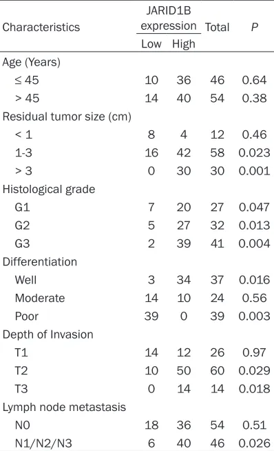
Table 1. Demographic characteristics of pa-tients with breast cancer
Characteristics
JARID1B
expression Total P
Low High Age (Years)
≤ 45 10 36 46 0.64
> 45 14 40 54 0.38
Residual tumor size (cm)
< 1 8 4 12 0.46
1-3 16 42 58 0.023
> 3 0 30 30 0.001
Histological grade
G1 7 20 27 0.047
G2 5 27 32 0.013
G3 2 39 41 0.004
Differentiation
Well 3 34 37 0.016
Moderate 14 10 24 0.56
Poor 39 0 39 0.003
Depth of Invasion
T1 14 12 26 0.97
T2 10 50 60 0.029
T3 0 14 14 0.018
Lymph node metastasis
N0 18 36 54 0.51
N1/N2/N3 6 40 46 0.026
[image:3.612.88.287.312.639.2]pNA) in 60 μl of assay buffer at 37°C for 2 h.
The absorbance was measured at 405 nm. Data analysis
Group data were expressed as the mean ±
SEM. and analyzed by SPSS17.0 software.
Student’s t-test was performed for two-group comparisons. One-way ANOVA followed by Dunnet’s t-test was used for multiple-group comparisons. Differences were considered to
be statistically significant when P < 0.05.
Figures were constructed by GraphPad Prism 5.0 software.
Results
Expression of JARID1B was upregulated in breast cancer tissues and increased JARID1B expression correlates with poor patient prog-nosis
To examine JARID1B expression in breast
cancer, we first compared the expression of
JARID1B in 30 breast cancer tissue samples to the expression in the adjacent normal tissues
using immunohistochemistry. Human normal
breast tissues did not show the JARID1B protein by immunostaining, but the ratio of JARID1B-positive cells in the breast cancer samples was higher than in the normal breast tissue samples (Figure 1A, 1B). These results
an important role in the tumorigenesis of breast cancer.
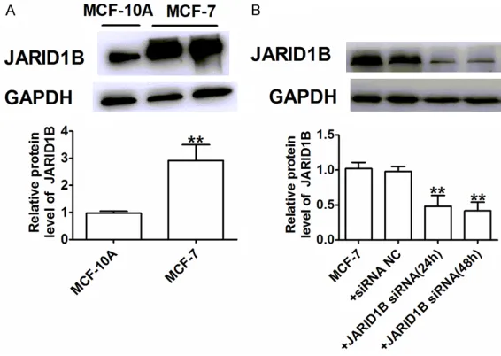
Establishment of stable JARID1B transfection in breast cancer cell lines
The JARID1B expression levels in MCF-7 cells and one normal breast cell line, MCF-10A cells, were measured by Western blot (Figure 2A). The results show that JARID1B was highly expressed in 7 cells compared to
MCF-10A cells. The efficiency of JARID1B knock-down by siRNA was verified at the protein level
(24, 48 h) relative to MCF-7 and siRNA-NC (Figure 2B). These results show that we could use siRNA to generate a stable JARID1B knock-down in MCF-7 cells.
The inhibition of JARID1B promoted apoptosis in breast cancer cells
[image:4.612.92.375.72.273.2]To investigate the possible effects and mecha-nisms of JARID1B in breast cancer cells, we used siRNA to silence JARID1B and then deter-mined caspase-3 activity and cell apoptosis. As illustrated in Figure 3A, cell viability was reduced in MCF-7 cells by the transfection of JARID1B siRNA for 24, 48, and 72 h in a time dependent manner. So JARID1B siRNA for 72 h in MCF-7 cells was used for subsequent experi-ments. JARID1B siRNA-induced apoptosis was Figure 2. The establishment of stable JARID1B transfection in breast
can-cer cell lines. A, B. Representative western blot bands of JARID1B protein. Values given were normalized to the band intensity of GAPDH as an internal control. A: **P < 0.01 vs. MCF-10A; B:**P < 0.01 vs. MCF-7 and siRNA NC groups. n = 4 each group.
indicate significant
overexpre-ssion of JARID1B in the breast cancer cells. To evaluate whe- ther increased JARID1B stain-ing in malignant breast cancer correlates with a worse prog-nosis, further analyses were conducted to discern the cor-relation of JARID1B expres-sion with a series of clinico-pathological parameters in 100 breast cancer cases. We demonstrated that JARID1B
expression had significant cor -relations with the depth of invasion, lymph node
metas-tasis and tumor size (Table 1).
However, there was no stati-stically significant connection
further confirmed using a TUNEL assay and flow cytometry; the number of apoptotic cells
was increased by 48 h after JARID1B siRNA treatment (Figure 3B, 3C). Finally, we measured the changes in caspase-3 activity. Caspase-3 is known to be a key downstream protease that executes the breast cancer apoptotic cascade; activation of caspase-3 is considered to be the last step in caspase dependent apoptosis [7, 17, 18]. As illustrated in Figure 3D, JARID1B siRNA increased the level of caspase-3 activity in MCF-7 cells. On the basis of our data analy-sis, we conclude that JARID1B siRNA triggers a potential apoptotic effect in breast cancer cells.
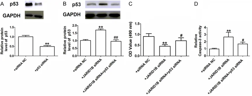
JARID1B siRNA promoted MCF-7 cells apopto-sis partly via p53
[image:5.612.91.526.72.357.2]p53 has important roles in the apoptosis of various cancer types, including breast cancer [15, 18, 19]. Thus, we examined whether p53
was involved in JARID1B-mediated tumor
apop-tosis. First, the efficiency of p53 knock-down by siRNA was verified at the protein level (Figure 4A). Next, we evaluated the effects of JARID1B siRNA on p53 expression in MCF-7 cells by Western blot. As shown in Figure 4B, knock-down of JARID1B dramatically upregulated the expression of p53 in MCF-7 cells. After we transfected the MCF-7 cells with JARID1B siRNA
Discussion
In the present study, we found that JARID1B was overexpressed in both breast cancer cell lines and breast cancer tissues but not in nor-mal breast tissues. The apoptosis of breast
cancer cells was significantly increased by the
interference of JARID1B by siRNA. Moreover, we also found that JARID1B siRNA dramatically increased p53 expression at the protein level. The inhibition of p53 reversed the JARID1B siRNA-induced apoptosis. Our results collec-tively suggest that JARID1B is overexpressed in breast cancer where it plays an important role in breast cancer cell apoptosis, perhaps partially by mediating p53 expression. Our
study has thus clarified a novel effect and
mechanism of JARID1B in response to breast cancer cell apoptosis.
Breast cancer is one of the most frequent malignancies and the second leading cause of cancer-related mortality in women. It is a complex disease with multiple deregulated signaling pathways, including apoptosis [3, 7]. Targeting apoptotic pathways has emerged as an attractive approach for cancer treatment. So far, numerous natural and synthetic com-pounds have been reported to possess anti-cancer activities through the induction of differ-ent apoptotic pathways [7, 20, 21]. A better understanding of the function of apoptosis in breast cancer may provide new therapeutic pathways for disease prevention or control. The JARID1 family of proteins can promote tran-scriptional activation, thus affecting important
processes such as hormone response, stem cell renewal, germ cell development, and cellu-lar proliferation and differentiation [16, 22]. Recent studies have shown that JARID1B is correlated with invasive ductal carcinoma of the breast [8-12]. For instance, high luminal JARID1B activity is associated with poor out-comes in patients with hormone receptor posi-tive breast tumors [8]. The histone demethyl-ase JMJD2B promotes hormonally responsive breast carcinogenesis [9-11]. JARID1B plays a key role in early embryonic development, in the development and differentiation of the nor-mal mammary gland, and in estrogen induced growth of estrogen receptor positive (ER+) breast cancer [12]. These results suggest that JARID1B may be involved in breast tumori-genesis. Some studies have shown that the depletion of JARID1B could induce apoptosis in head and neck squamous carcinoma cells and human acute lymphoblastic leukemia cells [13,
14]. However, the possible effect and mecha -nism of JARID1B in breast cancer cell apoptosis has remained scarcely investigated.
To unravel the function of JARID1B in breast
cancer cell apoptosis, we first examined the
levels of JARID1B in breast cancer samples and matched normal breast tissue samples. The
results showed that JARID1B was significantly
increased in cancers, but not in the normal breast tissues, which suggested that JARID1B was a candidate tumor oncogene in breast can-cer. To obtain a better understanding of the
[image:6.612.92.524.71.236.2]correlation of JARID1B expression with a series of clinicopathological parameters in 100 cases of breast cancer patients. We demonstrated
that JARID1B expression had significant corre -lations with the depth of invasion, lymph node
metastasis, and tumor size. Next, we
trans-fected JARID1B siRNA into MCF-7 breast can-cer cells to inhibit its expression. Our in vitro experiments demonstrated that the inhibition
of JARID1B significantly enhanced the apopto -sis of breast cancer cells and inhibited cell via-bility. These results indicate that JARID1B siRNA effectively promotes breast cancer cells apoptosis.
The p53 protein is a well-known tumor suppres-sor and has been demonstrated to have an essential role in breast cancer proliferation, apoptosis, and invasion [19, 22]. Previous research has reported that JARID1B promotes the proliferation, migration, and invasion activi-ties of lung cancer cells partially via downregu-lated p53 [16]. Therefore, we studied the con-nection between JARID1B and p53 in breast cancer cell apoptosis. Our results indicated
that the level of p53 was significantly increased
in JARID1B knockdown cells. The Inhibition of p53 by siRNA reversed the JARID1B siRNA induced promotion of apoptosis. All of these results demonstrated that JARID1B siRNA pro-motes the apoptosis of breast cancer cells par-tially via upregulated p53.
As a whole, our study reveals that JARID1B plays an important role in regulating breast cancer cell apoptosis and that this role may be mediated by p53. Thus, we propose that the candidate tumor oncogene JARID1B may be an effective novel therapeutic target in the treat-ment of breast cancer.
Disclosure of conflict of interest None.
Address correspondence to: Dr. Luchen Shan, Institute of New Drug Research and Guangzhou Key Laboratory of Innovative Chemical Drug Research in Cardio-cerebrovascular Diseases, Jinan University College of Pharmacy, Guangzhou 510632, China. Tel: +86 20 8522 5030; Fax: +86 20 8522 4766; E-mail: [email protected]
References
[1] Bombonati A, Sgroi DC. The molecular pathol-ogy of breast cancer progression. J Pathol 2011; 223: 307-317.
[2] Siegel RL, Miller KD, Jemal A. Cancer statistics, 2015. CA Cancer J Clin 2015; 65: 5-29. [3] Gao Y, Ma H, Gao C, Lv Y, Chen X, Xu R, Sun M,
Liu X, Lu X, Pei X, Li P. Tumor-promoting proper-ties of mir-8084 in breast cancer through en-hancing proliferation, suppressing apoptosis and inducing epithelial-mesenchymal transi-tion. J Transl Med 2018; 16: 38.
[4] Collins MK, Lopez Rivas A. The control of apop-tosis in mammalian cells. Trends Biochem Sci 1993; 18: 307-309.
[5] Hassan M, Watari H, AbuAlmaaty A, Ohba Y, Sakuragi N. Apoptosis and molecular targeting therapy in cancer. Biomed Res Int 2014; 2014: 150845.
[6] Tsuruo T, Naito M, Tomida A, Fujita N, Mashima T, Sakamoto H, Haga N. Molecular targeting therapy of cancer: drug resistance, apoptosis and survival signal. Cancer Sci 2003; 94: 15-21.
[7] Ding Y, Nguyen TA. Pq1, a quinoline derivative, induces apoptosis in t47d breast cancer cells through activation of caspase-8 and cas-pase-9. Apoptosis 2013; 18: 1071-1082. [8] Yamamoto S, Wu Z, Russnes HG, Takagi S,
Peluffo G, Vaske C, Zhao X, Moen Vollan HK, Maruyama R, Ekram MB, Sun H, Kim JH, Carver K, Zucca M, Feng J, Almendro V, Bessa- rabova M, Rueda OM, Nikolsky Y, Caldas C, Liu XS, Polyak K. Jarid1b is a luminal lineage-driv-ing oncogene in breast cancer. Cancer Cell 2014; 25: 762-777.
[9] Shi L, Sun L, Li Q, Liang J, Yu W, Yi X, Yang X, Li Y, Han X, Zhang Y, Xuan C, Yao Z, Shang Y. Histone demethylase jmjd2b coordinates h3k4/h3k9 methylation and promotes hor-monally responsive breast carcinogenesis. Proc Natl Acad Sci U S A 2011; 108: 7541-7546.
[10] Kawazu M, Saso K, Tong KI, McQuire T, Goto K, Son DO, Wakeham A, Miyagishi M, Mak TW, Okada H. Histone demethylase jmjd2b func-tions as a co-factor of estrogen receptor in breast cancer proliferation and mammary gland development. PLoS One 2011; 6: e17830.
[11] Toyokawa G, Cho HS, Iwai Y, Yoshimatsu M, Takawa M, Hayami S, Maejima K, Shimizu N, Tanaka H, Tsunoda T, Field HI, Kelly JD, Neal DE, Ponder BA, Maehara Y, Nakamura Y, Hamamoto R. The histone demethylase jm-jd2b plays an essential role in human carcino-genesis through positive regulation of cyclin-dependent kinase 6. Cancer Prev Res (Phila) 2011; 4: 2051-2061.
prolifera-tion in the mammary gland and in er+ breast cancer cells. Int J Oncol 2011; 38: 1267-1277. [13] Su H, Ma X, Huang Y, Han H, Zou Y, Huang W.
Jarid1b deletion induced apoptosis in jeko-1 and hl-60 cell lines. Int J Clin Exp Pathol 2015; 8: 171-183.
[14] Zhang P, Tu B, Wang H, Cao Z, Tang M, Zhang C, Gu B, Li Z, Wang L, Yang Y, Zhao Y, Luo J, Deng CX, Gao B, Roeder RG, Zhu WG. Tumor suppressor p53 cooperates with sirt6 to regu-late gluconeogenesis by promoting foxo1 nu-clear exclusion. Proc Natl Acad Sci U S A 2014; 111: 10684-10689.
[15] Shen X, Zhuang Z, Zhang Y, Chen Z, Shen L, Pu W, Chen L, Xu Z. Jarid1b modulates lung can-cer cell proliferation and invasion by regulating p53 expression. Tumour Biol 2015; 36: 7133-7142.
[16] Lopergolo A, Pennati M, Gandellini P, Orlotti NI, Poma P, Daidone MG, Folini M, Zaffaroni N. Apollon gene silencing induces apoptosis in breast cancer cells through p53 stabilisation and caspase-3 activation. Br J Cancer 2009; 100: 739-746.
[17] Dastjerdi MN, Rarani MZ, Valiani A, Mahmou- dieh M. The effect of adenosine a1 receptor agonist and antagonist on p53 and caspase 3, 8, and 9 expression and apoptosis rate in mcf-7 breast cancer cell line. Res Pharm Sci 2016; 11: 303-310.
[18] Okal A, Matissek KJ, Matissek SJ, Price R, Salama ME, Janat-Amsbury MM, Lim CS. Re-engineered p53 activates apoptosis in vivo and causes primary tumor regression in a dominant negative breast cancer xenograft model. Gene Ther 2014; 21: 903-912.
[19] Gali-Muhtasib H, Hmadi R, Kareh M, Tohme R, Darwiche N. Cell death mechanisms of plant-derived anticancer drugs: beyond apoptosis. Apoptosis 2015; 20: 1531-1562.
[20] Shimizu S. [development of anti-cancer drugs mediated by apoptosis and autophagy]. Nihon Rinsho 2015; 73: 1302-1307.
[21] Yang P, Du CW, Kwan M, Liang SX, Zhang GJ. The impact of p53 in predicting clinical out-come of breast cancer patients with visceral metastasis. Sci Rep 2013; 3: 2246.