Original Article
Nd: YAG surgical laser effects in canine prostate
tissue: ablation effect and damage distribution
Ren Wang1, Xing Wu1, Jin-Jin Chen2, Bing Hu1
1Department of Ultrasound in Medicine, Shanghai Jiao Tong University Affiliated Sixth People’s Hospital, Shanghai Institute of Ultrasound in Medicine, Shanghai 200233, China; 2Department of Children Care and Health, Shang-hai Children’s Hospital Affiliated to ShangShang-hai Jiao Tong University, ShangShang-hai 200040, China
Received August 7, 2016; Accepted December 13, 2016; Epub March 15, 2017; Published March 30, 2017
Abstract: Objective: To evaluate therapy effects and capability to preserve adjacent tissues of bilateral focal pros-tate ablation using Nd: YAG laser a canine model. Methods: 24 male Beagle dogs aged 3.9-7.4 years and weighting 9.4-12.1 kg were used in this study. The dogs underwent single focal Nd: YAG ablation. Applied the reasonable
prostatic ablation parameters screened in the study and set different intervals test of fiber lasers for ablation of
prostate-urethral tissue, then observed whether thermal damage occurred and therapy effect. Results: the single focal ablation zone area was 0.7 cm2 but when Distance of double needle laser was 3 mm, the ablation area was 1.25 cm2. With the distance increased, the ablation area significantly extended. The ablation area was 1.53, 1.62, 1.65 cm2 when focal distance was 6, 9, 12 mm, respectively. Although the area extended to 1.65 cm2 when the distance enlarged from 9 cm to 12 cm, the extension range was negligible. Hence, the rational focal distance was 9 mm. In the two-dimensional ultrasonographic examinations, the coagulation necrosis of ablation area showed the approximate oval area with no contrast agent and strong comparison with the surrounding prostate tissue in which enhancement effect of the contract agent exist, which made their boundary more distinct. Conclusion: Nd: YAG pros-tate ablation is shown to be a safe non-thermal ablative technique capable of preserving the adjacent structures and physiological functions. Further studies are needed to fully explore the advantages of Nd: YAG compared with other ablative techniques.
Keywords: Prostate, Nd: YAG, ablation, technology
Introduction
Benign prostatic hyperplasia (BPH), the non-cancerous enlargement of the prostate gland, is one of the most common disorders in aging men. Microscopic evidence of BPH is present in greater than 50% of men by the age of 60 years and in nearly 90% of men by the age of 85 years, with approximately one-fourth of these patients having symptoms during their lifetime that require treatment [1]. Demographic trends in the United States indicate an aging popula-tion in which the rate of prostatectomies would be expected to increase [2]. The recent intro-duction of nonsurgical and minimally invasive options for treatment of symptoms is anticipat-ed to affect this pranticipat-ediction by increasing the number of patients seeking treatment from among the eligible population by 15%-70% [3]. However, less than half of this greater patient population seeking treatment is anticipated to
undergo prostatectomy, with the remainder se- lecting between an increasing number of less invasive or medical methods of management. Destruction of prostate tissue by exposure to laser energy is a less invasive treatment me- thod that also may have an impact on the num-ber of prostatectomies performed. Advances in
optical fiber construction have improved tech
-niques to deliver light efficiently for treatment
understandably reluctant to apply this energy near adjacent structures such as the urinary sphincter or bladder neck, where damage is undesirable [10, 11]. In the present study, we sought to evaluate erectile function, side-ef- fects and preservation of adjacent tissues after a bilateral focal prostate ablation.
Materials and methods
Imaging evaluation of combination of different
power and time of single fiber laser ablation
canine prostate
24 male Beagle dogs aged 3.9-7.4 years and weighting 9.4-12.1 kg were used in this study [13, 14]. The dogs underwent single focal LE- DC ablation by the monopolar electrode Nano- Knife device. The dogs were equally assigned
to two groups for final pathological evaluation
at 7 days after ablation. The relevant parame-ters were shown as follow:
Group A: 3 w 5 min, 5 w 5 min, 7 w 5 min. Group B: 5 w 3 min, 5 w 5 min, 5 w 7 min.
After ablation, the ultrasonic imaging was con-ducted and prostate focal ablation zone sizes were measured. Then, pathologic specimens of the biggest ablation zone size were collected, then the depth and width of thermal coagula-tion damage were measured macroscopically. Lesion volume was estimated by assuming a half ellipsoid: volume =π/6* (depth) (width)2. HE dyeing was applied for evaluating urotheli-um change and urethra and prostate boundary situation followed by measuring and comparing
the changes of the single fiber ablation zone
size between the adjacent urethra prostate and inner prostate organization.
Combination of two bilateral focus
laser-melt-ing experiment
According to the reasonable prostatic ablation parameters of energy power and explore time
[image:2.612.87.524.83.126.2]screened in first part, we set a serial interval distance of bare-end bilateral fibers for abla -tion of prostate-urethral tissue adjacent to melt, then observed whether thermal damage occurred [15]. 24 male Beagle dogs aged 3.9-7.4 years and weighting 9.4-12.1 kg were used in the bilateral focus laser melting experiment.
Table 1. Influence of different single focus laser time and power on canine prostate ablation
Group A Group B
3 w 5 min 5 w 5 min 7 w 5 min 5 w 3 min 5 w 5 min 5 w 7 min Prostate zone size (ml) 4.32±0.62 6.83±0.34* 6.35±0.25* 5.11±0.52 6.83±0.34# 6.63±0.29#
*P<0.05, compared with Group 3 w 5 min. #P<0.05, compared with Group 5 w 3 min.
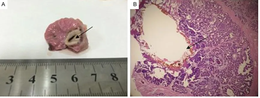
Figure 1. Canine prostate histopathologic examination after single focal Nd: YAG. A: Represents TTC staining of prostate gross cross-sectional slices where there was the narrow annular hyperemia edema zone of dark red band surrounding ablation zone and that four regions existed in outward area hyperemia from the center to the margin
which was the vaporizing cavity Area, brown carbonized area, dark necrotic area and inflammatory edema area in
[image:2.612.92.525.160.324.2]After 5 min ablation, the ultrasonic imaging or MRI enhancement were implemented fol-lowed observation of the melting focal zone, measurement of its size. Eventually, pathologic specimens were collected, and HE dyeing was conducted.
At termination, a comprehensive necropsy was performed to evaluate for potential injury to adjacent tissues, that is prostate, bladder,
ure-ters, urethra as well as rectal wall. Specific
attention was devoted to microscopic exami- nation of the prostatic capsule, urethra, ner-vous tissues and rectal wall. The study proto- col was reviewed and approved by the Insti- tutional Animal Care and Use Committee and carried out under Good Laboratory Practice conditions.
Statistics
Statistically derived experimental results are
given as means ± SD. Curve fits of the experi -mental results were made using the mean val-ues. Differences in results between depths
[image:3.612.89.526.70.405.2]were tested for significance using the Student
Figure 2. Two-dimensional ultrasonographic examinations of single focus laser ablation. A: Represents ultrasono-graphic examination in the frontage of tissue section before ablation. B: Represents ultrasonoultrasono-graphic examination in the back of tissue section before ablation. C: Represents ultrasonographic examination in the frontage of tissue section after ablation and there was remarkable micro-bubble in the laser ablation area which was absence of the
contract agent, but the surrounding normal prostate tissue filling contrast agent showed clear echogenicity in CEUS
examination after 5 min ablation. D: Represents ultrasonographic examination in the back of tissue section after ablation. There was also remarkable micro-bubble in the laser ablation area.
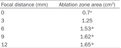
Table 2. Relation of focal distance and abla-tion zone area
Focal distance (mm) Ablation zone area (cm3)
0 0.7a
3 1.25
6 1.53*
9 1.62*
12 1.65*
[image:3.612.91.290.527.606.2]t-test and were considered significant when
P<0.05 (two-tailed).
Results
Imaging evaluation of combination of different
power and time of single fiber laser ablation
canine prostate
Different power and time of single fiber laser
ablation canine prostate were performed in which 24 male dog were evenly separated into two groups. As shown in Table 1, the biggest prostate zone size was 6.83±0.34 ml when the parameter was 5 w 5 min, and the second size were 6.63±0.29 ml when it was 5 w 7 min,
finally, the smallest size were 4.32±0.62 ml
when it was 3 w 5 min. In the Group A, the dis-tinct rising trend existed between 3 w 5 min and 5 w 5 min compared with the descending trend from 5 w 5 min to 7 w 5 min. The similar situation occurred in Group B. There was the apparent ascending trend between 3 w 5 min and 5 w 5 min and the declining trend from 5 w 5 min and 5 w 7 min. Therefore, 5 w 5 min was optimal combination parameter of single focus laser time and power in the study.
[image:4.612.90.523.71.424.2]center to the margin which was the vaporizing cavity Area, brown carbonized area, dark ne-
crotic area and inflammatory edema area in
sequence. The volume of focal ablation was 6.55 cm3 to 7.48 cm3, an average of 6.96 cm3±0.31 cm3.
As shown in Figure 1B, there was large coagula-tion necrosis area by microscope which was characterized by large evenly dyed red area in the HE staining. In this region, normal glandu- lar structure disappeared and necrosis zone was dotted with bleeding focal point. Moreover,
central gasification cavity area and carbonized
area both dyed red area in the coagulation ne- crosis area. The edge of coagulation necrosis, ultimately, was bleeding congestion edema se- parated with normal glandular structure. As shown in Figure 2A and 2B, it was obvious- ly that laser ablation process appeared the strong cloudiness echo gradually spreading to the margin in their two-dimensional ultrasono-graphic examinations before ablation.
As shown in Figure 2C and 2D, there was remarkable micro-bubble in the laser ablation area which was absence of the contract agent,
but the surrounding normal prostate tissue fill -ing contrast agent showed clear echogenicity in CEUS examination after 5 min ablation. The
tion zone area of single focus laser melt was 0.7 cm2 so the superposition of two single nee-dle ablation area, theoretically, was 1.4 cm2. As shown in Table 2, when distance of double nee-dle laser was 3 mm, the ablation area was 1.25 cm2. With the distance increased, the ablation
area significantly extended. The ablation area
was 1.53, 1.62, 1.65 cm2 when focal distance was 6, 9, 12 mm, respectively. Although the area extended to 1.65 cm2 when the distance enlarged from 9 cm to 12 cm, the extension scale was negligible. Hence, the rational bilat-eral focal distance was 9 mm.
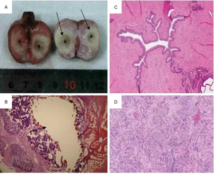
As shown in Figure 3A, there were two distinct vaporizing cavity area in the ablation zone. As same as appeared in the single focus ablation zone, it apparently presented symmetrically a double of four annular regions in outward hy- peremia zone from the center to the margin, that is vaporizing cavity Area, brown carboni-
zed area, dark necrotic area and Inflammatory
edema area in sequence.
Apparently, more significant coagulation necro -sis area by microscope than single focus abla-tion which was characterized by large evenly dyed red area in the HE staining, as shown in
Figure 3B. Despite instances of partially oc- cluded capsular arterioles adjacent to the area of ablation, larger arteries and veins or nervous coagulation necrosis of abla-tion area showed the approxi-mate oval area with no con-trast agent and strong com-parison with the surround- ing prostate tissue in which enhancement effect of the contract agent exist, which made their boundary more distinct. The volume of focal ablation was 5.93 cm3 to 7.83 cm3, an average of 6.64 cm3±0.66 cm3.
Combination of two bilateral
focus laser-melting experi -ment
According to results of single focus ablation examination, 5 min 5 w was chose as one of basic parameters of bilateral focus laser melting experi-ment. Meanwhile, the abla-Figure 4. Two-dimensional ultrasonographic examinations of bilateral focus
laser ablation. Nearly symmetric micro-bubbles existed in the laser ablation area which was absence of the contract agent, but the surrounding normal
[image:5.612.90.377.71.273.2]tissue were not adversely affected by LEDC in the Figure 3C and 3D.
Figure 4 revealed that there were nearly sym-metric micro-bubbles in the laser ablation area which was absence of the contract agent, but
the surrounding normal prostate tissue filling
contrast agent showed clear echogenicity in CEUS examination after 5 min ablation. The coagulation necrosis of ablation area showed the approximate oval area with no contrast agent and strong comparison with the sur-rounding prostate tissue in which enhance-ment effect of the contract agent exist, which made their boundary more distinct.
Discussion
Benign prostatic hyperplasia was a com- mon disease among the middle-aged and old males, of which relevant symptom seriously ef- fects on the quality of life of patients. Current drug treatment effect was unsatisfactory, so most patients eventually may need surgery, however the surgical trauma was severe [16]. Moreover, postoperative complications possi-bly were harmful to the quality of life [17]. Therefore, minimal invasion treatment charac-terized by safety and validity was still a topic in worth of inquiry [18]. In single needle ablation experiments of our study, changes of melting zone range depend on increase of the power and time. In the Group A, the distinct rising trend existed between 3 w 5 min and 5 w 5 min compared with the descending trend from 5 w 5 min to 7 w 5 min. The similar situation occurred in Group B. There was the apparent ascending trend between 3 w 5 min and 5 w 5 min and the declining trend from 5 w 5 min and 5 w 7 min. Therefore, 5 w 5 min was opti-mal combination parameter of single focus laser time and power in the study.
Though laser ablation was once applied in the prostate cancer treatment, the usage of laser ablation was decrease gradually because of related small ablation zone [19]. Bilateral
optical fibers for the in vitro prostate
abla-tion can be obtained a wide oval, three optical
fibers ablation can be obtained a roughly cir-cular structure and four optical fibers ablation
can be a square structure [20].
Hence it was feasible to choose multi-fibers
therapy program based on lesion size and po-
rous shape or according, which can obtain good conformal therapy effect. No matter when sin-gle or bilateral focus ablation, there was re- markable micro-bubble in the laser ablation area which was absence of the contract agent,
but the surrounding normal prostate tissue fill -ing contrast agent showed clear echogenicity in CEUS examination after 5 min ablation. The coagulation necrosis of ablation area showed the approximate oval area with no contrast agent and strong comparison with the sur-rounding prostate tissue in which enhance-ment effect of the contract agent exist, which made their boundary more distinct.
Disclosure of conflict of interest
None.
Address correspondence to: Bing Hu, Department of Ultrasound in Medicine, Shanghai Jiao Tong Uni-
versity Affiliated Sixth People’s Hospital,
Shang-hai Institute of Ultrasound in Medicine, 600 Yi- Shan Road, Shanghai 200233, China. Tel: +86-21-64369181; E-mail: [email protected]
References
[1] Rubinsky J, Onik G, Mikus P and Rubinsky B. Optimal parameters for the destruction of prostate cancer using Nd: YAG ablation. J Urol 2008; 180: 2668-2674.
[2] Onik G, Mikus P and Rubinsky B. Irreversible electroporation: implications for prostate abla-tion. Technol Cancer Res Treat 2007; 6: 295-300.
[3] Deodhar A, Monette S and Single GW Jr. Percutaneous Nd: YAG ablation: preliminary re-sults in a porcine model. Cardiovasc Intervent Radiol 2011; 34: 1278-1287.
[4] Thomson KR, Cheung W and Ellis SJ. Investi- gation of the safety of Nd: YAG ablation in hu-mans. J Vasc Interv Radiol 2011; 22: 611-621. [5] Bower M, Sherwood L, Li Y and Martin R. Nd:
YAG ablation of the pancreas: definitive local
therapy without systemic effects. J Surg Oncol 2011; 104: 22-28.
[6] Deodhar A, Monette S, Single GW Jr, Hamilton WC, Thornton R, Maybody M, Coleman JA and Solomon SB. Renal tissue ablation with irre-versible electroporation: preliminary results in a porcine model. Urology 2011; 77: 754-760. [7] Tracy CR, Kabbani W and Cadeddu JA. Nd: YAG
ablation: a novel method for renal tissue abla-tion. BJU Int 2011; 107: 1982-1987.
[9] Schoellnast H, Monette S, Ezell PC, Deodhar A, Maybody M and Erinjeri JP. Acute and sub-acute effects of irreversible electroporation on nerves: experimental study in a pig model. Int J Med Radiol 2011; 260: 421-427.
[10] Caso JR, Tsivian M, Mouraviev V, Kimura M and Polascik TJ. Complications and postoperative events after cryosurgery for prostate cancer. BJU Int 2012; 109: 840-845.
[11] Onik G, Porterfield B, Rubinsky B and Cohen J.
Percutaneous transperineal prostate cryosur-gery using transrectal ultrasound guidance: animal model. Urology 1991; 37: 277-281. [12] Scott W, Cilip C and Fried M. Nd: YAG laser
ab-lation of urinary stones through small-core
op-tical fibers. IEEE J Sel Top Quantum Electron
2009; 15: 435-440.
[13] Spore S, Teichman M, Corbin N, Champion P, Williamson N and Glickman D. Nd: YAG li- thotripsy: optimal power settings. J Endourol 2007; 13: 559-566.
[14] White MD, Moran ME, Calvano CJ, Borhan-Manesh A, Mehlhaff BA. Evaluation of retropul-sion caused by holmium: YAG laser with
vari-ous power settings and fibers. J Endourol
1998; 12: 183-6.
[15] Lee H, Ryan T, Teichman J, Kim J, Choi B, Arakeri N and Welch A. Stone retropulsion dur-ing Nd: YAG lithotripsy. J Urol 2003; 169: 881-885.
[16] Kang K, Lee H, Teichman J, Oh B, Kim J and Welch J. Dependence of calculus retropulsion on pulse duration during Nd: YAG laser Lithotripsy. Lasers Surg Med 2006; 38: 762-772.
[17] Finley D, Petersen J, Abdelshehid C, Ahlering M, Chou D, Borin J, Eichel L, McDougall E and Cayman K. Effect of Nd: YAG laser pulse width on lithotripsy retropulsion in vitro. J Endourol 2005; 19: 1041-1044.
[18] Marguet J, Sung J, Springhart W, L’Esperance
J, Zhou S, Zhong P, Albala P and Preminger D. In vitro comparison of stone retropulsion and fragmentation of the frequency doubled, dou-ble pulse Nd: YAG laser and the holmium: YAG laser. J Urol 2011; 173: 1797-1800.
[19] Li W, Fan Q, Ji Z, Qiu X, Li Z. The effects of irre -versible electroporation (IRE) on nerves. PLoS One 2011; 6: e18831.