ABSTRACT
MACKENZIE, SARAH HELEN. The Importance of the Dimer Interface in the Folding and Assembly of Procaspase-3. (Under the direction of Dr. A. Clay Clark).
Caspases are a family of cysteine proteases that are intimately involved in apoptosis and exist in the cell as inactive zymogens prior to activation. Initiator procaspases are monomers that must dimerize for activation. Executioner procaspases, such as procaspase-3, are dimers that must be processed for activation. The chemical properties of the dimer interface are different between the two subfamilies of caspases but their structures are similar, suggesting that the interface region is important for regulation. The goal of the studies presented here is to determine the importance of the dimer interface in folding and assembly of procaspase-3. A histidine mutation was introduced into the dimer interface region, which completely
abolished the activity of mature caspase-3. Equilibrium and kinetic folding studies were performed to elucidate how a mutation in the dimer interface prevents substrate turnover in the active site when the distance between the two regions is 20Å. The folding studies presented here coupled with the crystal structure show that the protein is entering a kinetic trap prior to dimerization because the histidine has to adopt an unusual rotomer to pack into a region that normally accommodates a much smaller valine residue. A hysteresis was
trapped in a conformation that is unstable and prone to aggregation prior to forming a dimerization competent species. The crystal structure of caspase-3 (V266H) revealed two separate pathways of inhibition starting from the dimer interface and culminating in the active site that could be responsible for the lack of activity in this mutant. These data, taken together, suggest that the dimer interface is a region that can be used to allosterically inhibit procaspase-3 because it is important for regulation of the enzyme. This is important because it could be used as a drug target for diseases that have too much cell death, such as
The Importance of the Dimer Interface in the Folding and Assembly of Procaspase-3
by
Sarah Helen MacKenzie
A dissertation submitted to the Graduate Faculty of North Carolina State University
in partial fulfillment of the requirements for the degree of
Doctor of Philosophy
Biochemistry
Raleigh, North Carolina 2009
APPROVED BY:
_______________________________ ______________________________ Dr. Dennis Brown Dr. Paul Agris
DEDICATION
To my mom, Kathleen, and my brother, Stuart for your love, support, and encouragement
BIOGRAPHY
Sarah MacKenzie was born in Aberdeen, Scotland. She moved to the United States
with her parents when her dad got his first faculty position at Texas A&M. She has lived all
over the country, following her mom as she advanced through the ranks of the
Pharmaceutical Industry. She moved to North Carolina in 1997 and found her home. She
graduated from North Carolina State University in May 2003 with a Bachelor of Science
degree in Biochemistry. In August 2004 she started graduate school in pursuit of a Ph.D. in
biochemistry at North Carolina State University under the direction of Dr. A. Clay Clark.
She developed her passion for teaching in the Preparing the Professoriate Program and won
three teaching awards during graduate school. Upon completion of her Ph.D., Sarah plans to
have a productive career in academics working on the cancer problem and teaching the next
ACKNOWLEDGMENTS
I would like to thank Dr. A. Clay Clark for his passion and commitment to making his
students the best they can be. You are truly an outstanding mentor, teacher and friend. I
would also like to thank my committee members Dr. Robert Rose, Dr. Paul Agris, and Dr.
Robert Kelly for your support and encouragement for the past five years. I appreciate your
commitment to making me understand the importance of telling a story through my results. I
would also like to thank Dr. Dennis Brown for giving his time and sitting in on my prelim
defense and my oral defense.
I would like to thank my family both near and far. A special thanks to my mom,
Kathleen, for never giving up on me and believing in me, no matter how stubborn I was.
Your unwavering support has enabled me to become the person that I am. To my brother,
Stuart, thanks for being the amazing and compassionate person that you are. I would like to
thank my grandparents, Muriel and Ralph, for their endless support and encouragement.
Your commitment to seeing me through my academic goals has driven me to be a more
diligent student. Lastly, but certainly not least I would like to thank my uncles, Richard and
Les for their support. The phone calls alone got me through graduate school especially when
I had to pull an all night experiment. I am blessed to have such an amazing family.
I would like to thank my friends for making the last five years some of the best times
in my life. Mialy, Karen, Stephanie, Katie, Maria, Garrett, Heba, Kyle, Dawn, John,
Michelle, Josh, Nikki and Mike, you are all incredible people and I don’t know what I would
Estella, Uma, Jad, Chris, Mialy and Sang-oh, it has gone by so quickly but you have all made
a profound impact on my life. To my fellow Claymates, Jad, Sara, Josh, Chunxiao and
TABLE OF CONTENTS
LIST OF TABLES ... xii
LIST OF FIGURES ... xiii
CHAPTER 1: Targeting cell death in tumors by activating caspases ...1
ABSTRACT ...2
A. Types of cell death ...3
B. Mechanisms of apoptosis ...4
C. Cancer drug targets in apoptosis ...7
C.1. Extrinsic pathway ...7
C.2. Caspases ...10
C.3. Intrinsic pathway ...10
C.4. IAPs ...12
D. Molecules that specifically bind to caspases...13
D.1. IAPs ...14
E. Caspase dimer interface ...15
F. A brief description of the dimer interface and active site formation ...17
G. Dimer interface experiments that suppress caspase-3 activity ...20
G.1. FICA and DICA ...20
G.2. Caspase-3 interface mutant Val266His ...22
G.3. Experiments that activate the caspase-3 zymogen ...22
REFERENCES ...25
CHAPTER 2: Role of dimerization in the regulation of caspase activity ...48
ABSTRACT ...49
A. Apoptosis leads to cell death ...50
B. Caspases are part of the cell death machinery ...51
C. Caspases are also part of the inflammatory response ...52
D. Caspases cleave with high specificity ...53
E. Caspases are produced initially as inactive zymogens ...53
F. Dimerization of caspase zymogens ...55
G. Caspase dimerization affects stability ...57
H. Comparison of caspase dimer interfaces ...58
I. Active site formation after chain cleavage ...63
J. Active site cooperativity ...68
K. Conclusions ...71
REFERENCES ...72
CHAPTER 3: Protein folding: thermodynamic and kinetic perspectives ...84
A. The protein folding problem ...85
B. Equilibrium and kinetic folding studies ...89
C. Examples of protein folding mechanisms ...90
C.1. Folding mechanism of procaspase-1 CARD ...90
C.2. Folding mechanism of DHFR ...91
C.4. Folding mechanism of SecA ...92
C.5. Summary ...93
D. Kinetic partitioning ...93
E. Examples of kinetic partitioning ...94
E.1. Serpins native conformation is metastable in the energy landscape ...94
E.2. The β-subunit of bacterial luciferase can form an alternative native state ...95
E.3. Summary...95
F. Project Aims ...96
REFERENCES ...97
CHAPTER 4: Materials and Methods ...100
Materials ...101
Stock solutions ...101
Methods...101
A. Protein purification ...101
B. Equilibrium folding studies ...103
C. Kinetic folding studies ...104
C.1. Single-mixing stopped-flow fluorescence and CD ...104
C.2. Emission scanning ...105
C.3. Hand-mixing fluorescence and CD spectroscopy ...106
C.4. Measuring folding kinetics ...107
C.5. Data analysis ...107
C.7. Exponential fits ...110
D. Size Exclusion Chromatography ...111
D.1. Column parameters and calibration ...112
D.2. Sample preparation ...112
D.3. Instrument procedure and data analysis ...113
REFERENCES ...117
CHAPTER 5: Equilibrium folding studies of procaspase-3 (C163S, V266H) ...119
ABSTRACT ...120
INTRODUCTION ...121
RESULTS ...126
A. Denaturation of procaspase-3 (C163S, V266H) ...126
B. Equilibrium unfolding of procaspase-3 (C163S, V266H) at pH 7.0 ...127
C. Reversibility of procaspase-3 (C163S, V266H) folding at pH 7.0 ...129
D. Protein concentration dependence of unfolding at pH 7.0 ...130
E. pH profiles of procaspase-3 (C163S, V266H), (C163S) by SEC ...131
F. Equilibrium unfolding of procaspase-3 (C163S, V266H) at pH 5.0 ...132
G. Reversibility of procaspase-3 (C163S, V266H) folding at pH 5.0 ...133
H. Protein concentration dependence of unfolding at pH 5.0 ...135
DISCUSSION ...137
REFERENCES ...142
CHAPTER 6: Kinetic Folding Studies of Procaspase-3 (C163S, V266H) ...161
INTRODUCTION ...163
RESULTS ...167
A. Multiple monomeric species are formed in the burst phase...169
B. Procaspase-3 (C163S, V266H) folds through multiple monomeric intermediates ....171
C. Aggregation of kinetically trapped monomeric intermediate species ...175
D. Proposed folding model for procaspase-3 (C163S, V266H) ...176
E. Unfolding data reveal two dimeric species ...176
F. Procaspase-3 (C163S, V266H) dissociated to form highly structured monomeric intermediates ...178
G. Monomeric procaspase-3 (C163S, V266H) unfolds slowly ...180
H. Proposed folding model for procaspase-3 (C163S, V266H) ...182
DISCUSSION ...183
REFERENCES ...188
CHAPTER 7: Discussion ...206
REFERENCES ...213
APPENDICES ...220
APPENDIX A: 5-19F-tryptophan labeling of procaspase-3 (C163S) and 19F-NMR preliminary studies ...221
INTRODUCTION ...222
RESULTS ...225
A. Procaspase-3 (C163S) expression in BL21 DE3 lys S cells ...225
C. Protein purification, percent incorporation and preliminary NMR studies ...228
D. NMR studies using fluorine cryporobe in Dr. Carl Frieden’s lab at
Washington University, St. Louis ...229
E. pQE80L-procaspase-3 (C163S) expression in W3110trpA33 cells ...231
LIST OF TABLES
CHAPTER 6
LIST OF FIGURES
CHAPTER 1
Figure1. Cell death pathways ...36
Figure 2. Sequence alignment of eleven human caspases ...37
Figure 3. Human caspase organization ...39
Figure 4. The caspase cascade ...40
Figure 5. Caspase-3 with XIAP BIR-2 bound ...42
Figure 6. Inhibited caspases ...43
Figure 7. Active site rearrangements of caspases-1,-3 and -9 ...45
CHAPTER 2 Figure 1. Human caspase organization ...76
Figure 2. The caspase cascade ...77
Figure 3. Comparison of caspase dimer interfaces ...79
Figure 4. Active site loop movements upon maturation of caspase-7 ...80
Figure 5. Active site rearrangements of caspases-1 and -3 ...81
Figure 6. Active site rearrangements of caspase-9 upon inhibitor binding ...82
Figure 7. Structure of caspase-7 with FICA bound in the dimer interface ...83
CHAPTER 4
Figure 1. SEC molecular weight standard chromatograms and standard curve ...118
CHAPTER 5
Figure 1. Structure of caspase-3...144
Figure 2. Equilibrium unfolding of procaspase-3 (C163S) ...145
Figure 3. Fluorescence emission spectra of procaspase-3 (C163S, V266H)
at pH 7.0 ...146
Figure 4. Equilibrium unfolding of procaspase-3 (C163S, V266H) at pH 7.0 ...147
Figure 5. Reversibility of equilibrium folding of procaspase-3 (C163S, V266H)
at pH 7.0 ...148
Figure 6. Overlay of refolding curves at pH 7.0 shows a concentration dependent
hysteresis ...150
Figure 7. Concentration dependence of procaspase-3 (C163S, V266H) unfolding
studies at pH 7.0 ...151
Figure 8. Denaturation profile of procaspase-3 (C163S, V266H) by
size exclusion chromatography at pH 7.0 ...152
Figure 9. pH profiles of procaspase-3 (C163S, V266H) and procaspase-3 (C163S)
by size exclusion chromatography ...153
Figure 10. Equilibrium unfolding of procaspase-3 (C163S, V266H) at pH 5.0 ...154
Figure 11. Reversibility of equilibrium folding of 2μM procaspase-3
Figure 12. Reversibility of equilibrium folding of 1μM procaspase-3
(C163S, V266H) at pH 5.0 ...156
Figure 13. Reversibility of equilibrium folding of 0.5μM procaspase-3 (C163S, V266H) at pH 5.0 ...157
Figure 14. Overlay of refolding curves at pH 5.0 shows a concentration dependent hysteresis ...158
Figure 15. Concentration dependence of procaspase-3 (C163S, V266H) unfolding studies at pH 5.0 ...159
Figure 16. Denaturation profile of procaspase-3 (C163S, V266H) by size exclusion chromatography at pH 5.0...160
CHAPTER 6 Figure 1. Far-UV CD measured at 228nm refolding data ...190
Figure 2. Complex burst phase during refolding ...191
Figure 3. Emission scanning of the burst phase species ...193
Figure 4. CD lag phase...195
Figure 5. Rate of the final urea concentration for fluorescence emission following excitation and by differential quenching by acrylamide ...196
Figure 6. Intermediate phase of refolding ...197
Figure 7. Slow phase of refolding shows procaspase-3 (C163S, V266H) enters a kinetic trap ...198
Figure 9. Proposed sequential refolding mechanism ...200
Figure 10. Unfolding fluorescence data ...201
Figure 11. Procaspase-3 (C163S, V266H) unfolding of the secondary structure in 9M urea detected by far-UV CD ...202
Figure 12. Procaspase-3 (C163S, V266H) unfolding in 8M urea detected by far-UV CD unfolding ...203
Figure 13. Procaspase-3 (C163S, V266H) unfolding of the tertiary structure detected by fluorescence emission and fluorescence anisotropy ...204
Figure 14. Proposed sequential unfolding mechanism ...205
Figure 15. Proposed overall folding mechanism for procapase-3 (C163S, V266S) ...206
CHAPTER 7 Figure 1. The initial model for caspase-3 (V266H) inactivity ...214
Figure 2. The crystal structure of the caspase-3 (V266H) dimer interface ...215
Figure 3. Structural details of intramolecular allosteric inhibition ...216
Figure 4. Structural details of intermolecular allosteric inhibition. ...217
Figure 5. The crystal structure of the caspase-3 (Y197C, V266H) dimer interface...218
Figure 6. Overlay of histidine residues from the caspase-3 (V266H) initial model, the caspase-3 (V266H) crystal structure and the caspase-3 (Y197C, V266H) crystal structure ...219
Figure 2. Procaspase-3 (C163S) optimal expression conditions in
BL21 DE3 lys S cells ...234
Figure 3. Procaspase-3(C163S) expression in the presence of either L-tryptophan
(L-Trp) or 5-19F-tryptophan (F-Trp) ...235
Figure 4. Extracted ion chromatogram for percent incorporation ...236
Figure 5. Preliminary fluorine NMR studies on procaspase-3 (C163S) after
19
F-tryptophan incorporation ...237
Figure 6. Comparison of native fluorine NMR peaks for procaspase-3 (C163S,
W214V), procaspase-3 (C163S, W206Y) and procaspase-3 (C163S) ...238
Figure 7. Denaturation profile of procaspase-3 (C163S) determined by
fluorine NMR ...239
Figure 8. Denaturation profile of procaspase-3 (C163S, W214V) determined
by fluorine NMR ...240
Figure 9. Denaturation profile of procaspase-3 (C163S, W206Y) determined
by fluorine NMR ...241
Figure 10. pQE80L-procaspase-3 (C163S) optimal expression conditions in
CHAPTER 1
Targeting Cell Death in Tumors by Activating Caspases
Sarah H. MacKenzieand A. Clay Clark
Department of Molecular and Structural Biochemistry, North Carolina State University,
Raleigh, NC 27695-7622
ABSTRACT
Cytotoxic approaches to killing tumor cells, such as chemotherapeutic agents, γ
-irradiation, suicide genes or immunotherapy, have been shown to induce cell death
through apoptosis. The intrinsic apoptotic pathway is activated following treatment with
cytotoxic drugs, and these reactions ultimately lead to the activation of caspases, which
promote cell death in tumor cells. In addition, activation of the extrinsic apoptotic
pathway with death-inducing ligandsleads to an increased sensitivity of tumor cells
toward cytotoxic stimuli, illustrating the interplay between the two cell death pathways.
In contrast, tumor resistance to cytotoxic stimuli may be due to defects in apoptotic
signaling. As a result of their importance in killing cancer cells, a number of apoptotic
molecules are implicated in cancer therapy. The knowledge gleaned from basic research
into apoptotic pathways from cell biological, structural, biochemical, and biophysical
approaches can be used in strategies to develop novel compounds that eradicate tumor
cells. In addition to current drug targets, research into molecules that activate procaspase–
3 directly may show the direct activation of the executioner caspase to be a powerful
therapeutic strategy in the treatment of many cancers.
Keywords: caspase, apoptosis, programmed cell death, protease, zymogen, dimerization, active site formation, human disease, cancer treatment, dimer interface, proteolytic
A. Types of Cell Death
Cell death can occur by many pathways, all of which have unique mechanisms
and morphologies. The three major types of cell death include necrosis, oncosis and
programmed cell death (Figure 1). Necrosis was defined originally as the accidental death
of cells and living tissue and generally is characterized by swelling of the cell, chromatin
condensation and the eventual nuclear and cellular lysis that leads to inflammation [1].
Later, Majno and Joris disputed the earlier definition by stating that necrosis is not a type
of cell death. Rather, it represents the end stage of the cell death process and can occur in
any pathway in the absence of phagocytosis [2] (see Figure 1). Instead, “oncosis” was
proposed as a term to refer to any type of cell death that is marked by cellular swelling.
The morphology of oncosis also includes chromatin condensation (pyknosis) and nuclear
fragmentation (karyorrhexis) with eventual nuclear fading (karyolysis) [3].
Programmed cell death (PCD) occurs through two distinct pathways that can be
differentiated by the morphology of the cell in response to death signals: condensation
prominent, called type I or apoptosis, and autophagy prominent, called type II. Type I
PCD often is found to occur during vertebrate development, especially in organ
morphogenesis [4-7], and was characterized first by Kerr in 1972 as having
morphological features that include cytoplasmic shrinkage, active membrane blebbing
and condensed chromatin [8]. Apoptosis eventually leads to cell segregation and the
formation of apoptotic bodies that contain intact organelles [8]. In addition, the nuclear
DNA is degraded, the cytoskeleton is dismantled and cell progression is halted [9, 10].
plasma membrane, then engulf the apoptotic bodies [8, 11]. In contrast, the most
prominent features of autophagic PCD are formation of autophagic vacuoles that
accompany the degradation of cytoplasmic materials, nuclear collapse and subsequent
phagocytosis.
B. Mechanisms of Apoptosis
A family of cysteine-dependent aspartate-directed proteases, known as caspases,
is intimately involved in apoptosis. The cleavage of key proteins in the cell by caspases
leads to the aforementioned morphological and biochemical changes observed during
apoptosis. Seven of the known eleven human caspases are involved in the execution of
apoptosis (Figures 2 and 3). The apoptotic caspases are divided into two groups, the
initiators and the effectors, depending on their time of entry into the apoptotic cascade
(Figure 3). For example, the initiator caspases, such as caspases–2, –8, –9 and –10,have
an early entry into the cascade, where they are responsible for activating the effector
caspases–3, –6 and –7 (Figure 4). The initiators are themselves activated either by
so-called extrinsic or intrinsic mechanisms, as described below.
The extrinsic pathway for initiator caspase activation ultimately is responsible for
the elimination of unwanted cells that are produced during development or that have
tumorogenic qualities. This pathway is initiated by ligation of a transmembrane death
receptor in response to an extracellular signal, where the death receptors belong to the
tumor necrosis factor receptor (TNFR) superfamily. Members of the TNFR family
contain an intracellular domain, called a death domain (DD), that binds to a number of
signal from the exterior surface of the cell to the cytosol. The most characterized TNF
receptors include CD95 (APO-1/Fas), TNF receptor 1 (TNFRI), R1 and
TRAIL-R2. Their respective death ligands are FasL, TNFα and TRAIL (TNF-related
apoptosis-inducing ligand) [12-14]. Upon ligation, the DD on the cytoplasmic tail of the death
receptor recruits a multiprotein complex called the DISC (death inducing signaling
complex). The DISC is thought to promote the dimerization of caspases–8 and –10 by
increasing the local concentration of procaspase monomers, thereby increasing the
probability of dimerization. This is referred to as the induced proximity model [15].
Following their activation, the initiator caspases cleave the zymogens of caspases–3, –6,
or –7 (Figure 4).
A fundamental difference exists between the caspase subfamilies, even though the
structures of the mature caspases are quite similar. The initiator caspases exist in the cell
as monomers that require a scaffold for dimerization. Once associated with the scaffold,
the procaspase dimer has sufficient enzymatic activity for autolysis [16-19]. In contrast,
the effector procaspases exist in the cell as stable dimers that have little enzymatic
activity [20, 21]. As described below, the effector procaspases are inactive due to
misaligned active sites that prevent efficient catalysis. Activation of the effector
procaspases occurs after proteolytic cleavage, which allows the active site loops to
rearrange, resulting in a large increase in activity (~200-fold) [21].
The caspase cascade also can be activated by the so-called intrinsic pathway,
which is a mitochondria-dependent pathway that is activated by a variety of stimuli,
[22] (see Figure 4). In response to the apoptotic stimuli, the permeability of the
mitochondrial membrane increases, and many apoptogenic factors are released, including
cytochrome c, apoptosis inducing factor (AIF), Smac (second mitochondria derived
activator of caspase)/DIABLO (direct IAP binding protein with low pI), Omi/HtrA2
(high temperature requirement A2) and endonuclease G [23] (Figure 4). The increased
permeability of the mitochondria is accomplished in part by the cleavage of Bid (BH3
interacting death agonist), a member of the Bcl-2 family of proteins, by caspase–2 [24]
and possibly by caspases–8 and –10 [23, 25]. Caspase–2 is activated by a death receptor
in a similar fashion as caspases–8 and –10 [26, 27]. The release of cytochrome c into the
cytosol results in the activation of caspases–3, –6 and –7 through the formation of the
apoptosome, which is a multiprotein complex comprised of cytochrome c, Apaf-1, and
caspase–9 (Figure 4). The apoptosome promotes dimerization and maturation of caspase–
9, which in turn activates caspase–3. One should note that the two caspase activation
pathways, intrinsic and extrinsic, are not mutually exclusive, and cross communication
between the two pathways leads to a massive response to the initial signal, regardless of
whether the initial signal was external or internal [28].
Smac/DIABLO and Omi/HtrA2 are pro-apoptotic molecules that are released
from the intermembrane space of the mitochondria. Both proteins neutralize the
inhibitory effects of inhibitors of apoptosis proteins (IAPs) [29, 30]. IAPs have evolved
to protect cells against unwanted self-execution that may occur as a result of the
premature activation of the caspase cascade. IAPs bind to and inhibit active caspases
by the binding of Smac/DIABLO or Omi/HtrA2 (see Figure 4). In contrast, AIF and
endonuclease G mediate caspase-independent cell death upon their release from the
mitochondria. Both proteins translocate into the nucleus and facilitate DNA
fragmentation [32, 33], although the precise role of AIF in apoptosis is still under debate
[34, 35].
The killing of tumor cells by cytotoxic approaches such as chemotherapeutic
agents, γ-irradiation, suicide genes or immunotherapy has been shown to induce cell death through apoptosis [22, 36-40]. Following treatment with cytotoxic drugs, the
intrinsic apoptotic pathway is activated and promotes cell death in tumor cells. In
addition, activation of the extrinsic pathway leads to increased sensitivity of tumor cells
toward the cytotoxic stimuli [41]. In contrast, tumor resistance to cytotoxic stimuli may
be due to defects in apoptotic signaling. In this review, we have focused on the apoptotic
molecules that are implicated in cancer therapy. In addition to current therapeutic targets
in the intrinsic and extrinsic apoptotic pathways, we suggest that research into molecules
that bind to and activate procaspase–3 directly may prove to be a viable therapeutic
strategy in cancer treatment.
C. Cancer Drug Targets in Apoptosis C.1.Extrinsic Pathway
Activation of the extrinsic apoptotic pathway has been shown to increase the
sensitivity of tumor cells to cytotoxic stimuli. For example, the CD95 receptor/ligand
system is found in many cell types, including those in the immune system as well as in
plays a major role in the regulation of the immune system by triggering autocrine suicide
or paracrine death in lymphocytes or other target cells. Since CD95 is expressed on the
surface of tumor cells, it has become a target of chemotherapy-induced tumor cell death
in many studies [43-45]. The results of those studies showed that the treatment of tumor
cells with anti-cancer drugs, such as doxorubicin or methotrexate (lymphoid leukemias)
or bleomycin (hepatoma cells), increased the level of FasL expression, which in turn
stimulated the CD95 pathway and activated the apoptotic cascade. In addition to
upregulating FasL expression, the expression of the CD95 receptor also increased upon
drug treatment [44]. Overall, these results indicate that the CD95 receptor/ligand system
amplifies apoptosis induced by cytotoxic anticancer drugs.
Similarly to the CD95 system, the TRAIL receptor system has been implicated as
a target in cancer therapy because TRAIL selectively kills tumor cells, but not normal
cells, by inducing apoptosis. For example, TRAIL was administered intravenously to
non-human primates, and no toxicity was observed, even at high doses [46]. In addition,
TRAIL was administered to several human cell lines, including endothelial cells, smooth
muscle cells, astrocytes, epithelial cells, endothelial cells and fibroblasts, and no
cytotoxicity was observed [47]. TRAIL selectivity is a vital finding in cancer therapy as
ligands for the CD95 and TNFR systems illicit severe toxicities in normal cells [14]. The
mechanism by which TRAIL selects tumor cells over normal cells has not been defined
but may be related to the ratios of pro- and anti-apoptotic molecules in normal cells
There is some concern, however, about potential toxicity to brain tissue and
hepatocytes in humans with increased doses of TRAIL [49, 50]. More recently, it has
been suggested that TRAIL actually promotes cell survival and proliferation in some
circumstances. For example, some cancer cells, such as acute leukemic cells, show
resistance to TRAIL-induced apoptosis by activating the transcription factor NF-κB [51],
which functions to activate the transcription of prosurvival genes, like cytokines or
growth factors involved in proliferation [52]. This finding is important because it
suggests that TRAIL could induce tumorogenic proliferation in TRAIL resistant tumors.
There are several possible reasons for the observed resistance to TRAIL-induced
apoptosis. For example, resistant tumors may have a downregulated expression of death
receptors. In addition, the decoy TRAIL receptor, TRAIL-R3, is overexpressed,
concurrent with the lack of expression of the agonists TRAIL-R1 and TRAIL-R2, as seen
in gastric carcinoma cells [53]. Interestingly, the loss of expression of the agonist TRAIL
receptors could be due to their location on chromosome eight, as they are found in a
region that has frequent loss of heterozygosity in tumors [48]. The lower expression of
CD95 and TRAIL receptors may be caused by the hypermethylation of their gene
promoters, which has been observed both in neuroblastoma and in colon carcinoma cells
[54-56]. Finally, histone deacetylation may block transcription of the genes encoding
these receptors by blocking transcription factor docking [57]. The addition of histone
deacetylase inhibitor to resistant tumor cells restored chemosensitivity and suppressed
C.2.Caspases
The expression level of effector caspases in tumor cells is a major determinant in
the effectiveness of anticancer agents. Expression levels of certain caspases in tumor cells
may have an impact on their activity, since a lower protein concentration may lead to a
decrease in apoptosis. For example, it has been observed that a frameshift mutation
within exon three in the caspase–3 gene in MCF-7 breast carcinoma cells leads to a
complete loss of functional protein for this vital executioner caspase [59]. Transfection of
MCF-7 tumor cells with procaspase–3 was shown to increase sensitivity to cytotoxic
drugs [60]. Indeed, there is a strong correlation between levels of procaspase–3
expression in several cancer cell lines (leukemia, lymphoma, melanoma, neuroblastoma,
breast, lung, adrenal, and renal cancers), where concentrations of the zymogen can vary
up to five-fold compared to those of normal cells, and sensitivity to a cytotoxic drug [61].
In addition to changes in the concentrations of effector caspases, effects also are observed
in initiator caspases. For example, hypermethylation of the caspase–8 promoter was
observed in a number of tumor cells, including neuroblastoma, malignant brain tumors,
Ewing tumors, and small lung cell carcinoma [62, 63]. Restoration of caspase–8 activity
was accomplished either by gene transfer or by demethylation treatment, which sensitized
resistant tumor cells to drug-induced apoptosis [63].
C.3. Intrinsic Pathway
The regulation of the intrinsic apoptotic pathway is governed by the Bcl-2 protein
family [64, 65]. Members of the Bcl-2 family localize to the mitochondria and function
Bcl-X and Mcl-1, while the proapoptotic members include Bax, Bak and Bad. Other members
are the BH3 domain only proteins, which are responsible for linking the extrinsic and the
intrinsic pathways, and include Bid, Bim, Puma and Noxa [66]. Bcl-2 family members
also have been implicated in autophagic cell death, which is a cellular response to
nutrient deprivation and which is suppressed during tumorigenesis [67]. Because of their
roles in apoptosis, autophagy and tumorigenesis, it has been suggested that Bcl-2 family
proteins act as a master switch in life and death decisions in the cell [65]. As a result,
Bcl-2 is the target of several cancer therapies [65].
Upon induction of apoptosis, the proapoptotic Bcl-2 proteins, such as Bax,
translocate from the cytoplasm to the mitochondrial outer membrane where they
oligomerize to form a pore that makes the mitochondria permeable [68, 69]. Formation of
the pore leads to the release of cytochrome c (see Figure 4). The BH3 domain only
proteins, like Bid, are responsible for triggering pore formation by Bax, Bak and Bad. In
contrast, the antiapoptotic Bcl-2 proteins are responsible for sequestering the BH3
domain only proteins to a stable mitochondrial complex so that Bax, Bak and Bad cannot
form the mitochondrial pores. The antiapoptotic Bcl-2 proteins also disrupt cytochrome c
release, thereby blocking apoptosis. This is accomplished by disrupting the mitochondrial
voltage-dependent anion channel (VDAC) or the permeability transition pore complex
(PTPC) made by proapoptotic members of the Bcl-2 family [70, 71]. In experimental
systems, mutating or changing the expression levels of proapoptotic or antiapoptotic
Bcl-2 family proteins drastically altered the drug response [7Bcl-2]. A chemoresistant phenotype
antiapoptotic Bcl-2 proteins or have a reduced level of the proapoptotic Bax protein. In
addition, a lack of Bax expression has been linked to TRAIL resistance in MMR (DNA
mismatch repair)-deficient colon cancer cells because the reduced permeability of the
mitochondria results in the deficient release of Smac/DIABLO from the mitochondria.
After restoring Bax expression to an optimal level, Wu and colleagues observed that the
tumor cells became sensitive to TRAIL induced apoptosis [73].
C.4. IAPs
Inhibitor of apoptosis proteins (IAPs) are a family of apoptosis inhibitors, one of
which (XIAP) specifically binds to and inhibits initiator and effector caspases to prevent
premature apoptosis, either by binding to the active site, in the case of caspases–3 and –7,
or the dimer interface, in the case of caspase–9 [74, 75](Figures 5 and 6). There are at
least eight members of this family of proteins found in humans including X-linked IAP
(XIAP), c-IAP1, c-IAP2, ILP-2, NAIP, ML-IAP, survivin and apollon. Of the
aforementioned family members, XIAP may be the only member that binds caspases[76],
and survivin and XIAP have the most potential as therapeutic targets in cancer cells [77].
IAPs, in general, are expressed at high levels in the majority of human cancers [75].
Several strategies have been employed to target IAP expression in cancer cells since
survivin is found to be dramatically overexpressed in a majority of cancer cells, such as
lung, breast, colon, stomach and pancreatic cancers, to name a few, implicating it in
tumor malignancy [78]. Changes in survivin concentrations may be caused by
dysregulation of transcription [78]. To this end, antisense oligonucleotides, ribozymes,
expression of survivin in cancer cells [79-82]. These studies, alone or coupled with
chemotherapy, resulted in the suppression of tumor growth in models both in vivo and in
vitro. Survivin is phosphorylated in vivo by the p34cdc2-cyclin B1 kinase complex during
mitosis [31]. Without this phosphorylation event, survivin has no antiapoptotic activity
[31]. The addition of cyclin-dependent kinase inhibitors, such as flavopiridol or
purvanolol A, increased tumor cell response to taxol [83, 84]. In addition, small molecule
antagonists of XIAP are in the forefront of current research [65]. One example involved
Smac peptides harboring the N-terminal region of Smac (Ala-Val-Pro-Ile) that is
essential for binding to IAPs. This molecule was shown to promote caspase activation
and to sensitize various tumor cell lines to cytotoxic drugs [85].
D. Molecules that Specifically Bind to Caspases
A particularly attractive area of cancer therapy focuses on antiapoptotic proteins that bind
to caspases and suppress caspase activity [65]. For example, c-FLIP (cellular FLICE-like
inhibitory protein) binds to caspases–8 or –10, typically suppressing the extrinsic
apoptotic pathway (see Figure 4). One drug that targets FLIP currently is in clinical trials
[65]. In addition, TUCAN/CARD8 (tumor-upregulated CARD-containing agonist of
caspase-9/caspase recruitment domain-containing protein 8) is overexpressed in some
tumor cells and has been shown to bind to procaspase–9, preventing its activation by the
apoptosome [86]. Using siRNA-mediated suppression of TUCAN expression in tumor
cells, Yamamoto and coworkers showed that the overexpression of TUCAN is likely to
D.1. IAPs
Structural studies of several IAPs reveal a conserved motif that is found to repeat
one to three times, depending on the IAP that is responsible for caspase inhibition
[88-91]. The repeat domains, termed BIR (baculovirus IAP repeat), are zinc-binding proteins
that are approximately eighty amino acids in length [77]. Most IAPs contain more than
one BIR domain, although each domain in the protein has a different inhibitory function
[77]. For example, XIAP is a 57 kDa protein that binds to and inhibits caspases–3, –7 and
–9 and contains three BIR domains (BIR 1-3). Structural and biochemical studies have
determined that the BIR-2 domain and the linker immediately N-terminal to BIR-2 are
responsible for inhibiting caspases–3 and –7 [77, 92]. The linker region binds to the
catalytic domain of caspase–3 and occludes the active site, thereby denying entry to
incoming substrates (Figure 5). The inhibition of effector caspases by the amino terminal
BIR-2 linker is weak, however, and requires the bulkiness of the BIR-2 domain to
promote strong inhibition. These findings, coupled with the crystal structure of the
XIAP-BIR-2 domain bound to caspase–3 [92], suggest that two binding events occur to inhibit
effector caspases by XIAP, the binding of the linker along with the binding of the BIR-2
domain [77]. Structural studies show that the active site loops of caspase–3 (Figure 5A)
are similar with a tetrapeptide inhibitor bound as with BIR-2 bound, with the exception of
active site loop 2’ (L2’) (Figures 5B and 5C). L2’ is sequestered by the C-terminal region
of BIR-2, which may be important for caspase inhibition because Asp179 (or Asp181) of
L2’ hydrogen bonds with the P4 site of the substrate. An analysis of caspase-3 structures
water molecule (Asp179) (Figure 5D), indicating that the L2’ region of the protein is
flexible. The structural insights suggest that one could create a novel inhibitor that
interacts with the L2’-binding region of BIR-2, thereby competing for the binding of
caspases–3 or –7. Indeed, McClendon and coworkers recently showed that peptide
mimics are effective at binding to a similar region of BIR-3 [91].
The BIR-3 domain of XIAP binds to and inhibits caspase–9 in a manner different
from the interactions of BIR-2 and caspase–3. The crystal structure of caspase–9 bound
to the BIR-3 domain shows that the interactions between the two molecules occur in the
dimer interface of caspase–9 [88] (Figure 6A). The binding of BIR-3 to the interface
effectively prevents the procaspase from forming a homodimer, which, in turn, prevents
formation of the active site. Indeed, two of the five active site loops (L2 and L3) are
either unstructured or misaligned in the caspase–9 monomer with BIR-3 bound (compare
Figures 5A and 6A). The structure also reveals a two-site mechanism with which BIR-3
inhibits caspase–9. In addition to the interface region, BIR-3 also interacts with the
N-terminus of the small subunit. This region is important for active site formation because it
interacts with the second active site in the dimeric structure, becoming active site loop 2’
(L2’, see Figure 5A, for example). Without the interactions of the so-called loop bundle
(L2, L4, and L2’), the active site cannot form properly [93]. As a result, BIR-3 locks
caspase–9 into the inactive monomeric form with a misaligned active site [88].
E. Caspase Dimer Interface
A primary goal in therapeutic strategies for the treatment of cancer is the
extrinsic or intrinsic apoptotic pathways, or both, by inhibiting antiapoptotic molecules,
such as IAPs or c-FLIP, or by elevating levels of proapoptotic factors, such as Bcl-2
antagonists. In addition, a number of signal transduction proteins involved in apoptosis,
such as p53 [94] and c-AKT [95], are attractive targets for inducing chemosensitivity by
activating the apoptotic pathways indirectly. Current research strategies target multiple
pathways to maximize the cytotoxic effects on tumor cells. For example, cocktails of
doxorubicin, bleomycin, vinblastine, and dacarbazine (called ABVD) and of bleomycin,
etoposide, doxorubicin, cyclophosphamide, vincristine, procarbazine and prednisone
(BEACOPP), as well as others, have been shown to be effective against Hodgkin’s
lymphoma [96, 97].
Interestingly, there have been no clinical drugs formulated that specifically target
procaspase–3 in cancer therapy. The intent of such studies would be to activate the
executioner caspase directly rather than relying on the aforementioned, more indirect,
strategies. This is an important consideration because the levels of procaspase–3 are
elevated in a number of cancer cell lines [61], but there are no deleterious effects on the
cancer cells since the procaspase has little or no enzymatic activity. A number of
inhibitors have been produced that bind to the active site of caspase–3 [98-100], and two
molecules, described below, bind to the dimer interface and inhibit enzymatic activity
[101]. However, as a general strategy for cancer therapy, the goal of drug development
would be to identify molecules that bind to the procaspase–3 zymogen and that result in
caspase–3 activation. This goal may be accomplished by identifying molecules that bind
site formation is coupled to dimerization via interactions between amino acids in the
active site and those in the dimer interface, in a manner that is not fully understood [101,
102]. Two examples are described below that illustrate the communication between the
dimer interface and the active site, in which molecules bound to the dimer interface
prevent active site formation. In addition, two examples are described which demonstrate
that targeting the dimer interface of procaspase–3 may be a viable strategy when
considering new targets for cancer drug development. Included in this description are
recent studies of a small molecule that binds to and activates procaspase–3, although at
present the binding site on the protein has not been established.
F. A Brief Description of the Caspase Dimer Interface and Active Site Formation
Structural studies of caspase–1, –7, and –9 provide clues to conformational
changes that occur during maturation [103-107]. In general, the active site is misaligned
in the zymogen, and cleavage of the intersubunit linker (see Figure 3) allows for
realignment of the active site loops. While the mechanistic details differ for caspases–7
and –1, both proteins follow the general scheme that the chain cleavage results in proper
formation of the substrate binding pocket as well as formation of the loop bundle among
L2, L4, and L2’ (Figure 5A, for example). Formation of the loop bundle stabilizes the
active site configuration and moves the catalytic cysteine into the S1 subsite where it is
positioned to attack the substrate at the P1 site.
One should note that a numbering system was established by Fuentes-Prior and
Salvesen using caspase-1 as a standard to which all other caspases are compared [108].
directly since the conversion from the caspase-1 numbering system is not always
straightforward. Without intending to establish a new numbering system, we provide a
direct comparison of the amino acids in each caspase (Figure 2), and these values are
used in Figures 5-8 and in the description below.
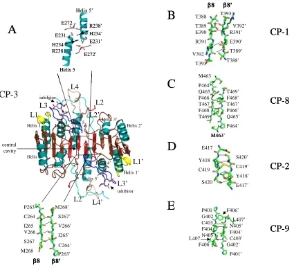
Maturation of caspase–3 is thought to be analogous to that of caspase–7. Prior to
cleavage of the polypeptide chain in the intersubunit linker, one of the two linkers of the
homodimer occupies the central cavity (between β strands 8 and 8’), whereas the other is
positioned outside of the cavity. This effectively sequesters active site loop 2’ (L2’) away
from the active site since it is part of the intersubunit linker in the zymogen. Following
cleavage at Asp175, residues in active site loop 3 (L3) move into the central cavity and
toward the dimer interface, resulting in formation of the substrate binding groove (Figure
5A). In the procaspase, L3 occupies the substrate binding pocket because the loop is
extended away from the protein. Upon cleavage of the intersubunit linker, the newly
formed N-terminus of the small subunit is called L2’, and the newly formed C-terminus
of the large subunit is called L2. L2’ leaves the central cavity and rotates approximately
180° to form new contacts in the loop bundle with L2, L3, and L4 from the opposing
monomer (Figure 5A).
Once L2’ rotates out of the central cavity, the N-terminal region of L3 can insert
into the cavity, and a functional active site forms. More specifically, the arginine
positioned next to the catalytic cysteine (Arg164 in caspase–3, see Figure 2) rotates from
a solvent exposed orientation in the substrate binding pocket toward the dimer interface
ordered lining to the central cavity (Figure 7A). The reorientation of Arg164 on L2
toward the dimer interface and the accompanying loop movements lock the S1–S4
subsites into place and position the backbone amide of the catalytic cysteine into the
correct orientation to form part of the oxyanion hole [103, 104]. Similar structural
rearrangements are observed for caspase–1 as well [106] except that Arg286 (equivalent
to Arg164 in caspase–3, see Figure 2) forms a salt-bridge with Glu390 in the dimer
interface (Figure 7B).
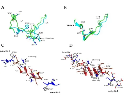
The effector caspases, such as caspases–3 and –7, do not have an equivalent salt
bridge at the dimer interface, but the internalization of the arginine does occur. Like
caspase–1, the comparable arginine in caspases–3 (Arg164) and –7 (Arg187), intercalates
between Tyr197 (β-strand 7) and Pro201 (L3) (caspase–3 numbering, see Figure 2) to
stabilize active site loops 2 and 3. However, the positive charge on Arg164 is neutralized
by Glu124, which is positioned on a loop just above the interface (Figure 7A). This loop
also contains the second residue involved in the catalytic dyad, His121, as well as
Gly122, which forms part of the oxyanion hole. Overall, the conformational changes in
active site loop 2 (L2) stabilize L2 and L3 and position L2, L2’, and L4 to form the loop
bundle (Figure 5A).
The formation of the active site of caspase–9 is unique from all other caspases
because only one active site has the correct conformation to bind substrate. The other
active site is not able to form due to steric constraints of residues in the dimer interface.
The two active sites are in contact with one another through two tyrosine residues and
Tyr345, on β-strand 7, as well as Phe404’ of the second monomer (Figure 7C). Upon
formation of one active site, Tyr345 rotates away from the active site and toward the
dimer interface, causing Phe404 also to rotate toward the dimer interface. This movement
creates a steric clash between the two phenylalanine residues across the dimer interface
(Phe404 and Phe404’), leading to the inactivation of one of the active sites [107]. In order
for the functional active site to form, the side chains of Ser344–Ser353, the so-called
“elbow loop,” from one monomer, contact residues in the dimer interface and insert into
the opposing monomer. Specifically, Phe348 and Phe351 from the elbow loop make
contacts with a hydrophobic pocket lined with Pro338’ and Phe406’ in the opposing
monomer (Figure 7D). By stabilizing the elbow loop, the contacts enable L3 to move into
the substrate binding conformation. Overall, the structural data for caspase–9 demonstrate
that packing forces in the dimer interface affect active site formation, primarily through
interactions between Tyr345 and Phe404 in each monomer and the equivalent
interactions across the dimer interface.
G. Dimer Interface Experiments that Suppress Caspase–3 Activity G.1. FICA and DICA
Disulfide trapping (called “tethering”) experiments were performed on caspases–
3, –7 and –1 to identify allosteric sites that potentially could be used to inhibit dimer
formation [101, 109]. Tethering uses a library of small thiol-containing compounds that
make disulfide bonds with naturally occurring cysteine residues in a protein. The
caspases–3 and –7. Representatives of these classes are FICA
(5-fluoro-1H-indole-2-carboxylic acid (2-mercapto-ethyl) amide) and DICA
(2-(2,4-dichlorophenoxy-N-(2-mercapto-ethyl)-acetamide). Both compounds bound to a single cysteine in the small
subunit on β-strand 8, Cys264 in caspase–3, which sits close to the dimer interface and is
14 Å from the catalytic cysteine. Binding of the inhibitors did not cause dissociation of
the dimer but did prevent binding of the substrate at the active site. Structural data
showed that, in caspase–7, FICA interacts with Tyr223’ (β-strand 7) on the opposing
monomer to which it binds and occupies the central cavity (Figure 6B), whereas DICA
interacts with Tyr223 in the same monomer to which it binds. Regardless, the same
conformational changes occur at the active site in the presence of either FICA or DICA.
By displacing Tyr223, Arg187, on L2, is pushed out of the central cavity and placed into
a position that occludes substrate binding to the active site. In fact, the large
conformational changes that occur as a result of inhibitor binding to the interface
resemble the conformation of the zymogen rather than that of the active enzyme.
In the case of caspase–1, it was found that a thienopyrazole bound to Cys331,
which is near the dimer interface on β-strand 7 and is in the same position as Tyr197 of
caspase–3 or Tyr223 of caspase–7 (see Figure 2) [109]. Like FICA in caspase–7, the
caspase–1 inhibitor interacted with residues on the neighboring monomer. Overall, the
binding of the tethered inhibitor resulted in a disorganized active site by causing the
catalytic cysteine to be rotated away from the S1 subsite, the substrate-binding loop to be
collapsed so that it could not interact productively with substrate, and the side chain of
cleft. As with caspase–7, caspase–1 adopted a conformation closely resembling its
ligand-free form when bound to the allosteric inhibitor. Current models suggest that
binding of the inhibitor to the dimer interface shifts the equilibrium of species found in
solution from the active form to the inactive form, underlining the coupling of the dimer
interface to the active site.
G.2. Caspase-3 Interface Mutant Val266His
In an effort to examine amino acids in the dimer interface and their
communication with active site residues, the central residue of the dimer interface of
caspase–3, Val266 (Figure 7A), was replaced with histidine or glutamate. While the
Val266Glu mutant is described below, replacing Val266 with histidine abolished activity
in both the zymogen and the mature caspase [102]. Limited proteolysis studies
demonstrated that active site loop 3 (L3) was hyperexposed in the mutant at a site over 20
Å from the site of the mutation, demonstrating that L3 was not properly inserted into the
substrate binding pocket in the mutant. Although the crystal structure of the mutant has
not been determined, the Val266His mutation is thought to present similar packing
problems as those found in caspase–9 (Figure 8). In this model, steric constraints in the
dimer interface prevent the side chain of Tyr197 from moving away from the active site,
effectively preventing L3 from forming the substrate binding pocket.
G.3. Experiments that Activate the Caspase-3 Zymogen
In addition to replacing the central valine of caspase–3 with histidine, the residue also
was replaced with glutamate. Remarkably, the Val266Glu mutation resulted in a
increased to a level comparable to that of the wild-type, mature, caspase–3, while
cleavage of the polypeptide chain was not required for the activity increase. Overall, data
from limited proteolysis studies indicate that the Val266Glu mutation in the dimer
interface affects the intersubunit linker in a manner that allows the loop bundle to form
between L2, L4 and L2’, independent of cleavage of the protein. Together, the dimer
interface mutations of caspase–3 are important because they demonstrate that one can
influence the activity of the zymogen, as well as that of the mature enzyme, by disrupting
the interactions between amino acids in the interface and those in the active site. FICA
and DICA, described above, likely mimic the packing effects of the Val266His mutation
to abrogate caspase–3 activity. In contrast, molecules that mimic the effects of the
Val266Glu mutation have yet to be identified, but the results show that the caspase–3
dimer interface should be considered a valid target when formulating drugs to activate the
caspase–3 zymogen.
Recently, Hergenrother and colleagues screened over twenty thousand
compounds for their ability to catalyze the activation of procaspase–3 to mature caspase–
3 [61]. Four compounds were identified that resulted in procaspase–3 activation, and one
compound demonstrated a dose-dependent effect. The compound is called PAC-1, for
first procaspase-activating compound. Importantly, PAC-1 was able to induce apoptosis
in a variety of cancer cell lines, and the data showed a strong correlation between the
cellular concentration of procaspase–3 and PAC-1 sensitivity. Finally, tumor growth was
retarded significantly in mice treated with PAC-1. Though these results are potentially
directly can be effective in cancers, more studies need to be done to determine the
mechanism of action.
H. Conclusions
Caspases are proteases that are intimately involved in the execution of apoptosis
in normal cells. Cancer cells typically have gained the ability to circumvent apoptosis, so
the proteins involved in the apoptotic cascades have become ideal targets for cancer
therapy. Targeting the upstream proteins in these pathways can lead to resistance in
cancer cells, thereby rendering cytotoxic drugs ineffective. On the other hand, directly
targeting effector caspases, such as caspase-3, could lead to reduced resistance, and thus
more effective therapy, since effector caspases ultimately are responsible for cellular
suicide upon their activation. Because dimerization is necessary for proper active site
formation of all caspases, the dimer interface should be considered a potential target for
cancer therapy, where the identification of small molecules that bind to the dimer
interfaces of procaspases, resulting in their activation, should be at the forefront of cancer
REFERENCES
1. Wyllie, A. H.; Kerr, J. F.; Currie, A. R. Cell Death: The Significance of
Apoptosis. International Review of Cytology 1980, 68, 251-306.
2. Majno, G.; Joris, I. Apoptosis, Oncosis and Necrosis. An Overview of Cell
Death. American Journal of Pathology 1995, 146, 3-15.
3. Sanders, E. J.; Wride, M. A. Programmed Cell Death in Development.
International Review of Cytology 1995, 163, 105-173.
4. Beaulaton, J.; Lockshin, R. The Relation of Programmed Cell Death to
Development and Reproduction: Comparative Studies and an Attempt at
Classification. International Review of Cytology 1982, 79, 215-235.
5. Schweichel, J.; Merker, H. The Morphology of Various Types of Cell Death in
Prenatal Tissues. Teratology 1973, 7, 253-266.
6. Zakeri, Z.; Bursch, W.; Tenniswood, M.; Lockshin, R. Cell Death: Programmed
Apoptosis, Necrosis or Other? Cell Death and Differentiation 1995, 2, 83-92.
7. Hinchliffe, J. In Cell Death in Biology and Pathology, I. Bowen and R. Lockshin,
Ed.; Chapman and Hall: New York, 1981, pp. 35-78.
8. Kerr, J. F. R.; Wyllie, A. H.; Currie, A. R. Apoptosis: A Basic Biological
Phenomenon with Wide-Ranging Implications in Tissue Kinetics. British Journal
of Cancer 1972, 26, 239-257.
9. Kaufmann, S. H. Induction of Endonucleolytic DNA Cleavage in Human Acute
Myelogenous Leukemia Cells by Etoposide, Camptothecin, and Other Cytotoxic
Anticancer Drugs: A Cautionary Note. Cancer Research 1989, 49, 5870-5878.
10. Canman, C. E.; Tange, H.-Y.; Normolle, D. P.; Lawrence, T. S.; Maybaum, J.
Variations in Patterns of DNA Damage Induced in Human Colorectal Tumor Cells by 5-Fluorodeoxyuridine: Implications for Mechanisms of Resistance and
Cytotoxicity. Proceedings for the National Academy of Sciences USA 1992, 89,
10474-10478.
11. Fadok, V. A.; Voelker, D. R.; Campbell, P. A.; Cohen, J. J.; Bratton, D. L.;
Henson, P. M. Exposure of Phosphatidylserine on the Surface of Apoptotic Lymphocytes Triggers Specific Recognition and Removal by Macrophages.
12. Ashkenazi, A.; Dixit, V. M. Death Receptors: Signaling and Modulation. Science
1998, 281, 1305-1308.
13. Ashkenazi, A.; Dixit, V. M. Apoptosis Control by Death and Decoy Receptors.
Current Opinion in Cell Biology 1999, 11, 255-260.
14. Walczak, H.; Krammer, P. H. The Cd95 (Apo-1/Fas) and the Trail (Apo-2l)
Apoptosis Systems. Experimental Cell Research 2000, 256, 58-66.
15. Muzio, M.; Stockwell, B. R.; Stennicke, H.; Salvesen, G. S.; Dixit, V. M. An
Induced Proximity Model for Capase-8 Activation. Journal of Biological
Chemistry 1998, 273, 2926-2930.
16. Stennicke, H. R.; Deveraux, Q. L.; Humke, E. W.; Reed, J. C.; Dixit, V. M.;
Salvesen, G. S. Caspase-9 Can Be Activated without Proteolytic Processing.
Journal of Biological Chemistry 1999, 274, 8359-8362.
17. Zou, H.; Li, Y.; Liu, X.; Wang, X. An Apaf-1.Cytochrome C Multimeric Complex
Is a Functional Apoptosome That Activates Procaspase-9. Journal of Biological
Chemistry 1999, 274, 11549-11556.
18. Medema, J. P.; Scaffidi, C.; Kischkel, F. C.; Shevchenko, A.; Mann, M.;
Krammer, P. H.; Peter, M. E. Flice Is Activated by Association with the Cd95
Death-Inducing Signaling Complex (Disc). The EMBO Journal 1997, 16,
2794-2804.
19. Srinivasula, S. M.; Hegde, R.; Saleh, A.; Datta, P.; Shiozaki, E.; Chai, J.; Lee,
R.-A.; Robbins, P. D.; Fernandes-Alnemri, T.; Shi, Y.; Alnemri, E. S. A Conserved Xiap-Interaction Motif in Caspase-9 and Smac/Diablo Regulates Caspase Activity
and Apoptosis. Nature 2001, 410, 112-116.
20. Pop, C.; Chen, Y.-R.; Smith, B.; Bose, K.; Bobay, B.; Tripathy, A.; Franzen, S.;
Clark, A. C. Removal of the Pro-Domain Does Not Affect the Conformation of
the Procaspase-3 Dimer. Biochemistry 2001, 40, 14224-14235.
21. Bose, K.; Pop, C.; Feeney, B.; Clark, A. C. An Uncleavable Procaspase-3 Mutant
Has a Lower Catalytic Efficiency but an Active Site Similar to That of Mature
Caspase-3. Biochemistry 2003, 42, 12298-12310.
23. van Loo, G.; Saelens, X.; van Gurp, M.; MacFarlane, M.; Martin, S.;
Vandenabeele, P. The Role of Mitochondrial Factors in Apoptosis: A Russian
Roulette with More Than One Bullet. Cell Death and Differentiation 2002, 9,
1031-1042.
24. Guo, Y.; Srinivasula, S. M.; Druilhe, A.; Fernandes-Alnemri, T.; Alnemri, E. S.
Caspase-2 Induces Apoptosis by Releasing Proapoptotic Proteins from
Mitochondria. Journal of Biological Chemistry 2002, 277, 13430-13437.
25. Roy, S.; Nicholson, D. W. Cross-Talk in Cell Death Signaling. Journal of
Experimental Medicine 2000, 192, 21-26.
26. Duan, H.; Dixit, V. M. Raidd Is a New 'Death' Adaptor Molecule. Nature 1997,
385, 86-89.
27. Baliga, B. C.; Read, S. H.; Kumar, S. The Biochemical Mechanism of Caspase-2
Activation. Cell Death and Differentiation 2004, 11, 1234-1241.
28. Fulda, S.; Debatin, K.-M. Extrinsic Versus Intrinsic Apoptosis Pathways in
Anticancer Chemotherapy. Oncogene 2006, 25, 4978-4811.
29. Du, C.; Fang, M.; Li, Y.; Li, L.; Wang, X. Smac, a Mitochondrial Protein That
Promotes Cytochrome C-Dependent Caspase Activation by Eliminating Iap Inhibition. Cell 2000, 102, 33-42.
30. Verhagen, A. M.; Ekert, P. G.; Pakusch, M.; Silke, J.; Connolly, L. M.; Reid, G.
E.; Moritz, R. L.; Simpson, R. J.; Vaux, D. L. Identification of Diablo, a Mammalian Protein That Promotes Apoptosis by Binding to and Antagonizing
Iap Proteins. Cell 2000, 102, 43-53.
31. Zangemeister-Wittke, U.; Simon, H.-U. An Iap in Action: The Multiple Roles of
Survivin in Differentiation, Immunity and Malignancy. Cell Cycle 2004, 3,
1121-1123.
32. Nagata, S. Apoptotic DNA Fragmentation. Experimental Cell Research 2000,
256, 12-18.
33. Li, L. Y.; Luo, X.; Wang, X. Endonuclease G Is an Apoptotic Dnase When
Released from Mitochondria. Nature 2001, 412, 95-99.
34. Klein, J. A.; Longo-Guess, C. M.; Rossman, M. P.; Seburn, K. L.; Hurd, R. E.;
Frankel, W. N.; Bronson, R. T.; Ackerman, S. L. The Harlequin Mouse Mutation
35. Hansen, T. M.; Nagley, P. Aif: A Multifunctional Cog in the Life and Death
Machine. Science STKE 2003, 193, pe31.
36. Debatin, K.-M. In Drug Resistance Updates, H. Broxterman, Ed.; Churchill
Livingstone: Edinburgh, 1999, pp. 85-90.
37. Debatin, K.-M. Anticancer Drugs, Programmed Cell Death and the Immune
System: Defining New Roles in an Old Play. Journal of the National Cancer
Institute 1997, 89, 750-753.
38. Herr, I.; Debatin, K.-M. Cellular Stress Response and Apoptosis in Cancer
Therapy. Blood 2001, 98, 2603-2614.
39. Kaufmann, S. H.; Earnshaw, W. C. Induction of Apoptosis by Cancer
Chemotherapy. Experimental Cell Research 2000, 256, 42-49.
40. Solary, E.; Droin, N.; Bettaieb, A.; Corcos, L.; Dimanche-Boitrel, M.; Garrido, C.
Positive and Negative Regulation of Apoptotic Pathways by Cytotoxic Agents in
Hematological Malignancies. Leukemia 2000, 14, 1833-1849.
41. Debatin, K.-M. Apoptosis Pathways in Cancer and Cancer Therapy. Cancer
Immunology, Immunotherapy 2004, 53, 153-159.
42. Krammer, P. H. Cd95's Deadly Mission in the Immune System. Nature 2000,
407, 789-795.
43. Friesen, C.; Herr, I.; Krammer, P. H.; Debatin, K.-M. Involvement of the Cd95
(Apo-1/Fas) Receptor/Ligand System in Drug-Induced Apoptosis in Leukemia
Cells. Nature Medicine 1996, 2, 574-577.
44. Muller, M.; Strand, S.; Hug, H.; Heinemann, E.-M.; Walczak, H.; Hofmann, W.
J.; Stremmel, W.; Krammer, P. H.; Galle, P. R. Drug-Induced Apoptosis in Hepatoma Cells Is Mediated by the Cd95 (Apo-1/Fas) Receptor/Ligand System
and Involves Activation of Wild-Type P53. Journal of Clinical Investigation
1997, 99, 403-413.
45. Muller, M.; Wilder, S.; Bannasch, D.; Israeli, D.; Lehlbach, K.; Li-Weber, M.;
Friedman, S. L.; Galle, P. R.; Stremmel, W.; Oren, M.; Krammer, P. H. P53 Activates the Cd95 (Apo-1/Fas) Gene in Response to DNA Damage by
46. Ashkenazi, A.; Pai, R. C.; Fong, S.; Leung, S.; Lawrence, D. A.; Marsters, S. A.; Blackie, C.; Chang, L.; McMurtrey, A. E.; Hebert, A.; DeForge, L.; Koumenis, I. L.; Lewis, D.; Harris, L.; Bussiere, J.; Koeppen, H.; Shahrokh, Z.; Schwall, R. H.
Safety and Antitumor Activity of Recombinant Soluable Apo2 Ligand. Journal of
Clinical Investigation 1999, 104, 155-162.
47. Lawrence, D. A.; Shahrokh, Z.; Marsters, S. A.; Achilles, K.; Shih, D.; Mounho,
B.; Hillan, K.; Totpal, K.; DeForge, L.; Schow, P.; Hooley, J.; Sherwood, S.; Pai, R. C.; Leung, S.; Khan, L.; Gliniak, B.; Bussiere, J.; Smith, C. A.; Strom, S. S.; Kelley, S.; Fox, J. A.; Thomas, D.; Ashkenazi, A. Differential Hepatocyte
Toxicity of Recombinant Apo2l/Trail Versions. Nature Medicine 2001, 7,
383-385.
48. LeBlanc, H. N.; Ashkenazi, A. Apo-2l/Trail and Its Death and Decoy Receptors.
Cell Death and Differentiation 2003, 10, 66-75.
49. Jo, M.; Kim, T.-H.; Seol, D.-W.; Esplen, J. E.; Dorko, K.; Billiar, T. R.; Strom, S.
C. Apoptosis Induced in Normal Human Hepatocytes by Tumor Necrosis
Factor-Related Apoptosis-Inducing Ligand. Nature Medicine 2000, 6, 564-567.
50. Nitsch, R.; Bechmann, I.; Deisz, R. A.; Haas, D.; Lehmann, T.-N.; Wendling, U.;
Zipp, F. Human Brain-Cell Death Induced by Tumour-Necrosis-Factor-Related
Apoptosis-Inducing Ligand (Trail). Lancet 2000, 356, 827-828.
51. Ehrhardt, H.; Fulda, S.; Schmid, I.; Hiscott, J.; Debatin, K.-M.; Jeremias, I. Trail
Induced Survival and Proliferation in Cancer Cells Resistant Towards
Trail-Induced Apoptosis Mediated by Nf-Κb. Oncogene 2003, 22, 3842-3852.
52. Karin, M.; Cao, Y.; Greten, F. R.; Li, Z.-W. Nf-Κb in Cancer: From Innocent
Bystander to Major Culprit. Nature Reviews Cancer 2002, 2, 301-310.
53. Sheikh, M. S.; Huang, Y.; Fernandez-Salas, E. A.; El-Deiry, W.; Friess, H.;
Amundson, S.; Yin, J.; Meltzer, S. J.; Holbrook, N. J.; Fornace, A. J. J. The Antiapoptotic Decoy Receptor Trid/Trail-R3 Is a P53-Regulated DNA Damage Inducible Gene That Is Overexpressed in Primary Tumors of the Gastrointestinal
Tract. Oncogene 1999, 18, 4153-4159.
54. Baylin, S. B. Mechanisms Underlying Epigenetically Mediated Gene Silencing in
Cancer. Seminars in Cancer Biology 2002, 12, 331-337.
55. van Noesel, M. M.; van Bezouw, S.; Voute, P.; Herman, J. G.; Pieters, R.;
Verstee, R. Clustering of Hypermethylated Genes in Neuroblastoma. Genes,