DOI : https://doi.org/10.32628/CSEIT195231
An Efficient Clustering Approach for Automatic
Detection of Calcification in Low Dose Chest CT
Dr. P. Tamijiselvy, N. Kavitha, K. M. Keerthana, D. Menakha
Department of Computer Science and Engineering, Sri Krishna College of Technology, Coimbatore Tamil Nadu, India
ABSTRACT
The degree of aortic calcification has been appeared to be a risk pointer for vascular occasions including cardiovascular events. The created strategy is fully automated data mining algorithm to segment and measure calcification using Low-dose Chest CT in smokers of age 50 to 70 .The identification of subjects with increased cardiovascular risk can be detected by using data mining algorithms. This paper presents a method for automatic detection of coronary artery calcifications in low-dose chest CT scans using effective clustering algorithms with three phases as Pre-Processing, Segmentation and clustering. Fuzzy C Means algorithm provides accuracy of 80.23% demonstrate that Fuzzy C means detects the Cardio Vascular Disease at early stage. Keywords : Calcium Scoring, Clustering, low-dose chest CT, lung cancer screening.
I. INTRODUCTION
Coronary Artery Disease (CAD) is one of the leading causes of mortality nowadays. Screening with low-dose chest CT has been found effective in reducing mortality from lung cancer in current or former heavy smokers. However, heavy smoking is not only a major risk factor for lung cancer, but also for heart disease. The presence of CVD is detected using CT scans by measuring the amount of coronary artery calcification (CAC) CVD [1]. Being prolonged and heavy smoker is a strong etiologic factor in the development of lung cancer and Cardiovascular Disease (CVD), simultaneously simultaneous screening for both diseases in a high-risk population of heavy smokers and former smokers is an attractive strategy for maximizing the beneficial effects on
survival.There are different types of calcifications found on coronary artery.
A. Metastatic calcification
Metastatic calcification with hypercalcemia happens when calcium stores in normal tissue .Metastatic calcification can be isolated into harmful and non threatening causes. The system of metastatic calcification isn't clear. The procedure appears to influence for the most part interstitial tissues of the gastric mucosa, kidneys, lungs, systemic arteries and pneumonic veins [2,3].
B. Dystrophic calcification
Dystrophic calcification occurs in previously damaged tissue. When the membrane of a vesicle gets damaged, calcium binds to the phospholipid in the membrane. Some lesions with dystrophic calcification include atherosclerotic plaques, aging or damaged heart valves, and tuberculous lymph nodes. Dystrophic calcification looks like tiny, white, gritty granules [2,3].
II. RELATED WORKS
Nikolas Lessmanna, IvanaIsgumadescribesthe measure of calcifications in the Coronary Arteries is a ground- breaking and autonomous indicator of cardiovascular occasions and is utilized to recognize subjects at high risk who may benefit by preventive treatment. Routine evaluation of coronary calcium scores can supplement screening programs utilizing low-portion chest CT, for example, lung malignant growth screening or Cardiovascular illness. The paper present a framework for programmed coronary calcium scoring dependent on profound convolutional neural systems (CNNs) [4] .
Wehab Ahmed , Michiel A. de Graaf says A fully automatic detection and quantification of Agatston CAC score on differentiation CTA is attainable and demonstrates an relationship with the Agatston CAC score inferred from non-differentiate CT. Moreover, a great understanding was gotten between the non-gated CT and the non-gated CT inside the Agatston CAC
score chance classifications. The accuracy to rule-out CAC on contrast CTA compared to non-contrast CT was excellent [5].
Wei Liu, Yue Zhang, Cheuk-Man Yu describes Coronary vein calcification (CAC) is exceedingly predominant in patients with coronary illness (CHD) and is related with major unfriendly cardiovascular occasions. There are two perceived kind of CAC intimal and average calcification, and every one of them have explicit risk factors.Several theories about the mechanism of vascular calcification have been put forward, and currently believe that vascular calcification is an active, regulated process. CAC be found in patients with extreme CHD, and this asymptomatic phenomenon make early analysis of CAC essential [6].
III. PROPOSED SCHEME
The proposed method detects and cluster the calcification from LDCT. The basic block diagram consists of four steps. Each step uses algorithms to classify the frames in the image . The first method is pre-processing with images where both input and output are intensity images.The second method is segmentations to simplify and change the representation of an image into something that is more meaningful and easier to analyze. The third is clustering by k-means clustering and fuzzy c means clustering logic.
A.Dataset
For training and validation, Low-dose Chest CT scans acquired in the National Lung Screening Trial (NLST) are used. The NSLT was a large lung cancer screening trial in the United States that enrolled 254 current or former heavy smokers aged 50 to 70 [7]. To establish a reference standard, calcifications were manually labeled in all scans. Manual calcium calculation usually requires the observer to select only a single voxel per lesion [8]. The lesion is automatically segmented with region growing using the standard intensity threshold of 130HU. Depending on the amount of calcification and the image quality, the annotation effort varied from 5-10 minutes for images with k-means clustering with large amounts of calcium [9].The scans were acquired at breath-hold after inspiration in helical scanning mode without contrast enhancement and without ECG synchronization. Tube voltage was set to 120 kVp, or 140 kVp for large subjects (5%). In-plane resolution ranged from 0.49mm to 0.98mm, slice thickness from 1mm to 3mm and slice spacing from 0.6mm to 3.0mm. Since calcium scoring is typically performed on 3mm thick slices, we reconstructed 3mm thick axial slices with 1.5mm slice spacing from all scans.
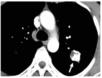
Fig.2.1. This picture shows the Low Dose Chest CT image with Calcification.
B. Preprocessing
Pre-processing is an improvement of the image
information that performs smoothning of
undesirable distortions or upgrade some image features for further handling. Pre-processing is to improve image
information that stifles undesirable bending or upgrades some image highlight essential for further preparing. Contrast Limited Histogram Equalization (CLAHE) [11] is a technique to enhance the visibility of local subtleties of a image by expanding the complexity of nearby regions. Contrast Limited Adaptive Histogram Equalization (CLAHE) was originally developed for medical imaging.CLAHE is the advancement of Adaptive
Histogram Equalization(AHE) .CLAHE was
created to keep the over enhancement of noise that adaptive histogram equalization give rise to [12]. It works on little information locales (tiles), instead of the whole image. It presents substantial changes in the pixel dark dimensions. CLAHE limits the intensification by cut- out the histogram at a predefined esteem (called clip limit) before processing the CDF. Contrast Limited Histogram Equalization (CLAHE) [13] varies from ordinary histogram equalization out in its contrast limiting.The enhancement of image is performed using CLAHE
C.Segmentation
Image segmentation is to segment an image into totally different teams [14].The goal of segmentation is to simplify and change the illustration of an image into one image that is more meaning and easier for research. Before applying K-means algorithmic rule, partial stretching enhancement is applied to the image to boost the standard of the image.In k-means clustering, it partitions a collection of information into a k variety group of information.
Fig.2.3. Segmentation Using K Means on Low Dose CT Image
D. Clustering
Clustering is that grouping of similar data together. The clustering method applied on the segmented image isfuzzy c means clustering [13].
(i)
Fuzzy C-Means ClusteringClustering involves assigning data points to clusters such that items in the same cluster are as similar as possible, while items belonging to different clusters are as dissimilar as possible. Clusters are identified via similarity measures. These similarity measures include distance, connectivity, and intensity.Fuzzy c-means clustering is used for the chest CT images for clustering the calcium in the coronary artery [15]. The gray scale image of the chest CT scan is given as input image for fuzzy c-means clustering. Each time
the required image is read and the output image is given using fuzzy c-means clustering [16].
The steps in the algorithm
Initialize the value to the matrix generated. At k- steps calculate the centers vectors then STOP. If the condition is not satisfied then keep doing the loops.
Fig.2.4. Clustering using Fuzzy C Means algorithm on low dose CT image
Advantages of Fuzzy C Means Clustering
● Fuzzy C Means algorithm gives the best result of overlapped data.It is better than K Means algorithm.
● In K Means algorithm the cluster point is present in one cluster centre but in the Fuzzy C Means algorithm the cluster points are present in more than one clustering.
IV.EXPERIMENTAL RESULTS
The accuracy and sensitivity are calculated from the Fuzzy C Means algorithm. The performance analysis of Fuzzy C Means can be calculated using the formula depicted in equation (1) and (2)
Accuracy=(((TP+TN))/(TP+TN+FP+FN))*100 (1) Sensitivity=(TP/(TP+FN))*100
(2) Where,
is YES and the actual output is also YES)
TN=True Negative (The cases in which the prediction is NO and the actual output is also NO)
FP=False Positive (The cases in which the prediction is YES and the actual output is NO)
FN=False Negative (The cases in which the prediction is NO and the actual output is YES)
Fig.3.This picture describes the performance analysis for Fuzzy C Means algorithm
The above bar charts shows that Fuzzy C Means Clustering algorithms has more sensitivity and accuracy. This shows that the calcification content in the coronary artery are effectively predicted in the early stage by using Fuzzy C Means algorithm with the accuracy of 80.23%
V. CONCLUSION
This paper presents a method for automatic detection of CAC (Coronary Artery Calcifications) in Low-dose Chest CT at an early stage.Fuzzy C Means algorithm is more effective in finding the Calcium deposition in coronary artery. The future enhancement is done by optimizing the output produced by the clustering algorithm for exact output prediction of calcium content.
VI. REFERENCES
[1]. Rasmussen T, Kober L, Abdulla J, et al. Coronary artery calcification detected in
lung cancer screening predicts cardiovascular death. ScandCardiovasc J 2015;49:159–67 [2]. M. Senba, K. Kawai, S. Chiyoda, and O.
Takahara, “Metastatic liver calcification in
adult T-cell leukemia- lymphoma associated
with hypercalcemia,” American Journal of
Gastroenterology, vol. 85, no. 9, pp. 1202–
1203, 1990. View at Google Scholar · View at Scopus
[3]. M. Senba and K. Kawai, “Metastatic
calcification due to hypercalcemia in adult T-cell leukemia-lymphoma (ATLL),” Zentralblatt
für Pathologie, vol. 137, no. 4, pp. 341–345, 1991. View at Google Scholar · View at Scopus [4]. Nikolas Lessmanna, IvanaIsguma, Arnaud A.A.
Setiob, Bob D. de Vosa,FrancescoCiompib, Pim A. de Jongc, MatthijsOudkerkd, Willem P.Th.M. Malic,Max A. Viergevera, and Bram
van Ginnekenb,”Deep convolutional neural
networks for automatic coronary calcium scoring in a screening study with low-dose chest CT.
[5]. Wehab Ahmed Michiel A. de Graaf Alexander Broersen Pieter H. Kitslaar Elco Oost Jouke Dijkstra
[6]. Jeroen J. Bax Johan H. C. Reiber Arthur J.
Scholte” Automatic detection and
quantification of the Agatston coronaryartery calcium score on contrast computed
tomography angiography”2014.
[7]. Mahesh V.Madhavan , Madhusudhantarigopul
, Gary S.Mintz , Akikomaehara “Coronary
Artery Calcification: Pathogenesis And
Prognostic Implications“(2014).
[8]. The National Lung Screening Trial Research
Team, “Reduced lung-cancer mortality with
low-dose computed tomography screening.”
New England J. Med., vol. 365, pp. 395-409, Aug. 2011.
the National Lung Screening Trial. AJR. 2011;197:1165–1169. doi:
10.2214/AJR.11.6533. PMid:22021510.
[10]. Blanchon T, Bréchot JM, Grenier PA, Ferretti GR, Lemarié E, Milleron B, et al. Baseline results of the Depiscan study: a French randomized pilot trial of lung cancer screening comparing low dose CT scan (LDCT) and chest X-ray (CXR). Lung Cancer2007;58:50–
8.10.1016/j.lungcan.2007.05.009.
[11]. Oken MM, Hocking WG, Kvale PA, Andriole GL, Buy s SS, Church TR, Crawford ED, Fouad MN, Isaacs C, R eding DJ, et al.; PLCO Project Team. Screening by chest radiograph and lung cancer mortality: the Prostate, Lung, Colorectal, and
Ovarian(PLCO)randomizedtrial. JAMA2011;306:1865– 1873.
[12]. MadhukarBhat,TarunPatil M S.Adaptive clip limit for contrast limited adaptive histogram equalization (CLAHE) of medical images using least mean square algorithm 2014.INSPEC
Accession Number:
14870319,DOI:10.1109/ICACCCT.2014.70193 00
[13]. K. H. Memon and D. Lee, "Generalised fuzzy c-means clustering algorithm with local information," in IET Image Processing, vol. 11, no. 1, pp. 1-12, 1 2017.doi: 10.1049/iet-ipr.2016.0282
[14]. Zuiderveld, Karel. (1994). Contrast Limited Adaptive Histogram Equalization. Graphics Gems IV. 474-485. 10.1016/B978-0-12-336156-1.50061-6.
[15]. Office for National Statistics. Adult Smoking Habits in the UK: 2015. Newport: Office for National Statistics; 2017.
[16]. Alan Jose, S. Ravi and M. Sambath, Brain Tumor Segmentation using K -means Clustering and Fuzzy C- means Algorithm and its Area Calculation. In International Journal of Innovative Research in Computer and
Communication Engineering, vol. 2, issue 2, March (2014).
[17]. T. Kanungo, D. M. Mount, N. S. Netanyahu, C. D. Piatko, R. Silverman and A. Y. Wu, "An efficient k- means clustering algorithm: analysis and implementation," in IEEE Transactions on Pattern Analysis and Machine Intelligence, vol. 24, no. 7, pp. 881-892, July 2002. doi: 10.1109/TPAMI.2002.1017616. [18]. K. H. Memon and D. Lee, "Generalised fuzzy
c-means clustering algorithm with local information," in IET Image Processing, vol. 11, no. 1, pp. 1-12, 1 2017.doi: 10.1049/iet-ipr.2016.0282.
[19]. Shmueli A, FraifeldS, Peretz T, Gutfeld O, Gips M, Sosna J, et al. Cost-effectiveness of baseline low-dose computed tomography screening for lung cancer: the Israeli experience. Value Health [Internet] 2013 Sep;16(6):922–931. [cited 2015 Aug 28]; [20]. Office for National Statistics. Adult Smoking
Habits in the UK: 2015. Newport: Office for Menakha, "An Efficient Clustering Approach For Automatic Detection of Calcification in Low Dose Chest CT", International Journal of Scientific Research in Computer Science, Engineering and Information Technology (IJSRCSEIT), ISSN : 2456-3307, Volume 5 Issue 2, pp. 163-168, March-April
2019. Available at doi :
https://doi.org/10.32628/CSEIT195231