DOI: 10.1534/genetics.105.052258
FluG-Dependent Asexual Development in
Aspergillus nidulans
Occurs via
Derepression
Jeong-Ah Seo,* Yajun Guan
†,1and Jae-Hyuk Yu*
,†,2*Department of Food Microbiology and Toxicology and†Molecular and Environmental Toxicology Center, University of Wisconsin, Madison, Wisconsin 53706
Manuscript received October 10, 2005 Accepted for publication December 30, 2005
ABSTRACT
The asexual spore is one of the most crucial factors contributing to the fecundity and fitness of fila-mentous fungi. Although the developmental activator FluG was shown to be necessary for activation of asexual sporulation (conidiation) and production of the carcinogenic mycotoxin sterigmatocystin (ST) in the model filamentous fungus Aspergillus nidulans, the molecular mechanisms underlying the devel-opmental switch have remained elusive. In this study, we report that the FluG-mediated conidiation inA. nidulansoccurs via derepression. Suppressor analyses of fluGled to the identification of thesfgAgene encoding a novel protein with the Gal4-type Zn(II)2Cys6binuclear cluster DNA-binding motif at the N terminus. Deletion (D) and 31 other loss-of-function sfgA mutations bypassed the need for fluG in conidiation and production of ST. Moreover, bothDsfgAandDsfgADfluGmutations resulted in identical phenotypes in growth, conidiation, and ST production, indicating that the primary role of FluG is to remove repressive effects imposed by SfgA. In accordance with the proposed regulatory role of SfgA, overexpression of sfgA inhibited conidiation and delayed/reduced expression of conidiation- and ST-specific genes. Genetic analyses demonstrated that SfgA functions downstream of FluG but upstream of transcriptional activators (FlbD, FlbC, FlbB, and BrlA) necessary for normal conidiation.
R
EPRODUCTION of fungi results in the formation of enormous numbers of spores that are extremely efficient for genome protection, survival, and propaga-tion. Spores are also the primary means of infecting host organisms for many human and plant pathogenic fungi. Asexual sporulation is a highly successful and ef-fective reproductive mechanism for a diverse group of fungi because vast numbers of spores (called conidia for higher fungi) can be produced from a single colony through repetitive cycles of mitosis (reviewed in Adams 1994).Members of the genus Aspergillus are the most com-mon fungi and many are beneficial to humans. However, they also include serious human and plant pathogens. Furthermore, certain aspergilli have the capacity to pro-duce one or more toxic secondary metabolites called mycotoxins, which contaminate various plant-based foods/ feeds and cause adverse health effects to humans and animals such as liver or kidney damage, cancer, and even death (reviewed in Seoand Yu2004). All aspergilli pro-duce asexual spores (conidia) as the main means of
dispersion and infection. Asexual development (con-idiation) and production of certain carcinogenic myco-toxins are intimately associated (reviewed in Calvoet al. 2002; Yuand Keller2005).
Aspergillus nidulanshas served as an excellent model system for studying multicellular development and sec-ondary metabolism (Timberlake 1990; Martinelli 1994; Yuand Keller2005). Conidiation inA. nidulans involves many common developmental themes includ-ing spatial and temporal regulation of gene expression, specialized cellular differentiation, and intercellular communication. The asexual reproductive cycle of A. nidulansbegins and ends with the asexual spore called the conidium and can be divided into two distinct phases, growth and development. The vegetative growth phase involves germination of the conidium and formation of an undifferentiated network of interconnected hyphal (vegetative) cells that form the mycelium. After a certain period of vegetative growth, under appropriate con-ditions, some of the hyphal cells stop normal growth and begin development by forming complex multicel-lular structures called conidiophores that bear multiple chains of conidia (reviewed in Adams1994).
A key step in conidiophore development is activa-tion of thebrlAgene, which encodes a C2H2zinc finger
transcription factor (Adams et al. 1988; Chang and Timberlake 1992). A series of studies have identified six genes (fluG,flbA,flbB,flbC,flbD, andflbE) that are Sequence data from this article have been deposited with the EMBL/
GenBank Data Libraries under accession no. AAY99779.
1Present address:Department of Environmental Health Sciences, Univer-sity of Michigan, 109 Observatory St., 1506 SPH I, Ann Arbor, MI 48109. 2Corresponding author:Food Research Institute, University of Wisconsin, 1925 Willow Dr., Madison, WI 53706-1187. E-mail: [email protected]
required for proper brlA expression and conidiation. Mutations in any of these genes result in ‘‘fluffy’’ colonies that are characterized by undifferentiated cotton-like masses of vegetative cells (reviewed in Adamset al. 1998). The possible genetic interactions between various flb genes were examined and the two genetic regulatory cascades have been proposed (Figure 1), in which the fluGgene functions most upstream (Wieserand Adams 1995; Leeand Adams1996).
Loss offluGfunction results in the blockage in both conidiation and production of the carcinogenic myco-toxin sterigmatocystin (ST), the penultimate precursor of the better-known potent carcinogen aflatoxin (Lee and Adams1994b; Hickset al. 1997). Conidiation in the
fluGmutants can be rescued by growing them next to
wild-type or other developmental mutants. This obser-vation has led to the proposal that FluG is responsible for the production of an extracellular diffusible con-idiation factor (Leeand Adams1994b). The C-terminal half of FluG contains a glutamine synthetase I (GSI)-like domain (Lee and Adams 1994b) and the N-terminal half is similar to the product of the early nodulin gene MtN6 inMedicago truncatula(Mathiset al. 1999). Over-expression of the full-length or the C-terminal half of fluGin vegetative hyphae was sufficient to cause devel-opment of conidiophores in liquid submerged culture, where conidial development is normally suppressed (Leeand Adams1996; D’Souzaet al. 2001). Collectively, it has been proposed that FluG is involved in the con-stitutive synthesis of an extracellular sporulation-inducing factor that is related to glutamine or glutamate (reviewed in Adamset al. 1998). The two primary consequences
of FluG activity were thought to be: (1) activation of development-specific regulatory cascades and (2) posi-tive modulation of the regulator of G-protein signaling (RGS) protein FlbA, which in turn inactivates vegetative growth signaling mediated by a heterotrimeric G pro-tein (see Figure 1; Leeand Adams1994a; Yuet al. 1996; Adamset al. 1998). For asexual development to occur both processes must occur. However, the molecular mechanism underlying the FluG-mediated initiation of conidiation remains to be uncovered.
To dissect the molecular events responding to FluG activity, we isolated 40 suppressor mutations (s uppres-sor of fluG, sfg) that bypass the need for fluG in co-nidiation and ST production (Seoet al. 2003). In this study, we have identified and characterized the key suppressorsfgAthat is defined by 31sfgmutations. The sfgAgene is predicted to encode a novel 601-amino-acid (aa) protein containing the Gal4-type Zn(II)2Cys6
binuclear cluster DNA-binding motif at the N terminus, suggesting that SfgA is likely a transcription factor. Various mutations have been identified within the sfgA -coding region from all 31 SFGA mutants. Deletion of sfgA completely bypasses fluG in conidiation and ST production. Further studies ofsfgAhave elucidated that conidiation inA. nidulansoccurs via derepression and the primary role of FluG is to remove intracellular repressive effects imposed by SfgA. Our findings eluci-date a new theory for the central regulatory mechanism of conidiation and ST biosynthesis inA. nidulans.
MATERIALS AND METHODS
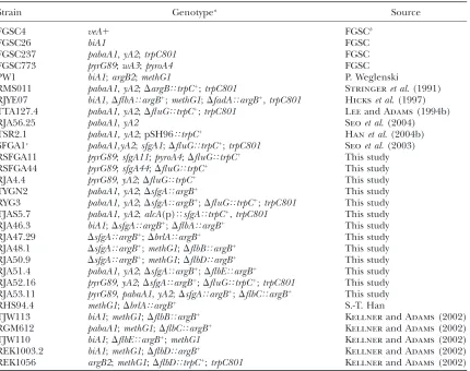
A.nidulans strains, media, growth conditions, and genetic analysis:A. nidulansstrains used in this study are listed in Table 1. Standard culture and genetic techniques were employed (Pontecorvo et al. 1953; Ka¨ fer 1977). All liquid cultures
were inoculated with 53107spores in 100 ml of liquid glucose (1%) minimal medium½pH 6.5; 10 g/liter glucose, 6 g/liter NaNO3,0.52 g/liter MgSO47H2O, 0.52 g/liter KCl, 1.52 g/ liter KH2PO4, and 1 ml/liter of 10003trace element solution composed of 22 g/liter ZnSO47H2O, 11 g/liter H3BO3, 5 g/ liter MnCl24H2O, 5 g/liter FeSO47H2O, 1.6 g/liter CoCl25H2O, 1.6 g/liter CuSO45H2O, 1.1 g/liter (NH4)6Mo7O244H2O, 50 g/liter Na2EDTAwith supplements (simplified as MMG) or MMG with 0.1% (w/v) yeast extract (YE) and incubated at 37°, 250 rpm. Submerged development of individual strains was observed under a microscope at 1-hr intervals after an initial 18-hr growth period in liquid culture. Asexual developmental induction was performed as previously described (Adamset al.
1988). As a prerequisite for the identification of thesfgAgene,
DfluG sfgA11 pyrG89 (RSFGA11) and DfluG sfgA44 pyrG89
(RSFGA44) strains were generated by meiotically crossing
DfluG sfgA11andDfluG sfgA44strains with the developmen-tally wild-type strain FGSC773 (Table 1) and by examining 20 sporulating progeny that required uracil for growth and carried the DfluG genotype. Such conidial strains with the
DfluGSouthern blot hybridization pattern are expected to have theDfluG sfgA11 pyrG89orDfluG sfgA44 pyrG89genotypes.
To examine the effect of overexpression of sfgA by an ectopic copy of sfgA under the alcA promoter, control (TSR2.1) and alcA(p)TsfgA (TJAS5.7; Table 1) strains were Figure1.—Genetic model for growth and developmental
control inA. nidulans.Activities of two antagonistic regulatory pathways control growth, development, and ST (structure shown) production. Activation of G-protein-mediated vegeta-tive signaling represses both development and ST biosynthe-sis (Yuet al.1996; Hickset al.1997). FlbA is an RGS protein
that rapidly turns off this growth signaling (Leeand Adams
1994a; Yuet al.1996). At least partial inhibition of growth
sig-naling is required for development and ST production. Acti-vation of conidiation requires the activities of FluG and other developmental genes,flbB,flbC,flbD,flbE, andbrlA(reviewed in Adamset al.1998). FluG is thought to have dual functions,
grown in liquid MMG at 37°, 250 rpm for 14 hr. Subsequently, mycelia were collected, rinsed with liquid medium without a carbon source, divided into two equal parts, transferred into solid (1.6% agar added to the above composition) MMG (noninducing conditions) or solid MM with 100 mm
threo-nine as a sole carbon source (MMT) (inducing overexpression ofsfgA; composition the same as above except for adding 11 g/ liter threonine instead of 10 g/liter glucose), and further incubated at 37°. Progression of asexual development of control andalcA(p)TsfgAstrains was examined under a mi-croscope, and samples were collected at 3, 6, 9, 12, and 24 hr post-transfer and subjected to total RNA isolation.
Cloning ofsfgA:Cloning ofsfgAwas carried out employing the pRG3-AMA1-based wild-type genomic DNA plasmid li-brary withpyr4ofN. crassaas the selective marker (Osherov
and May2000). EachDfluG sfgA11 pyrG89andDfluG sfgA44
pyrG89mutant strain was transformed with the library and the fluffy progeny were screened visually. After screening.5000 transformants for each case, two to three DfluG-like fluffy transformants were isolated. Total DNA was isolated from individual transformants and used to transformEscherichia coli
DH10B by electroporation. From each transformant, the pRG3-AMA1 plasmids carrying an7.0-kb insert were isolated. These plasmids were then reintroduced into DfluG sfgA11 pyrG89 and DfluG sfgA44 pyrG89 recipient strains and were
confirmed to restore the fluffy phenotype at a high (60%) frequency. The responsible gene within the 7.0-kb insert was identified by a transposon (Tn)-mediated mutagenesis using anin vitrotransposon kit (GPS-1 system; New England Biolabs, Beverly, MA). Twenty Tn-tagged clones were analyzed for Tn integration within the 7.0-kb insert and such Tn-tagged clones were introduced intoDfluG sfgA11 pyrG89and DfluG sfgA44 pyrG89recipient strains. About 70% of the Tn-tagged clones failed to restore the fluffy phenotype, indicating that Tn had disrupted the responsible gene. Most of the Tn-tagged plasmids were sequenced, and the assembled sequences were used for blastn search of theA. nidulansgenome (the Broad Institute: http://www.broad.mit.edu/annotation/fungi/aspergillus/index. html).
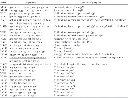
Construction ofsfgAmutants:The double-joint PCR method was used to generate thesfgAdeletion mutant (Yuet al. 2004).
Briefly, using wild-type (FGSC4) genomic DNA as a template 59- and 39-flanking regions (1 kb) ofsfgAwere amplified with the primer pairs of OYG7-9 and OYG10-11, respectively. The
argB1
marker was amplified with OKH60-61 (Hanet al. 2004a).
The three amplicons were fused as described (Yuet al.2004).
The finalsfgAdeletion construct was amplified with OYG8 and OYG12 (Table 2) and introduced into PW1. Resulting trans-formants were randomly screened for deletion ofsfgAby PCR analysis using OYG21-22 followed by Southern blot. TheDsfgA TABLE 1
A. nidulansstrains used in this study
Strain Genotypea Source
FGSC4 veA1 FGSCb
FGSC26 biA1 FGSC
FGSC237 pabaA1,yA2;trpC801 FGSC
FGSC773 pyrG89;wA3;pyroA4 FGSC
PW1 biA1;argB2;methG1 P. Weglenski
RMS011 pabaA1,yA2;DargBTtrpC1;trpC801 Stringeret al. (1991)
RJYE07 biA1,DflbATargB1;methG1;DfadATargB1,trpC801 Hickset al. (1997)
TTA127.4 pabaA1,yA2;DfluGTtrpC1;trpC801 Leeand Adams(1994b)
RJA56.25 pabaA1,yA2 Seoet al. (2004)
TSR2.1 pabaA1, yA2; pSH96TtrpC1
Hanet al. (2004b)
SFGA1c pabaA1,yA2;sfgA1;DfluG
TtrpC1;trpC801 Seoet al. (2003)
RSFGA11 pyrG89;sfgA11;pyroA4;DfluGTtrpC1 This study
RSFGA44 pyrG89;sfgA44;DfluGTtrpC1 This study
RJA4.4 pyrG89,yA2;DfluGTtrpC1 This study
TYGN2 pabaA1,yA2;DsfgATargB1 This study
RYG3 pabaA1,yA2;DsfgATargB1;DfluGTtrpC1;trpC801 This study TJAS5.7 pabaA1,yA2;alcA(p)TsfgATtrpC1,trpC801 This study RJA46.3 biA1;DsfgATargB1;DflbATargB1 This study
RJA47.29 DsfgATargB1;DbrlATargB1 This study
RJA48.1 DsfgATargB1;methG1;DflbBTargB1 This study RJA50.9 DsfgATargB1;methG1;DflbDTargB1 This study RJA51.4 pabaA1,yA2;DsfgATargB1;DflbETargB1 This study RJA52.16 pyrG89,yA2;DsfgATargB1;DfluGTtrpC1;trpC801 This study RJA53.11 pyrG89,pabaA1,yA2;DsfgATargB1;DflbCTargB1 This study
RHS94.4 methG1;DbrlATargB1 S.-T. Han
TJW113 biA1;methG1;DflbBTargB1 Kellnerand Adams(2002)
RGM612 pabaA1;methG1;DflbCTargB1 Kellnerand Adams(2002)
TJW110 biA1;DflbETargB1;methG1 Kellnerand Adams(2002)
REK1003.2 biA1;methG1;DflbDTargB1 Kellnerand Adams(2002)
REK1056 argB2;methG1;DflbDTtrpC1;trpC801 Kellnerand Adams(2002)
aAll strains carry theveA1mutation except FGSC4. bFungal Genetics Stock Center.
mutant was meiotically crossed with RJA4.4 (pyrG89, yA2;
DfluGTtrpC1) and theDsfgADfluGdouble mutant was sub-sequently identified among the progeny. ThesfgAoverexpression mutant was generated by a PCR-assisted method as described (Yuet al.2004). Briefly, thealcApromoter (Gwynneet al. 1987)
and thesfgAORF with terminator were amplified using the primer pairs OJA106-107 and OYG11-28, respectively, and fused as described (Yuet al.2004). ThealcA(p)TsfgAfusion
product was amplified with OJA108 and OYG29, and the final amplicon was digested withBamHI and ligated into theBamHI cut pSH96 plasmid (Wieserand Adams1995). The sequence
verified that thealcA(p)TsfgA plasmid was introduced into FGSC237 (Table 1). The single-copy integration of the plasmid into thetrpClocus was confirmed by PCR. MultiplealcA(p)T
sfgAtransformants were isolated and further examined.
ST analysis: Conidia (106) of relevant strains (Table 1) were inoculated into 2 ml liquid complete medium in 8-ml test tubes. The stationary cultures were incubated at 37°for 7 days and ST was extracted as described (Yuand Leonard1995).
Approximately 5ml of a concentrated sample was applied onto a thin-layer chromatography (TLC) silica plate containing a fluorescence indicator (Kiesel gel 60; Merck, St. Louis). ST standard was purchased from Sigma. The TLC plate was developed in toluene:ethyl acetate:acetic acid (80:10:10, v/ v/v). Enrichment of ST visualization was achieved by spraying the TLC plate with 20% AlCl2in 95% ethanol followed by incubation at 80°for 5 min (Stackand Rodricks1971).
Nucleic acid isolation and manipulation: Genomic DNA and total RNA isolation and Northern blot analyses were
carried out as described (Seoet al. 2003). To examine thesfgA
mRNA levels in wild type, samples from liquid submerged and postdevelopmental induction cultures were collected as de-scribed (Hanet al.2004a). Briefly, the conidia (53107/ml) of
FGSC4 were inoculated in 100 ml liquid MMG with 0.1% YE in 250-ml flasks and incubated at 37°, 250 rpm. For the vegetative growth phase, samples were collected from liquid submerged cultures at 14, 18, and 22 hr, squeeze dried, and stored at80°
until subjected to total RNA isolation. For sexual and asexual developmental induction, 18-hr vegetatively grown mycelia were transferred to solid MMG and the plates were either air exposed for asexual developmental induction or tightly sealed and shielded from light to induce sexual development. Samples were collected at various time points after developmental in-duction and subjected to total RNA isolation. Approximately 8mg per lane of total RNA was separated by electrophoresis using a 1.1% agarose gel containing 6% formaldehyde and ethidium bromide and the nucleic acids were transferred to the MagnaProbe Nylon membrane (0.45 mm; Osmonics, Minnetonka, MN).
To examine the effects of overexpression ofsfgAon mRNA levels of developmental or ST biosynthetic genes, total RNA (8
mg/lane) of control and alcA(p)TsfgA strains was isolated from the samples mentioned above. Probes forflbD,flbC,brlA,
yA, andstcUmRNA examination were prepared by amplifying coding regions of individual genes (see Table 2 for primers) from wild-type (FGSC4) genomic DNA. Individual amplicons were labeled with32P-dCTP and used for Northern blot hy-bridization as described (Yuand Leonard1995).
TABLE 2
Oligonucleotides used in this study
Primer Sequence Position/purpose
OKH60 gac tct ata cca ccg tac gcc gat at Forward primer forargB1 OKH61 cac cgg gtg cga ttt gcc cca ttt cc Reverse primer forargB1
OYG7 ggt gct cat ccc gtg tca aac a 59-Flanking forward primer ofsfgA
OYG8 gct aat tgg tat ttt gcc tct a 59-Flanking nested forward primer ofsfgA
OYG9 agt caa atg agg cct cta aac tgg tc a 59-Flanking reverse primer ofsfgAwithargBtail (underlined) aga gct gta agg gag aga cat g
OYG10 agc caa ggt aga tcc agg cct aac ac a 39-Flanking forward primer ofsfgAwithargBtail (underlined) gac cat aca cct cgc act ttc t
OYG11 cat gga ctg aat aat gtg gct c 39-Flanking reverse primer ofsfgA
OYG12 cta tct ata att aag cca agc a 39-Flanking nested reverse primer ofsfgA
OYG13 acc tcg ccc ctc cat gtc tct c Sequencing ofsfgA,RT–PCR OYG21 cta tat aga cgc cgt aac caa c Confirmation ofDsfgA, RT–PCR OYG22 atc tca gat cat ctc atc aag a Confirmation ofDsfgA
OJA107 aca atc aat gtt caa tgt ac 59end ofalcA(p) OJA106 ttt gag gcg agg tga tag gat tgg a 39end ofalcA(p)
OJA108 cg gga tccagt ggt tcg gta atc alcA(p) 59nested withBamHI tail (boldface italic) OYG28 tcc aat cct atc acc tcg cct caa a 39end ofalcA(p) (underlined)159forward ofsfgAORF
atg gaa gct cgt acg atg gt
OYG29 cg ggatc cta tct ata att aag cca agc a 39nested ofsfgAwithBamHI (boldface italic) OYG30 atg tgc tac cag tat gct 59forward offlbD
OYG31 tcg tag cgt gcc tag gta 39reverse offlbD
OYG38 acttgatcatcgctccaa 59forward offlbC
OYG39 gaaagccgaatcgtacac 39reverse offlbC
OJA133 gag att cga gcc tgt gc 59forward offlbA
OJA134 ctg tca tga acg ttg tg 39reverse offlbA
OJA142 ctg gca ggt gaa caa gtc 59forward ofbrlA
OJA143 aga agt taa cac cgt aga 39reverse ofbrlA
OJA152 aac tct cga gct gac atg 59forward ofyA
Microscopy: The colony photographs were taken using a Sony DSC-F707 digital camera. Photomicrographs were taken using an Olympus BH2 compound microscope installed with an Olympus DP-70 digital imaging system.
RESULTS
The sfgA gene encodes a novel protein with the Zn(II)2Cys6 DNA-binding motif: Previously we found that 31 of 40sfgmutations mapped to linkage group A (sfgA) and thereby proposed that SfgA might play a key role in controlling conidiation inA. nidulans(Seoet al. 2003). The recessive nature of the sfgA suppressor mutations suggests that suppression of DfluG results from loss-of-function mutations in the sfgAlocus. The sfgAgene was isolated by transformation-based comple-mentation employing a wild-type genomic DNA library (seematerials and methods). Blastn search of theA.
nidulansgenome database (the Broad Institute: http:// www.broad.mit.edu/annotation/fungi/aspergillus/ index.html) using the sequences derived from the Tn-tagged plasmids has identified a contig that maps to chromosome II. We also searched theA. nidulansEST database (http://www.genome.ou.edu/asper_blast.html) and found an EST clone that could define the 39end of thesfgAmRNA coding region. Positions of Tn integra-tion, sequence analyses of the RT–PCR product, and the EST information (10b04a1.f1) led to the identification of thesfgAORF composed of 1908 bp with 49- and 53-bp introns (Figure 2A). The 2.7-kbsfgAmRNA accumulates at high levels during the vegetative growth phase and decreases at later stages in asexual and sexual develop-ment (Figure 2B), implying that sfgA is subjected to transcriptional regulation.
The predicted SfgA protein is a 68-kDa (601-aa) pro-tein with the fungal-specific Zn(II)2Cys6(or C6)
DNA-binding domain at the N terminus. This DNA-DNA-binding domain is found in a number of regulatory proteins in-cluding Gal4p (Laughonand Gesteland1984), AlcR (Felenboket al.1988), Ume6p (Strichet al.1994), and Nit4 (Yuan and Marzluf 1992; see Figure 2C). The highly conserved six cysteine residues bind two zinc
atoms, forming a structure (Zn2Cys6) that is required for
the recognition of specific DNA sequences (Pan and Coleman 1990; Marmorstein et al. 1992). The pres-ence of such a conserved DNA-binding domain indi-cates that SfgA may function as a transcription factor (TF). Furthermore, SfgA contains a nuclear localization signal at the N terminus (31st–48th aa) and has a 82.6% probability of nuclear localization½predicted by PSORT II (psort.nibb.ac.jp/form2.html). While no dimer probability was detected, potential (homo/hetero) di-merization of SfgA cannot be excluded.
Analyses of sfgA alleles:To verify that the identified gene truly definessfgA, the coding region ofsfgAfrom the original 31 sfgA mutants (sfgAS DfluG) was PCR amplified and each amplicon was sequenced directly. Analyses of sequences revealed that all thesfgAmutants contain mutations, which are distributed throughout the sfgAORF (Figure 3). In summary, thesfgA mutant alleles are derived from 18 missense and 9 nonsense mutations, two insertions and two deletions followed by frameshift and early termination (Figure 3, fs*). In-terestingly, some mutant alleles (2 and 4; 15, 35, and 37) are identical, suggesting possible mutation hot spots. All of the nine nonsensesfgAmutant alleles resulted in the fully restored conidiation. The fact that even thesfgA34 andsfgA36mutations causing truncation of only 14 and 30 aa from the C-terminal end, respectively, could re-store conidiation to the wild-type level in the absence offluGindicates that the full-length SfgA is required for its proper function.
Twelve (6 nonsense and 6 missense) sfgA mutants produced conidiophores in liquid submerged culture, which normally suppresses sporulation (for reference see Figure 4C). Among these, 5 mutants have mutations clearly affecting the C6 domain. ThesfgA46andsfgA48 mutant alleles have nonsense mutations prior to the C6 domain, which essentially nullify sfgA. The sfgA45, sfgA44, andsfgA50mutant alleles have missense muta-tions within the C6 domain that are predicted to abolish the DNA-binding ability. The sfgA45 allele has a sub-stitution of the 40th aa Gly to Cys (sfgAG40C), where Gly40 is just prior to the first Cys (the 41st aa), forming the zinc Figure2.—ThesfgAgene structure, expression,
and alignment. (A) ThesfgAORF (shaded box) of 1908 bp with 49- and 53-bp introns (shown by dis-continuity in the arrow) is presented. Approxi-mate position of the Zn(II)2Cys6 domain is marked as C6. (B) The steady-state mRNA levels of sfgA during the life cycle of A. nidulans are shown. Numbers indicate the time (hours) of in-cubation in liquid submerged culture (Veg) and postasexual (Asex) or sexual (Sex) developmental induction. Equal loading of total RNA was eval-uated by ethidium-bromide-stained rRNA. (C) Alignment of the C6 region of An-SfgA (accession no. AAY99779) and Sc–Gal4 (Laughon and
Gesteland1984), An–AlcR (Felenboket al. 1988), Nc–Nit4 (Yuanand Marzluf1992), and Sc–Ume6 (Strichet al. 1994) is shown.
clustered binuclear structure. Although the amino acid conservation of that position is low (see Figure 2C), it is possible that such G40C substitution resulting in the two repeated Cys residues might disturb proper binuclear formation. ThesfgA50allele has a G to C transversion, causing a substitution of the 61st aa Cys to Ser (sfgAC61S). The 61st aa Cys of SfgA is the last one of the six Cys residues, suggesting that thesfgA50mutation may cause an effect similar to that caused by the null allele (see Figure 4). ThesfgA44allele is defined by two nucleotide changes (TG/ AT) affecting two amino acids at the 48th and 49th positions, sfgAH48QV49F. Crystallographic analysis of Gal4p has shown that the Lys-18 (equivalent to His-48 in SfgA) residue is necessary for formation of multiple sequence-specific bonds with the Gal4-binding
site (Marmorsteinet al. 1992). Thus, such His (basic) to Gln (uncharged polar) substitution alone would likely abolish the DNA-binding ability of SfgA, resulting in a (complete) loss of function. Both SFGA50 and SFGA44 mutants elaborated complete conidiophore structures within 20 hr of liquid culture (Figure 4C; Seoet al. 2003). The fact that the sfgA30, -42, and -47 mutations defined by R221P, R32P, and C478F substi-tutions, respectively, resulted in development of conid-iophores in liquid submerged culture suggests that such amino acid changes may completely abolish SfgA function.
Those sfgA mutants with partially restored conidia-tion exhibited delayed sporulaconidia-tion phenotypes on solid medium, indicating that each mutant allele might have Figure 3.—Summary of
the 31 sfgA mutant alleles. ThesfgAORF composed of 1908 nucleotides and each
sfgA mutation is presented. In the table, allele number, position, and nature of the mutation and resulting amino acid change(s) for eachsfgA
mutant allele are indicated. Phenotypes of thesfgA mu-tants are divided into three groups on the basis of the levels of restored conidia-tion on solid medium: wild type (W), hypersporulation (HS), and delayed sporula-tion (DS). Twelve mutants that elaborated conidio-phores in liquid submerged culture are marked by red dots under (or before) the allele number.
Figure4.—Suppression ofDfluGbyDsfgA
and sfgA50. The conidia of wild-type (WT; RJA56.25),DfluG(TTA127.4),DsfgA(TYGN2),
DfluG DsfgA (RYG3), and DfluG sfgA50
(SFGA50) strains were point inoculated on solid medium (A and B) and colony photo-graphs were taken at 3 days incubation at 37°
(see B for close-up views). All strains carried a spore color marker (yA2) and, thus, pro-duced yellow conidia. (C) Conidiophore for-mation in liquid submerged culture was photographed at 22 hr of incubation. Note that DsfgA, DfluG DsfgA, and DfluG sfgA50
different remaining repressive activity. All four partial suppressor mutants (SFGA6, -11, -26, and -51) have missense mutations. ThesfgA26andsfgA6alleles have T39I and G40R substitutions within the C6 domain, respectively, that might not be as detrimental as the above-mentionedsfgA45G40Cmutation. ThesfgA11andsfgA51 alleles have N247K and S386N missense mutations, respectively.
Deletion of sfgAeliminates the need forfluGin co-nidiation and ST production:To verify that elimination ofsfgAfunction is sufficient to bypass thefluGfunction, we generated thesfgAdeletion mutant withargB1as a selective marker. SeveralDsfgAtransformants were iso-lated and subsequently crossed with a DfluG strain (RJA4.4). Three independentDfluGDsfgAstrains were isolated and were found to behave identically. The
DfluGDsfgAdouble mutant exhibited conidiation levels similar to those of wild type and was indistinguishable from thefluG1D
sfgAsingle mutant (Figure 4, A and B). As did thefluG1 D
sfgAand DfluG sfgA50mutants, the
DfluGDsfgAmutant produced conidiophores within 20 hr in liquid submerged culture (Figure 4C). Moreover, thefluG1D
sfgA,DfluG sfgA50, andDfluGDsfgAmutants accumulatedbrlAmRNA as early as 18 hr in liquid sub-merged culture (not shown). Finally, the DfluG DsfgA mutant regained the ability to produce ST and restored the expression of ST-specific genes (Brownet al. 1996) at wild-type levels (not shown). These results indicate that SfgA plays a key role in negative regulation of co-nidiation and ST production, and that the primary role of FluG is to remove SfgA-mediated repression of conidiation.
To further address a potential additional positive role of FluG in conidiation, we examined levels of conidiation (and growth) of wild-type,DsfgA fluG1
, andDsfgADfluG strains in various ways. If FluG plays an additional role in
conidiation,DsfgA fluG1
would likely cause higher levels of sporulation than DsfgA DfluG. Conversely, if FluG’s main role were to remove the SfgA-mediated repressive effects, both mutants would exhibit the same pheno-types. We compared multiple strains for: (i) timing and levels of conidiation in liquid submerged culture, (ii) timing of the first conidiophore formation from a single conidium on various solid media, and (iii) number of conidia per square centimeter and per colony on var-ious solid media. The two mutants showed no differ-ences in levels (or timing) of conidiation or vegetative growth (radial growth and dry mass), confirming that FluG’s primary role is to remove the repressive effects imposed by SfgA.
Overexpression ofsfgAinhibits conidiation:To further verify the repressive role ofsfgAin conidiation and ST production we generated the sfgAoverexpression mu-tant by fusing the sfgA ORF with the inducible alcA promoter (Gwynneet al. 1987). Multiple strains with a single-copy integration of the alcA(p)TsfgA fusion
construct were generated and expression of sfgA was controlled by varying carbon sources:e.g., glucose (non-inducing) or threonine ((non-inducing). Phenotypic changes caused by overexpression ofsfgAwere evaluated in two ways and thealcA(p)TsfgAmutant exhibited repressed
conidiation levels under all inducing conditions (Figure 5, A and C). To correlate phenotypic changes caused by overexpression of sfgA with the molecular events, we examined mRNA levels of selected developmental or ST regulators (or indicators) including brlA, yA, andstcU (reviewed in Adamset al. 1998). Overexpression ofsfgA resulted in delayed and/or reduced levels of mRNA of all genes examined (Figure 5B). This result indicates that the elevated level of sfgA mRNA is sufficient to inhibit the expression of developmental and ST genes. Constitutive level accumulation of flbD (Wieser and Figure5.—Overexpression ofsfgAinhibits
conidiation. (A) Control (TSR2.1) and al-cA(p)TsfgA (TJAS5.7) strains were grown in liquid glucose medium for 14 hr and then transferred onto solid medium with glucose (MMG; noninducing) or threonine (MMT; in-ducing) as a sole carbon source. Photographs were taken at 24 hr post-transfer. (B) Samples were collected from control andalcA(p)TsfgA
strains at designated time points postinduc-tion on solid threonine medium (MMT; shown in A). Total RNA was isolated and sub-jected to Northern blot analyses for the levels ofsfgA,brlA,yA, andstcUmRNA. Equal load-ing of total RNA was evaluated by ethidium bromide staining of rRNA. (C) The conidia of control and alcA(p)TsfgAstrains were di-rectly inoculated (streaked) onto solid MMT and incubated at 37°for 3 days. Each colony was derived from a single conidium. Note that
Adams 1995) and flbC (this study) mRNA was not af-fected by overexpression ofsfgA(not shown), indicating thatsfgA-mediated repression of conidiation does not occur by altering mRNA levels offlbDorflbC.
Genetic position of sfgAin the FluG-dependent co-nidiation pathway:To determine the genetic position of sfgAin the FluG-initiated asexual developmental regu-latory cascade, a series of double-mutant analyses was carried out. As shown in Figure 6, deletion ofsfgAcould not bypass the need forflbA,flbD,flbB,flbC, orbrlAin conidiation, indicating thatsfgAfunctions upstream of these genes. In contrast,DsfgAsuppressedDflbEto some degree (note the fluffy nonsporulating region at the growing edge of the colony in Figure 6), implying that SfgA functions downstream or at the same level as FlbE. The flbE gene is predicted to encode a novel protein (201 aa) that is conserved in many filamentous fungal species ( J. K. Wieserand T. H. Adams, personal com-munication). On the basis of the genetic data, two new genetic models are proposed (see discussion and Figure 7).
DISCUSSION
While asexual sporulation is a common reproductive mode for many filamentous fungi, the mechanisms underlying the initiation of asexual spore formation are largely unknown. In this study, based on the useful framework established by the previous studies (see Figure 1; reviewed in Adamset al. 1998; Seoet al.2003), we unveiled that the FluG-dependent activation of asex-ual development in the model fungus A. nidulans re-quires removal of negative regulation imposed by the novel protein SfgA.
SfgA belongs to a family of Zn binuclear cluster pro-teins (or C6 factors) that are transcriptional regulators of genes involved in a wide variety of cellular/physio-logical processes including carbon/nitrogen utilization, secondary metabolism, and multicellular development (reviewed in Toddand Andrianopoulos1997). This family includes .50 and 100 different Saccharomyces cerevisiae (Sc) and A. nidulans (An) proteins, respec-tively. The classical examples are Sc–Gal4p and An–AlcR that activate the genes of the galactose and alcohol utilization pathway, respectively, permitting the use of galactose and alcohol as the sole source of carbon (Laughonand Gesteland1984; Felenboket al. 1988). Biosynthesis of certain secondary metabolites including aflatoxins, sterigmatocystin (reviewed in Yuand Keller 2005), and fumonisins (Flaherty and Woloshuk 2004) is also controlled by C6 factors. In most cases, C6 factors are known to regulate (activate) a set of genes specific for a given metabolic or physiological pathway and thus are often called pathway-specific transcription factors. Recent studies have identified C6 factors that are involved in regulation of sexual development inA. nidulans(Vienkenet al. 2005) or asexual development inNeurospora crassa(Rerngsamranet al. 2005). Figure 6.—Phenotypes of various double mutants.
Colo-nies of designated single- and double-deletion mutants grown on solid glucose medium for 3 days at 37°are shown. Note partial suppression ofDflbEbyDsfgA.
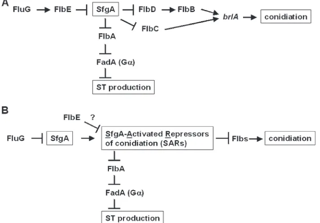
Figure 7.—Genetic models for regulation
of conidiation and ST production inA. nidu-lans. (A) A simple genetic model in which SfgA functions downstream of FlbE. This model is not sufficient to explain the partially restored conidiation phenotype of the DflbE DsfgAmutant or the production of ST by the
DflbEmutant (Hickset al. 1997). (B) A
com-plex working model. The fact that most C6 factors function as transcriptional activators (reviewed in Todd and Andrianopoulos
1997) led to the proposal that SfgA activates a group of genes (SfgA-Activated Repressors
It is evident that SfgA has multiple functional do-mains and the full-length SfgA is required for its func-tionality. As revealed by allele analyses, the integrity of the C6 domain is crucial for SfgA function. Mutations predicted to affect the C6 domain result in hyperactive conidiation phenotypes essentially identical to those caused by deletion ofsfgA. Furthermore, any mutations causing at least partial loss ofsfgAfunction(s) seem to be sufficient to restore conidiation to certain levels (Figure 3). Deletion ofsfgAeliminates the requirement of FluG in conidiation and ST production. These data clearly suggest that the primary role of FluG is to remove re-pressive effects imposed by SfgA.
One of the key hypotheses in A. nidulans asexual developmental control is that FluG is responsible for the production of an extracellular factor that is required for developmental initiation. Earlier studies suggested that this factor is ,6000–8000 Da in size (Leeand Adams 1994b). Levels of the fluG mRNA and protein are relatively constant throughout the life cycle (Lee and Adams1994b), implying that the amount of the FluG factor may increase gradually in vegetative cells. As overexpression of fluG did (Lee and Adams 1996; D’Souzaet al. 2001), deletion and 12 othersfgAmutant alleles resulted in formation of conidiophores in liquid submerged cultures that have all the cell types including stalks, vesicles, metulae, phialides, and conidia even in the absence offluG. Taken together, it is speculated that during the early vegetative growth phase the level of the FluG factor in cells is low and SfgA-mediated repression of conidiation dominates. Once accumulation of the FluG factor reaches a certain level in cells, it removes SfgA-mediated negative control of conidiation (possibly involving downregulation ofsfgAtranscription), which triggers the initiation of conidiophore development.
Another key role of FluG is thought to be post-transcriptional activation of FlbA (Lee and Adams 1996), which in turn attenuates vegetative proliferation signaling mediated by a heterotrimeric G protein composed of FadA (Ga), SfaD (Gb), and GpgA (Gg) (Yu et al. 1996; Rose´ n et al. 1999; Seo et al. 2005). Constitutive activation of this FadA signaling blocks development and production of ST (see Figure 1; Yu
et al. 1996, 1999; Hickset al. 1997). It was demonstrated previously that mutational inactivation of FadA by-passed the requirement of FluG in ST biosynthesis but not in conidiation (Hickset al. 1997). This observation has led to the hypothesis that the primary role of FluG in activating ST is to activate FlbA, which in turn inactivates FadA signaling, shifting the physiological balance in favor of development. However, the role of FluG in activating FlbA has remained elusive. Our findings that deletion and all 31 sfgA mutations restored ST pro-duction in theDfluGmutant may also provide a missing link between FluG and FlbA. It can be speculated that the end result of SfgA activity is negative regulation of conidiation and FlbA, and removal of SfgA-mediated
repression by FluG confers both activation of conidia-tion and inhibiconidia-tion of FadA signaling (see Figure 7). Fur-ther characterization of SfgA coupling with G-protein signaling will elucidate a regulatory network coordinat-ing the balance between growth and development.
Genetic data clearly demonstrate thatsfgAfunctions downstream offluGbut upstream offlbD,flbB,flbC, and brlA (Figure 6). In contrast, the genetic interaction betweensfgAandflbEcannot be easily resolved. While a simple model for sfgAfunctioning downstream of flbE can be proposed (Figure 7A), this model cannot explain the incomplete suppression ofDflbEbyDsfgAas well as the ability of theflbEmutant to produce ST (Hicks
et al. 1997). Thus, we propose an alternative model that FlbE and SfgA function at the same level and compete for the expression of downstream genes (Figure 7B). Regard-less of the position ofsfgA, our study elucidates a new concept for the FluG-dependent central regulatory mechanism of conidiation in the model fungus A. nidulans.
The authors are grateful to the Broad Institute for the genome database and to Amy Wong and Ellin Doyle for critically reviewing this manuscript. Special thanks are given to Thomas Adams, Jenny Wieser, Ellen Kellner, BeeNa Lee, Sang-Tae Han, and other fungal develop-mental biologists. This work was supported by Hatch grant WIS-04667 and National Science Foundation grant MCB-0421863 to J.H.Y.
LITERATURE CITED
Adams, T. H., 1994 Asexual sporulation in higher fungi, pp. 367– 382 inThe Growing Fungus, edited by N. A. R. Gowand G. M. Gadd. Chapman & Hall, London.
Adams, T. H., M. T. Boylanand W. E. Timberlake, 1988 brlAis necessary and sufficient to direct conidiophore development inAspergillus nidulans.Cell54:353–362.
Adams, T. H., J. K. Wieserand J.-H. Yu, 1998 Asexual sporulation in
Aspergillus nidulans.Microbiol. Mol. Biol. Rev.62:35–54. Brown, D. W., J.-H. Yu, H. S. Kelkar, M. Ferdandes, T. C. Nesbitt
et al., 1996 Twenty-five co-regulated transcripts define a sterig-matocystin gene cluster inAspergillus nidulans.Proc. Natl. Acad. Sci. USA93:1418–1422.
Calvo, A. M., R. A. Wilson, J. W. Bok and N. P. Keller, 2002 Relationship between secondary metabolism and fungal development. Microbiol. Mol. Biol. Rev.66:447–459.
Chang, Y. C., and W. E. Timberlake, 1992 Identification of
Aspergil-lus brlAresponse elements (BREs) by genetic selection in yeast. Genetics133:29–38.
D’Souza, C. A., B. N. Leeand T. H. Adams, 2001 Characterization of the role of the FluG protein in asexual development ofAspergillus nidulans.Genetics158:1027–1036.
Felenbok, B., D. Sequeval, M. Mathieu, S. Sibley, D. I. Gwynne
et al., 1988 The ethanol regulon inAspergillus nidulans: charac-terization and sequence of the positive regulatory gene alcR.
Gene73:385–396.
Flaherty, J. E., and C. P. Woloshuk, 2004 Regulation of fumo-nisin biosynthesis in Fusarium verticillioidesby a zinc binuclear cluster-type gene, ZFR1. Appl. Environ. Microbiol. 70: 2653– 2659.
Gwynne, D. I., F. P. Buxton, S. Sibley, R. W. Davies, R. A. Lockingtonet al., 1987 Comparison of thecis-acting control regions of two coordinately controlled genes involved in ethanol utilization inAspergillus nidulans.Gene51:205–216.
Han, K.-H., J.-A. Seoand J.-H. Yu, 2004b Regulators of G protein signaling in Aspergillus nidulans: RgsA downregulates stress re-sponse and stimulates asexual sporulation through attenuation of GanB (Ga) signaling. Mol. Microbiol.53:529–540.
Hicks, J. K., J.-H. Yu, N. P. Kellerand T. H. Adams, 1997 Aspergillus sporulation and mycotoxin production both require inactivation of the FadA Gaprotein-dependent signaling pathway. EMBO J.
16:4916–4923.
Ka¨ fer, E., 1977 Meiotic and mitotic recombination in Aspergillus and its chromosomal aberrations. Adv. Genet.19:33–131. Kellner, E. M., and T. H. Adams, 2002 Mutations insfdAandsfdB
suppress multiple developmental mutations inAspergillus nidu-lans.Genetics160:159–168.
Laughon, A., and R. F. Gesteland, 1984 Primary structure of the
Saccharomyces cerevisiaeGAL4 gene. Mol. Cell. Biol.4:260–267. Lee, B. N., and T. H. Adams, 1994a Overexpression offlbA, an early
regulator ofAspergillusasexual sporulation leads to activation of
brlAand premature initiation of development. Mol. Microbiol.
14:323–334.
Lee, B. N., and T. H. Adams, 1994b TheAspergillus nidulans fluG gene is required for production of an extracellular developmen-tal signal. Genes Dev.8:641–651.
Lee, B. N., and T. H. Adams, 1996 fluGandflbAfunction interde-pendently to initiate conidiophore development in Aspergillus nidulansthroughbrlAbactivation. EMBO J.15:299–309. Marmorstein, R. M., M. Carey, M. Ptashneand S. C. Harrison,
1992 DNA recognition by GAL4: structure of a protein-DNA complex. Nature356:408–414.
Martinelli, S. D., 1994 Aspergillus nidulansas an experimental or-ganism, pp. 33–58 inAspergillus: 50 Years On(Progress in Indus-trial Microbiology, Vol. 29), edited by S. D. Martinelliand J. R. Kinghorn. Elsevier Science, Amsterdam.
Mathis, R., C. Grosjean, F.de Billy, T. Huguetand P. Gamas, 1999 The early nodulin gene MtN6 is a novel marker for events preceding infection ofMedicago truncatularoots bySinorhizobium meliloti.Mol. Plant-Microbe Interact.12:544–555.
Osherov, N., and G. May, 2000 Conidial germination inAspergillus
nidulansrequires RAS signaling and protein synthesis. Genetics
155:647–656.
Pan, T., and J. E. Coleman, 1990 GAL4 transcription factor is not a ‘‘zinc finger’’ but forms a Zn(II)2Cys6binuclear cluster. Proc.
Natl. Acad. Sci. USA87:2077–2081.
Pontecorvo, G., J. A. Roper, L. M. Hemmons, K. D. Macdonaldand A. W. J. Bufton, 1953 The genetics ofAspergillus nidulans.Adv. Genet.5:141–238.
Rerngsamran, P., M. B. Murphy, S. A. Doyleand D. J. Ebbole, 2005 Fluffy, the major regulator of conidiation inNeurospora crassa, directly activates a developmentally regulated hydropho-bin gene. Mol. Microbiol.56:282–297.
Rose´ n, S., J.-H. Yuand T. H. Adams, 1999 TheAspergillus nidulans
sfaDgene encodes a G proteinbsubunit that is required for nor-mal growth and repression of sporulation. EMBO J.18:5592– 5600.
Seo, J.-A., and J.-H. Yu, 2004 Toxigenic fungi and mycotoxins, pp. 689–721 inMycology 22: Handbook of Industrial Mycology, edited by Z. An. Marcel Dekker, New York.
Seo, J.-A., Y. Guan and J.-H. Yu, 2003 Suppressor mutations bypass the requirement offluGfor asexual sporulation and sterig-matocystin production in Aspergillus nidulans. Genetics 165:
1083–1093.
Seo, J.-A., K.-H. Hanand J.-H. Yu, 2005 Multiple roles of a hetero-trimeric G proteingsubunit in governing growth and develop-ment ofAspergillus nidulans.Genetics171:81–89.
Stack, M., and J. V. Rodricks, 1971 Method for analysis and chem-ical confirmation of sterigmatocystin. J. Assoc. Off. Anal. Chem.
54:86–90.
Strich, R., R. T. Surosky, C. Steber, E. Dubois, F. Messenguyet al., 1994 UME6 is a key regulator of nitrogen repression and mei-otic development. Genes Dev.8:796–810.
Stringer, M. A., R. A. Dean, T. C. Sewalland W. E. Timberlake, 1991 Rodletless, a new Aspergillus developmental mutant in-duced by directed gene inactivation. Genes Dev.5:1161–1171. Timberlake, W. E., 1990 Molecular genetics ofAspergillus
develop-ment. Annu. Rev. Genet.24:5–36.
Todd, R. B., and A. Andrianopoulos, 1997 Evolution of a fungal regulatory gene family: the Zn(II)2Cys6binuclear cluster DNA
binding motif. Fungal Genet. Biol.21:388–405.
Vienken, K., M. Schererand R. Fischer, 2005 The Zn(II)2Cys6 pu-tativeAspergillus nidulanstranscription factor repressor of sexual development inhibits sexual development under low-carbon con-ditions and in submersed culture. Genetics169:619–630. Wieser, J., and T. H. Adams, 1995 flbDencodes a myb-like DNA
binding protein that controls initiation ofAspergillus nidulans co-nidiophore development. Genes Dev.9:491–502.
Yu, J.-H., and N. P. Keller, 2005 Regulation of secondary metabo-lism in filamentous fungi. Annu. Rev. Phytopathol.43:437–458. Yu, J.-H., and T. J. Leonard, 1995 Sterigmatocystin biosynthesis in
Aspergillus nidulansrequires a novel type I polyketide synthase. J. Bacteriol.177:4792–4800.
Yu, J.-H., J. Wieserand T. H. Adams, 1996 TheAspergillusFlbA RGS domain protein antagonizes G-protein signaling to block prolif-eration and allow development. EMBO J.15:5184–5190. Yu, J.-H., S. Rose´ nand T. H. Adams, 1999 Extragenic suppressors of
loss-of-function mutations in the Aspergillus FlbA RGS domain protein. Genetics151:97–105.
Yu, J.-H., Z. Hamari, K.-H. Han, J.-A. Seo, Y. Reyes-Domı´nguezet al., 2004 Double-joint PCR (DJ-PCR): a PCR-based molecular tool for gene manipulations in filamentous fungi. Fungal Genet. Biol.
41:973–981.
Yuan, G. F., and G. A. Marzluf, 1992 Molecular characterization of mutations of nit-4, the pathway-specific regulatory gene which controls nitrate assimilation inNeurospora crassa.Mol. Microbiol.
6:67–73.