M E T H O D O L O G Y
Open Access
Development of an ELISA-array for simultaneous
detection of five encephalitis viruses
Xiaoping Kang, Yuchang Li, Li Fan, Fang Lin, Jingjing Wei, Xiaolei Zhu, Yi Hu, Jing Li, Guohui Chang, Qingyu Zhu,
Hong Liu and Yinhui Yang
*Abstract
Japanese encephalitis virus(JEV), tick-borne encephalitis virus(TBEV), and eastern equine encephalitis virus (EEEV) can cause symptoms of encephalitis. Establishment of accurate and easy methods by which to detect these viruses is essential for the prevention and treatment of associated infectious diseases. Currently, there are still no multiple antigen detection methods available clinically. An ELISA-array, which detects multiple antigens, is easy to handle, and inexpensive, has enormous potential in pathogen detection. An ELISA-array method for the simultaneous detection of five encephalitis viruses was developed in this study. Seven monoclonal antibodies against five encephalitis-associated viruses were prepared and used for development of the ELISA-array. The ELISA-array assay is based on a“sandwich”ELISA format and consists of viral antibodies printed directly on 96-well microtiter plates, allowing for direct detection of 5 viruses. The developed ELISA-array proved to have similar specificity and higher sensitivity compared with the conventional ELISAs. This method was validated by different viral cultures and three chicken eggs inoculated with infected patient serum. The results demonstrated that the developed ELISA-array is sensitive and easy to use, which would have potential for clinical use.
Background
Japanese encephalitis virus(JEV), tick-borne encephalitis virus(TBEV), eastern equine encephalitis virus (EEEV), sindbis virus(SV), and dengue virus(DV) are arboviruses and cause symptoms of encephalitis, with a wide range of severity and fatality rates [1]. Establishment of an accurate and easy method for detection of these viruses is essential for the prevention and treatment of asso-ciated infectious diseases. Currently, ELISA and IFA are the methods which are clinically-available for the detec-tion of encephalitis viral antigens, but they could only detect one pathogen in one assay [2,3].
There are a variety of different methods available for identifying multiple antigens in one sample simulta-neously, such as two-dimensional gel electrophoresis (2-DE), protein chip, mass spectrometry, and suspension array technology [4-6]. However, the application of these techniques on pathogen detection is still in an early phase, perhaps due to the complicated use and high cost.
Antibody arrays for simultaneous multiple antigen quantification are considered the most accurate methods [7-10]. Liew [11] validated one multiplex ELISA for the detection of 9 antigens; Anderson [12] used microarray ELISA for multiplex detection of antibodies to tumor antigens in breast cancer, and demonstrated that ELISA-based array assays had the broadest dynamic range and lowest sample volume requirements com-pared with the other assays.
However, the application of ELISA-based arrays is cur-rently limited to detection of cancer markers or inter-leukins; no detection of pathogens has been reported. In this study, we developed an ELISA-based array for the simultaneous detection of five encephalitis viruses. Seven specific monoclonal antibodies were prepared against five encephalitis viruses and used to establish an ELISA-array assay. The assay was validated using cul-tured viruses and inoculated chicken eggs with patient sera. The results demonstrated that this method com-bined the advantage of ELISA and protein array (multi-plex and ease of use) and has potential for the identification of clinical encephalitis virus.
* Correspondence: [email protected]
Department of Virology, State Key Laboratory of Pathogen and Biosecurity, Beijing Institute of Microbiology and Epidemiology, Beijing 100071, China
Methods
Monoclonal antibody preparation
Monoclonal antibodies were prepared from hybridoma cell lines constructed by Prof. Zhu et al. Purification was conducted by immunoaffinity chromatography on pro-tein G affinity sepharose [13]. Specific monoclonal anti-bodies (4D5 against JEV, 2B5 against TBEV, 1F1 against SV, 2B8 against serotype 2 DV, 4F9 against serotype 4
DV, 4E11 against EEEV, and 2A10 againstFlavivirus)
were selected for this study. All of the antibodies were raised according to standard procedures.
Using 4D5, 2B5, 1F1, 2B8, 4F9, and 4E11 as capture antibodies, detection antibodies (2A10, 1 F1, and 4E11) were coupled to biotin-NHS ester(Pierce, Germany) at
4°C for 3 h according to the manufacturer’s instructions.
Unincorporated biotin was removed by Desalt spin col-umn (Pierce). Immunologic reactions were reported by Streptavidin-HRP (CWBIO, Beijing, China) and Super Signal ELISA Femto Maximum sensitive substrate. Puri-fied goat-anti mouse antibody was used as a positive control.
Virus culture
JEV and DV were cultured in C6/36 cells; SV, TBEV, and EEEV were cultured in BHK-21 cells. The culture of TBEV and EEEV was conducted in biosafety level 3 facility, however, JEV, DV and SV were conducted in biosafety level 2 facility. Viral titers were determined by
the 50% tissue culture infectious dose (TCID50) method.
All the cultures were inactivated by 0.025% b
-propiono-lactone at 4°C overnight, then 37°C for 1 h to
decom-poseb-propionolactone.
Antibody spotting and optimization
Antibodies were spotted using a BIODOT machine (BD6000;California, USA) on ELISA plates (30 nl/dot). The plates were blocked with 3% BSA-PBS in 37°C for 1 h, followed by washing 3 times with PBS containing 0.1% Tween-20 for 2 min each. Then, the plates were dried, sealed, and stored at 4°C before use [11].
When spotting, different spotting buffers and concen-trations of capture monoclonal antibodies were
evalu-ated to optimize the ELISA-array assay. The
optimization was evaluated by dot morphology and sig-nal intensity. The tested spotting buffers included 1 × phosphate buffer saline (PBS), PBS +20% glycerol, and 1 × PBS + 20% glycerol+0.004% Triton-X100. A range of monoclonal antibody concentrations (0.0125, 0.025, 0.05, 0.1, and 0.2 mg/ml) were compared.
Following a double antibody sandwich format, printed plates were incubated sequentially with inactivated viral cultures, biotin-labeled detecting antibody, HPR-labeled avidin, and substrate, followed by signal evaluation.
ELISA-array analysis
Antigen binding was performed in PBS(containing 0.1% Tween-20 and 5% FCS) at 37°C for 2 h, followed by washing 3 times(1 × PBS containing 0.1% Tween-20). Incubation of ELISA plates with biotinylated detecting antibody cocktails was performed in PBS (containing 0.1% Tween-20 and 5% FCS) at 37°C for 2 h. After washing, specific binding of the detecting antibodies was reported by streptavidin-HRP and stained with Super Signal ELISA Femto Maximum sensitive substrate (Thermo scientific, Rockford, USA) [11,14,15]. Visualiza-tion of the plate was performed in AE 1000 cool CCD image analyzer(Beijing BGI GBI Biotech Company., LTD, China). The signal intensity and background of
each spot was read out and recorded with“
Monster"-software. The positive signals were defined as a signal value > 400 and a signal value (sample)/signal value (negative) > 2.
Conventional ELISA
The identical antibodies used in the ELISA-array format were also tested in a conventional ELISA format to determine the difference in sensitivity and specificity of the two methods. The conventional ELISAs were per-formed at the same time as the ELISA-array assays to ensure similar reaction conditions. The conventional ELISAs were performed in an identical maner to the ELISA-array, except that antibodies were coated at a
concentration of 2μg/mL in PBS (pH 7.4), and substrate
TMB was used instead of Super Signal ELISA Femto Maximum sensitive substrate [16,17].
Clinical sample treatment and chicken egg inoculation Three serum samples were collected from patients with nervous system symptoms and histories of tick bites. The serum samples were treated with penicillin and streptomycin, then inoculated into the allantoic cavities of chicken eggs. 3 days later, the liquid was collected and divided into two portions (one for inactivation and one for RNA extraction). The RNA and inactivated sam-ples were stored at -70°C before use.
Real-time RT-PCR assay
RNA was extracted from the inoculated chicken eggs using a RNeasy mini kit (Qiagen Inc., Valencia, CA,
USA) according to the manufacturer’s instructions. All
RNA extraction procedures were conducted at BSL-3 facilities. The primers and probes were used as pre-viously described [18]. The real-time RT-PCR was con-ducted with a Quti-teck q-RT-PCR Kit (Qiagen Inc,).
The reaction consisted of 10 μL of 2 × reaction buffer
(0.2 μL reverse transcription enzyme, and 250 nmol/l
added to a final volume of 20 μl. PCR was performed with a LightCycler 2.0 (Roche, Switzerland) [19].
Results and discussion
Optimization of the ELISA-array assay
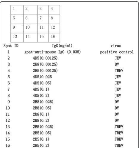
The spotted array layout is depicted in Figure 1 and the efficacy of three different spotting buffers on the quality of the printed ELISA-arrays were investigated by spot
morphology observation and signal intensity
comparison.
The spotting concentration of the capture antibodies varied from 0.2 to 0.0125 mg/ml (each was serially diluted 2-fold). The efficacy of the spotting concentra-tion of the capture antibodies was evaluated by virus culture detection, the proper spotting concentration was determined by a combination of minimized cross reac-tion and higher signal intensity. Figure 1 illustrates the array layout and Figure 2 demonstrates the result of the three spotting buffers and spot concentration of anti-body 2B5 by TBE virus culture detection. Cross reaction detection was also conducted by applying JEV, YF, and DV cultures.
Spot morphology observation (Figures 2a, b, and 2c) demonstrated that spotting buffer containing PBS with 20% glycerol produced tailed spot morphology; buffers containing PBS alone and PBS with 20% glycerol +0.004% Triton-X100 gave good spot morphology (round and full). Buffers containing PBS with 20% gly-cerol and PBS with 20% glygly-cerol+0.004% Triton-X100
produced higher signal intensities than PBS alone. Thus, PBS with 20% glycerol+0.004% Triton-X100 was adopted as the optimized spotting buffer for subsequent experiments. Simultaneously, the spot concentration evaluation suggested that 0.05 mg/ml was optimal. At this concentration, the signal intensity was higher and the cross-reaction did not appear (Figure 2d). Conse-quently, spotting concentration optimization of other capture antibodies (4D5, 1F1, 4E11, and 2B8) demon-strated that 0.05 mg/ml was also suitable(data not shown).
The optimized ELISA array layout is shown in Figure 3, which was applied in the following experiments.
Figure 1Print layout of antibodies spotted on ELISA plates, spot volume = 30 nl.
Figure 2Evaluation of different spotting buffers and antibody concentrations in ELISA-array application.aspot morphology visualization by using 20% glycerol-PBS buffer;bspot morphology visualization by using PBS + 20% glycerol+0.004%Triton-X100 buffer; cspot morphology visualization by using PBS buffer.dSignal intensity comparisons of different solutions and spotting concentration of 2B5.
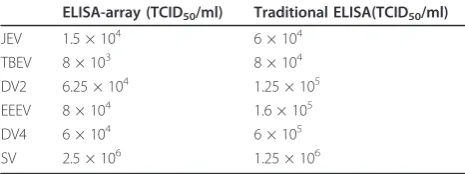
Sensitivity and specificity of ELISA-array compared with conventional ELISAs
Successful detection of viral pathogens requires a test with high sensitivity and specificity. To evaluate the per-formance of the designed antibody arrays, the specificity and sensitivity of the individual analytes were examined. By testing serially-diluted viral cultures, including DV-2, DV-4, JEV, TBE, SV, and EEEV, the sensitivity of ELISA-array and the identical conventional ELISA were com-pared (Table 1). The detection limit of the two methods was compared and demonstrated. The cross-reactivity test was conducted using BHK-21 and vero cell lysate,
Yellow fever virus (YFV) cultures (5 × 10⁵TCID50/ml,
West Nile virus(WNV) cultures(2 × 10⁶TCID50/ml), and
Western equine encephalitis virus(1 × 10⁷TCID50/ml).
The results demonstrated that neither the ELISA-array nor traditional ELISA displayed cross-reactivity.
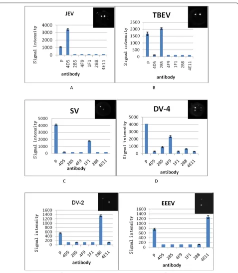
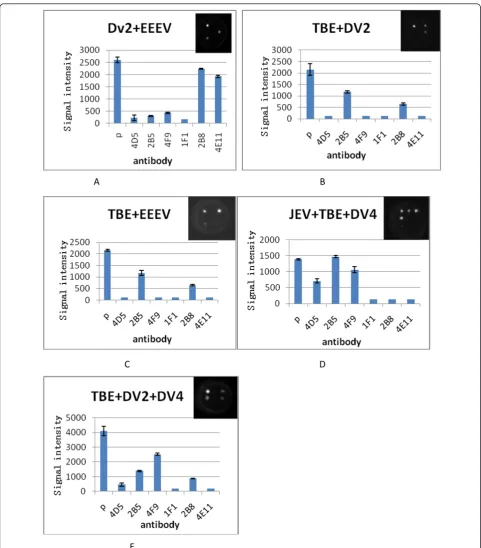
Single and multiplex detection of TBEV, JEV, DV, SV, and EEEV
Equal volumes of cultured TBEV, JEV, DV-2, DV-4, SV, and EEEV were prepared for single sample detection; two or three of the cultures were mixed for multiplex detection. A cocktail of biotin conjugated antibody (2A10, 4E11, and 1F1) was used in all tests. The results demonstrated that for all virus combinations, each virus was detected specifically, with no false-positive or-nega-tive results (Figures 4 and 5).
Validation by mock clinical samples
Chicken eggs inoculated with infected human serum were used for validation of the ELISA-array assay. All samples showed high reaction signals with capture anti-body 2B5, which was specific for TBEV (Figure 6b). The ELISA-array assay suggested that the three patients were all infected with TBEV.
To verify the results tested by ELISA-array, RNA extracted from chicken eggs was applied to a real time-RT-PCR assay using primers and probes targeting TBEV. The results were also positive (Figure 6a). The consensus detection results confirmed that the ELISA-array assay was reliable.
Discussion
To be widely used in the clinical setting, the detection system should be easy to use and can be performed by untrained staff with little laboratory and experimental experience. Moreover, when the volume of the clinical samples is limited and an increasing number of patho-gens per sample needs to be tested, the detecting system should be high-throughput to allow detection of multi-ple pathogens simultaneously [6,20,21]. Multimulti-ple detec-tion, easy to use, and affordability are requirements for detection methods in the clinical setting. Thus, an ELISA-array, which combines the advantages of ELISA and protein array, meets the above requirements.
It has been reported that an ELISA-array has been used in the diagnosis of cancer and auto-allergic disease [7,12]; however, No study has reported the detection of viral pathogens. In this study, we developed a multiplex ELISA-based method in a double-antibody sandwich format for the simultaneous detection of five encephali-tis-associated viral pathogens.
The production of a reliable antibody chip for identifi-cation of microorganisms requires careful screening of capture of antibodies [14]. Cross-reactivity must be minimized and the affinity of the antibody is as impor-tant as the specificity. First, we prepared and screened 23 monoclonal antibodies against eight viruses and veri-fied the specificity and affinity to the target viruses by an immunofluorescence assay. Then, the antibodies were screened by an ELISA-array with a double-anti-body sandwich ELISA format. The antibodies which produced cross-reactivity and low-positive signals were excluded. Finally, six antibodies were selected as capture antibodies. Another monoclonal antibody, 2A10, which
could specifically react with all viruses in the genus
Fla-vivirus was used for detecting antibody against DV, JEV,
and TBEV. For the detection of EEEV and SV, although the detecting and trapping antibodies were the same (1F1 and 4E11, respectively), the antibodies produced excellent positive signals. The epitope was not defined; however, we suspect that the antibodies both target the surface of the virions. As one virion exits as, many with the same epitope appear, thus no interference occurred using the same antibody in the double-antibody sand-wich format assay.
Currently, the availability of antibodies suitable for an array format diagnostic assay is a major problem. In the ELISA-array assay, this problem exists as well. Because of the limitation of available antibodies, this assay could only detect 5 pathogens. In the future, with increasing numbers of suitable antibodies,
espe-cially specific antibodies againstFlavivirus, this
ELISA-array might be able to test more pathogens and be of greater potential use.
Table 1 Detection limit comparison between ELISA-array and traditional ELISA
ELISA-array (TCID50/ml) Traditional ELISA(TCID50/ml)
To make the assay more amenable to multiple virus detection, the assay protocol was optimized. In addition to the dotting buffer, the capture antibody concentration and the different virus inactivation methods (heating
andb-propiolactone) were also compared and evaluated.
Heat inactivation was performed by heating the viral
cultures at 56°C for 1 h, and b-propiolactone
inactiva-tion was performed by adding b-propiolactone into the
A B
C D
E F
EEEV
viral cultures to obtain a final concentration of 0.25‰,
incubating at 4°C overnight, then decomposing b
-pro-piolactone at 37°C for 1 h. The performance was evalu-ated based on the infectivity experiments and detecting
the limit of virus titers (data not shown). The infectivity experiments demonstrated that both of the two methods could totally inactive the viruses, but the sensitivity limit
assay suggested that virus treated by b-propiolactone
A B
C D
E
retains better antigenicity than the heat-inactivation
method. Thus,b-propiolactone treatment was chosen as
the virus-inactivation method.
A conventional ELISA is a standard method in many diagnostic laboratories. We compared the ELISA-array with a conventional ELISA and confirmed that the advantage of the ELISA-array was evident with com-parable specificity and higher sensitivity than ELISA. The time required for the ELISA-array is significantly less than for conventional ELISA (4 h vs. a minimum of 6 h, respectively). Furthermore, less IgG is required for printing than for coating ELISA plates. Coating of
a single well in microtiter plate requires 100 μl of a 1
μg/ml antibody solution, which is equivalent to 100 ng
of IgG. For the ELISA-array, only 30 nl of a 50μg/ml
antibody solution is required for each spot, which is equivalent to 1.5 ng of IgG. With the characteristics of ease of use, sensitivity, specificity, and accuracy, the ELISA-array assay would be widely accepted for clini-cal use.
Acknowledgements
This study was supported by the National Natural Science Foundation of China (Grant no. 81171663) and National Key Research Special Foundation of China (Grant no. 2011ZX10004-001).
Authors’contributions
XK: designed the study, performed the laboratory testing, analyzed the test results, and co-wrote and edited the manuscript. H L and YL performed the virus cultures. LF performed laboratory testing. XC, FL, and GC performed real time-PCR. QZ and YY organized the overall project and helped edit the manuscript. All of the authors read and approved the final manuscript.
Authors’information
State Key Laboratory of Pathogen and Biosecurity, Beijing Institute of Microbiology and Epidemiology, Beijing 100071, China
Competing interests
The authors declare that they have no competing interests.
Received: 26 August 2011 Accepted: 27 February 2012 Published: 27 February 2012
References
1. Eshoo MW, Whitehouse CA, Zoll ST, Massire C, Pennella TT, Blyn LB, Sampath R:Direct broad-range detection of alphaviruses in mosquito extracts.Virology2007,368:286-295.
2. Stephen FK:Multiplexed protein measurement: technologies and applications of protein and antibody arrays.Nat Rev Drug Discov2006,
5:310-320.
3. Ulrik BN, Bernhard HG:Multiplexed sandwich assays in microarray format. J Immunological Methods2004,290:107-120.
4. Townsend MB, Smagala JA, Dawson ED, Deyde V, Gubareva L, Klimov AI, Kuchta RD, Rowlen KL:Detection of adamantane-resistant influenza on a microarray.J Clin Virol2008,42:117-123.
5. Aytur T, Foley J, Anwar M, Boser B, Harris E, Beatty PR:A novel magnetic bead bioassay platform using a microchip-based sensor for infectious disease diagnosis.J Immunol Methods2006,314:21-29.
6. Sandor B, Peter T, Neil L, Gerrit V:Advances in viral disease diagnostic and molecular epidemicological technologies.Expert Rev Mol Diagn2009,
9:367-381.
7. De Ceuninck F, Marcheteau E, Berger S, Caliez A, Dumont V, Raes M, Anract P, Leclerc G:Assessment of some tools for the characterization of the human osteoarthritic cartilage proteome.J Biomol Tech2005,
16:256-265.
8. Mendoza LG, McQuary P, Mongan A, Gangadharan R, Brignac S, Eggers M:
High-throughput microarray-based enzyme-linked immunosorbent assay (ELISA).BioTechniques1999,27:778-788.
9. Knight PR, Sreekumar A, Siddiqui J, Laxman B, Copeland S, Chinnaiyan A, Remick A:Development of a sensitive microarray immunoassay and comparison with standard enzyme-linked immunoassay for cytokine analysis.Shock2004,21:26-30.
10. Nielsen UB, Geierstanger BH:Multiplexed sandwich assays in microarray format.J Immunol Methods2004,290:107-120.
11. Michael L, Matthew CG, James ET, Sara LC, Joann EM, Justin DH, Karl V, Carl W, Rex SS:Validating a custom multiplex ELISA against individual commercial immunoassays using clinical samples.BioTechniques2007,
42:327-333.
12. Anderson KS, Ramachandran N, Wong J, Raphael JV, Hainsworth E, Demirkan G, Cramer D, Aronzon D, Hodi FS, Harris L, Logvinenko T, LaBaer J:Application of protein microarrays for multiplexed detection of antibodies to tumor antigens in breast cancer.J Proteome Res2008,
7:1490-1499.
13. Xiaoping K, Bao-an Y, Yanli L, Yuyang H, Hui Z, Wei X, Qing-yu Z:Human neutralizing SARS-CoV specific Fab molecules generated by phage display.Clinical and Vaccine Immunology2006,13:953-958.
14. Rick W, Yuri B, Tom P, Patricia T, Mike H:Simultaneous multianalyte ELISA performed on a microarray platform.Clinical Chemistry2001,
47:1451-1457.
15. Stewart TGB, Fiona K, Nichola O’L, Alan JR, Marisa CK, John SB, Juraj P, Peter G, Colin JC:A multiplexed protein microarray for the simultaneous
A
serodiagnosis of human immunodeficiency virus/hepatitis C virus infection and typing of whole blood.Analy Bioch2008,382:9-15. 16. Birgit H, Ralf E, Hans-Juergen M:A simple and rapid protein array based
method for simultaneous detection of biowarfare agent.Proteomics2006,
6:2972-2981.
17. Abdullahi MR:Microarray immunoassay for the detection of grapevine and tree fruit viruses.J Virol Meth2009,160:90-100.
18. Michaela S, Pascal C:Development of a quantitative real-time RT-PCR assay with internal control for the laboratory detection of tick borne encephalitis virus (TBEV) RNA.Journal of Clinical Virology2003,27:136-145. 19. Xiaoping K, Yuchang L, Hong L, Fang L, Xuyu C, Tingting S, Guohui C,
Qingyu Z, Yinhui Y:A duplex real-time reverse transcriptase polymerase chain reaction assay for detecting western equine and eastern equine encephalitis viruses.Virology J2010,7:284-288.
20. Kwon JA, Lee H, Lee KN, Chae K, Lee S, Lee DK, Kim S:High diagnostic accuracy of antigen microarray for sensitive detection of hepatitis C virus infection.Clin Chem2008,54:424-428.
21. Pejcic B, De Marco R, Parkinson G:The role of biosensors in the detection of emerging infectious diseases.Analyst2006,131:1079-1090.
doi:10.1186/1743-422X-9-56
Cite this article as:Kanget al.:Development of an ELISA-array for simultaneous detection of five encephalitis viruses.Virology Journal2012
9:56.
Submit your next manuscript to BioMed Central and take full advantage of:
• Convenient online submission
• Thorough peer review
• No space constraints or color figure charges
• Immediate publication on acceptance
• Inclusion in PubMed, CAS, Scopus and Google Scholar
• Research which is freely available for redistribution