Purinergic signalling in bone cells
ASTRID HOEBERTZ
A thesis submitted fo r the degree o f
Doctor o f Philosophy at the University o f London
2001
Department o f Anatomy and Developmental Biology
University College London
ProQuest Number: U643406
All rights reserved
INFORMATION TO ALL USERS
The quality of this reproduction is dependent upon the quality of the copy submitted. In the unlikely event that the author did not send a complete manuscript and there are missing pages, these will be noted. Also, if material had to be removed,
a note will indicate the deletion.
uest.
ProQuest U643406
Published by ProQuest LLC(2016). Copyright of the Dissertation is held by the Author. All rights reserved.
This work is protected against unauthorized copying under Title 17, United States Code. Microform Edition © ProQuest LLC.
ProQuest LLC
789 East Eisenhower Parkway P.O. Box 1346
ABSTRACT
There is increasing evidence that ATP and other extracellular nucleotides, signalling through P2 receptors, play an important role in bone remodelling. I used immunohistochemistry and in situ hybridisation to study the expression o f P2 receptors in rat bone sections and on cultured bone cells: osteoclasts, the multinucleated cells responsible for bone resorption, osteoblasts, the bone-forming cells, and chondrocytes were all shown to express a number o f P2 receptors, both of the ionotropic P2X family and the metabotropic P2Y family.
ATP has previously been shown to be a potent stimulator o f bone resorption, however, it was not known which receptor subtypes mediate this stimulatory effect. Three different models were used to study the effects o f a wider range of P2 receptor agonists on osteoclast recruitment and resorption: the 26 hour disaggregated rat osteoclast assay, 72 hour whole organ mouse calvaria cultures, and 10 day mouse marrow cultures. My main discovery is that extracellular ADP, the first degradation product of ATP, is a powerful stimulator, at nanomolar concentrations, o f osteoclast activation and recruitment. Furthermore, evidence is provided, using subtype-selective agonists and antagonists, that these ADP effects are probably mediated via the P2Yi receptor. It is suggested that in vivo, ADP could be involved in the mediation o f inflammatory bone loss.
CONTENTS
ABSTRACT... 2
CONTENTS... 4
TABLES AND FIGURES... 9
List of Tables...9
List of figures...10
ACKNOWLEDGEMENTS... 13
PREFACE... 14
CHAPTER 1: GENERAL INTRODUCTION... 17
BONE...17
Bone composition...17
Osteoblasts... 18
Origin and differentiation... 18
Bone deposition... 21
Control o f osteoblastic function... 23
Osteocytes... 24
Osteoclasts... 25
Origin and differentiation... 25
How do osteoclasts resorb bone?... 29
Attachment... 29
Degradation o f bone matrix... 32
Maintenance o f intracellular p H... 35
Regulation o f resorption... 35
Mature osteoclast isolation...37
Bone growth and remodelling...38
Regulation o f bone remodelling... 39
Systemic hormones... 39
Parathryoid hormone (PTH)... 40
Calcitonin... 41
Sex steroids... 41
Other systemic hormones... 42
Local regulators... 44
Pathophysiology o f hone remodelling / Bone diseases... 46
Cartilage... 47
Receptors for extracellular nucleotides...49
History... 49
P I receptors... 50
P2X receptors...50
P2Xi receptor...52
P2X2 receptor...53
P2X] receptor...53
P2X4 receptor...54
P2X5 receptor...54
P2Xô receptor...54
P2Xy receptor...55
P2Y receptors...55
P2Yi receptor...57
P2Y2 receptor...57
P2Y4 receptor...58
P2Yô receptor...58
P2Yii receptor...58
P2Y]2 receptor... 59
Sources and fate o f nucleotides... 59
Physiological roles o f ATP...61
Short-term purinergic signalling... 61
Long-term purinergic signalling... 62
CHAPTER 2: EXPRESSION OF P2 RECEPTORS IN BONE AND CULTURED
BONE CELLS...66
Introduction...66
Materials and Methods... 68
Tissue Preparation...68
Cell Culture...68
Immunocytochemistry... 69
Western blot...71
In situ hybridisation... 72
R esults... 74
Immunocytochemical localisation o f P2X].? receptors...74
In situ hybridisation... 75
Immunocytochemical localisation o f P2Y]^ 2 , 4 receptors... 76
Discussion... 83
CHAPTER 3: EXTRACELLULAR ADP IS A POWERFUL OSTEOLYTIC AGENT: EVIDENCE FOR SIGNALLING THROUGH THE P2Yi RECEPTOR ON BONE CELLS...88
Introduction... 88
Materials and Methods... 90
Materials and solutions... 90
Resorption p it formation assay... 90
Mouse calvarial bone resorption assay...92
Osteoclast formation assays...94
Statistics... 95
R esults... 96
Relationship between pH, PCD2 and [HCO3] in tissue culture media...96
Effect o f ADP on mature rat osteoclasts...97
Effect o f ADP on Ca^^ release from mouse calvariae...109
Effect o f ADP in mouse marrow cultures (mixed cell population)...113
Discussion...120
CH APTER 4: EFFECTS O F NUCLEOTIDES ON OSTEOBLASTIC FUNCTION ...127
In tro d u ctio n ... 127
P2 receptors and osteoblasts... 127
Mineral formation in cell culture...129
M E T H O D S... 131
Bone nodule assay... 131
Proliferation assays... 132
[^H]thymidine incorporation...132
DNA quantification using Hoechst 33258...133
RESULTS...135
Bone nodule assay... 135
Proliferation assays... 140
[^HJthymidine incorporation...140
DNA quantification using Hoechst 33258...140
Discussion... 143
CHAPTER 5; INTERACTION OF LOW PH AND OSTEOLYTIC A GENTS 151 In tro d u ctio n ... 151
Acid-base balance... 151
Acidosis and the skeleton... 153
M E T H O D S ... 157
Human osteoclastoma cultures...157
Disaggregated rat osteoclast assay... 158
Immunocytochemistry fo r ASIC...159
R esu lts...160
Acid activation o f osteoclasts derived from human osteoclastoma...160
Interaction between RANKL and low p H... 162
Expression o f ASIC in osteoclasts... 165
CHAPTER 6: GENERAL DISCUSSION AND FUTURE W O RK ... 174
REFEREN CES... 185
APPENDIX I: ABBREVIATIONS... 226
TABLES AND FIGURES
LIST OF TABLES
1.1 Local factors affecting bone remodelling...45
2.1 Expression of P2X and P2Y receptors on bone cells...76
3.1 Lack o f effect o f ADP on the number of mononuclear cells / disc...97
3.2 Lack of effect of ADP and/ or acid on the number of cells / disc... 98
3.3 Lack o f effect of ADP and 2-meSADP on the number of cells / disc... 101
3.4 Lack of effect o f adenosine, AMP and ADP on the number o f cells / disc...102
3.5 Lack of effect of ADP, a,p-meATP and p,y-meATP on the number of cells / disc...102
3.6 Lack of effect of ADP and MRS 2179 (MRS) on the number of cells / disc... 106
3.7 Lack of effect of ADP and ATP on the number of mononuclear haematopoietic cells / disc... 116
4.1 Effects of nucleotides on rat osteoblast proliferation 1... 142
LIST OF FIGURES
1.1 Regulation of osteoblast growth and differentiation... 19
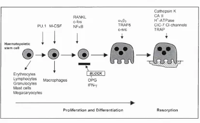
1.2 Osteoclast differentiation pathway...28
1.3 How does the osteoclast resorb bone?...34
1.4 Structure of P2X receptors...52
1.5 Structure of P2Y receptors... 56
2.1 Localisation o f P2X] receptor subtype on bone sections and bone cells... 77
2.2 Western blotting o f rat calvarial osteoblastic cell lysates...78
2.3 Localisation o f P2X4 receptor subtype on rat osteoclasts... 78
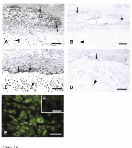
2.4 Localisation o f P2Xs receptor subtype on rat bone sections and cultured rat osteoblasts... 79
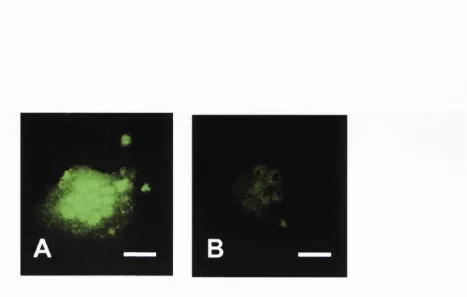
2.5 Localisation of P2X? receptor subtype on cultured rat osteoclast, visualised with fluorescein... 80
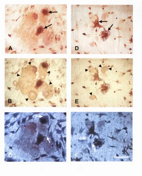
2.6 Localisation o f P2Yi receptor subtype in rat long bone sections and on cultured bone cells...81
2.7 Localisation o f P2Y] receptor subtype in rat long bone sections and on cultured bone cells...82
3.1 Relationship between pH, PCO2 and HCO3' in tissue culture media... 96
3.2 Effect of ADP on resorption pit formation by rat osteoclasts... 99
3.3 Comparison o f the effects of ADP on resorption pit formation by rat osteoclasts cultured in unmodified MEM or in acidified MEM for 26 h ...100
3.4 Effect of the selective P2Yi agonist 2-methylthioADP (2-meSADP) on resorption pit formation by rat osteoclasts...103
3.5 Lack of effect of further degradation products o f ADP, namely AMP and adenosine, on resorption pit formation by rat osteoclasts... 104
3.7 Inhibition o f ADP-stimulated resorption pit formation by the P2Yi antagonist
MRS 2179... 107
3.8 Typical appearance o f a disaggregated rat osteoclast assay... 108
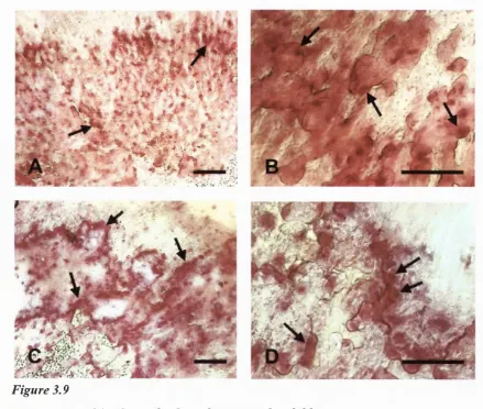
3.9 Whole-mount histology of cultured mouse calvarial bones...109
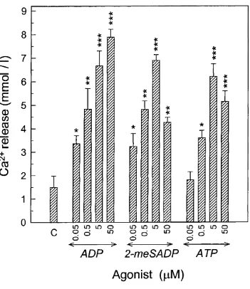
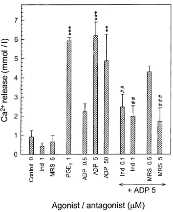
3.10 Stimulatory effect of ADP, 2-meSADP and ATP on Ca^^ release from mouse half-calvaria... I l l 3.11 Inhibition of the stimulatory action o f ADP on Ca^^ release from mouse half- calvariae by MRS 2179 (MRS) and indomethacin (Ind)... 112
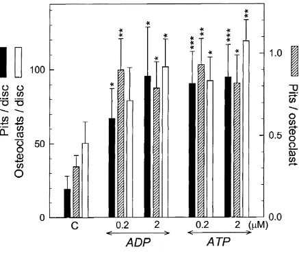
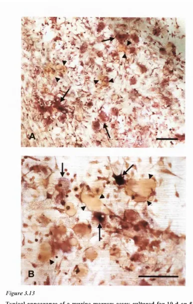
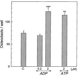
3.12 Effect o f ADP and ATP on osteoclast formation and excavation o f resorption pits in mouse marrow cultures maintained for 10 d on 5 mm dentine discs...114
3.13 Typical appearance of a murine marrow assay cultured for 10 d on 5 mm dentine discs...115
3.14 Effect of ADP and ATP on osteoclast formation and resorption in stromal cell-free mouse marrow cultures maintained for 10 d on 5 mm dentine discs... 117
3.15 Stimulatory effect o f ADP and ATP on osteoclast formation in stromal cell-free mouse marrow cultures maintained for 10 d in 48-well culture plates...118
3.16 Typical appearance o f stromal cell-free marrow cultures...119
4.1 Standard curve for DNA quantification using Hoechst 33258 dye... 134
4.2 Appearance o f bone nodules in culture, and stained with alizarin red...136
4.3 Inhibitory effect of ATP on bone nodule formation... 137
4.4 Inhibitory effect o f UTP on bone nodule formation... 138
4.5 Lack of effect of adenosine on bone nodule formation... 139
4.6 Lack of effect o f ADP on bone nodule formation...139
4.7 [^HJthymidine incorporation...141
5.1 Stimulation of resorption pit formation by osteoclasts derived from human osteoclastoma as extracellular pH is reduced...160
5.2 Resorption pits excavated by human osteoclastoma cells... 161
5.4 Effects o f RANKL and low pH on resorption areas...164 5.5 Large ‘snail trail’ pit excavated by osteoclasts treated with RANKL... 165 5.6 Immunostaining for H^-gated ion channels in osteoclasts cultured on plastic or
ACKNOWLEDGEMENTS
I am grateful to the many people who have contributed to this thesis, and who made my time at UCL an unforgettable experience. My first thanks go to my two supervisors, Dr Tim Arnett and Prof Geoffrey Bumstock, who both provided excellent guidance and help throughout these three years. With their different approaches, knowledge and experiences in science, they made a perfect team.
I would also like to thank present and former members o f the Autonomic Neuroscience Institute and Department o f Anatomy and Developmental Biology, UCL, who helped me greatly by ‘getting me started’, teaching me methods, and giving me ideas and advice in helpful discussions: Ute Groschel-Stewart, Michelle Bardini, Tim Robson, Eamonn Moules, Philippe Bodin, Brian King, Rainer Glass, and Mina Ryten, amongst many others.
Without a number of collaborations, I would have missed out on some good results: Dr Sajeda Meghji at the Eastman Dental Hospital, UCL, performed the ‘Mouse calvarial organ cultures’ for me, for which I am very grateful. Some sets of experiments were done in collaboration with two BSc students in our lab: Nichola Zanellato and Siva Mahendran. Andrea Townsend-Nicholson designed the oligonucleotide probes against P2X and P2Y receptors for in situ hybridisation. Rainer Glass performed the Western blotting for me. Gudrun Stenbeck from the Bone and Mineral Centre, UCL, introduced me to ‘Confocal Microscopy’, and I am grateful to this group and Prof Mike Horton for ‘adopting’ me as a regular member of their group meetings, tennis matches and pub trips.
My love and thanks to my parents and family, and to my friends in Germany, Switzerland, and London for their support, and to Brian for simply being there for me during that last two years.
PREFACE
In this preface, I would like to give a short outline o f the different chapters in this thesis. The work presented here has focused mainly on purinergic signalling in osteoblasts and osteoclasts, in addition to investigating some new aspects o f the effects of acidosis on bone structure and function.
Extracellular nucleotides are now recognised as important signalling molecules mediating a wide range of functions. They act via two types of receptors: P2X receptors, which are ligand-gated ion channels, and P2Y receptors, which are G protein-coupled receptors raising cytosolic free calcium ( [Ca^'^Jj ).
My project was based on a large number of pharmacological and electrophysiological studies suggesting the presence of P2 receptors on bone cells, and on relatively few functional studies reporting that adenosine 5’-triphosphate (ATP) had an effect on both formation and activation o f osteoclasts, the multinucleated cells responsible for bone resorption, and also on bone formation by osteoblasts. However, ATP is a potent agonist at most P2 receptor subtypes, and it was neither known which P2 receptor subtypes were present on bone cells, nor which of these receptors might be mediating effects on osteoclast and osteoblast function.
In the first experimental chapter (Chapter 2), the expression of P2 receptors was studied on frozen sections of rat bone, and on cultured rat osteoclasts and osteoblasts, using immunohistochemistry and in situ hybridisation. Evidence is presented that osteoblasts, osteoclasts and chondrocytes express a wide range o f P2 receptors, both of the P2X and P2Y families.
The second experimental chapter (Chapter 3) explored which receptor subtype(s) might be involved in mediating the osteolytic effects o f ATP on bone, using P2 receptor subtype- selective agonists and antagonists in three different assays: disaggregated rat osteoclast resorption assays, whole organ cultures o f mouse calvariae, and mouse marrow cultures.
Chapter 4, the third experimental chapter, explored which receptors might play a role in bone formation, again using a range of P2 receptor agonists, and an in vitro model of bone formation (bone nodule assay), in addition to two different assays to assess changes in proliferation of osteoblastic cells after nucleotide application.
Chapter 5, the fourth experimental chapter, investigated a few aspects o f acidosis and bone, since extracellular acidification has long been known to have a powerful stimulatory effects on osteoclasts. However, it is still unclear by which mechanism these deleterious effects are mediated. First, it was investigated whether human osteoclasts are as acid-sensitive as rat, rabbit and chick osteoclasts, then the interaction between RANKL and low pH was studied, and I looked for the expression of acid-sensing ion channels on osteoclasts.
CHAPTER 1
GENERAL INTRODUCTION
BONE
Bone composition
Bone is a highly specialised form o f connective tissue that, together with cartilage, makes up the skeletal system. It is composed of: (1) inorganic mineral salts deposited within an organic collagen matrix; (2) thin layers o f non-mineralised collagen matrix (called osteoid); and (3) three major cell types: osteoclasts, osteoblasts and osteocytes.
The skeleton’s major functions are to provide structural support for the body, to protect vital internal organs and house the bone marrow, and to act as the main reservoir of mineral salts for the body, thus playing a crucial role in calcium (Ca^^) and phosphate homeostasis. 99% of the body’s calcium and 85-90% of the body’s phosphorus are stored in the skeleton.
The organic matrix makes up about 30% o f the total skeletal mass, and consists mainly of type I collagen embedded in a glycosaminglycan gel containing non- collagenous proteins such as osteocalcin, osteonectin, osteopontin and bone sialoprotein. The organic component also contains traces of growth factors and cytokines that may have an important local regulatory role in bone remodelling. Deposition o f mineral salts into the organic matrix gives bone its characteristic rigidity and functional strength. The major inorganic components are hydroxyapatite (Caio(P0 4)6(OH)2) crystals. Sodium (Na^) and small amounts of magnesium and carbonate are also present in bone.
trabeculae often occupied by bone marrow. Compact bone, composing about 80% of the skeleton and mainly found in the shafts of long bones and surfaces of flat bones, is much denser and less metabolically active than spongy bone; nutrients are provided via Haversian canals containing blood vessels, lymphatic tissue and nerves. Mineralised matrix is arranged in concentric layers around each canal and forms cylinders called osteons or Haversian systems. In contrast, trabecular bone is found mainly at the ends of long bones and in the inner parts of flat bones. The potential for metabolic activity is much higher, since more bone surface is freely exposed; nutrients can diffuse from the extracellular fluid into the trabeculae.
There are three major cell types that maintain the bone structure: bone-forming osteoblasts, bone-resorbing osteoclasts and osteocytes.
Osteoblasts
Origin and differentiation
A: Developmental progression
Self renewing
Q
--- ►Proliferative Proliferative
o — — ►
Proliferative ►
Post-Proliferative
► Stem cell Stromal mesen
chymal cell Osteoprogenitor Commlted
(inducible osteoprogenitor) (determined) Pre-Osteoblast Osteoblast Osteocyte
B; Factors contributing to phenotype
M---► BMPs TGF-ps FGFs PDGF PTH/PTHrP
1 ,2 5 (0 H )2 D 3
Glucocorticoids
PGEg
IGF-1/2 TGF-ps
C: Temporal expression levels of phenotype-related genes
Proliferation Matrix maturation Mineralization
&
Collagen Cbfa1 Fra2/Jun D Alkaline P hos
phatase Collagen TGF.p1 Msx-2 c-fos/c-jun Osteopontin O steocalcin Bone sialoprotein
Figure 1.1
Regulation of osteoblast growth and differentiation.
Interestingly, only very few osteoblast-specific genes, that do not exist in other cell types, have been identified to date: one example is osteocalcin, a vitamin K-dependent protein secreted exclusively by osteoblasts. The heterodimeric transcription factor ‘core binding factor a l ’ (Cbfal), also called Runx2, was first thought to be specific for osteoblasts, but it has now also been identified as a hypertrophic chondrocyte differentiation factor (Takeda et a l, 2001). However, this role is independent from its role as osteoblast an differentiation factor (Ducy et a l, 1997; Otto et a l, 1997). Cbfal is expressed in all mesenchymal condensations before osteoblast differentiation has been initiated. Later in development, Cbfal expression increases in cells of the osteoblast lineage. Study of Cbfal-deficient mice revealed its crucial role in skeletogenesis: these mice develop a normally patterned skeleton that is made exclusively o f cartilage because osteoblast differentiation never occurs (Otto et a l, 1997).
Additionally, Cbfal-deficient mice lack osteoclasts since, as discussed later, osteoclastogenesis requires the presence o f osteoblasts. Overexpression of Cbfal leads to ectopic endochondral bone formation. Cbfal is involved in the regulation of several osteoblastic genes such as osteocalcin, osteopontin and type 1 collagen, and many growth factors controlling the osteoblast differentiation pathway seem to interact with Cbfal (Ducy et a l, 1997). A recent study reported that Cbfal can also regulate the expression, and thus secretion, of the osteoclast-inhibiting factor osteoprotegerin (OPG) in osteoblasts, favouring a role for Cbfal in inhibiting bone resorption, and providing a molecular link between bone formation and bone resorption (Thirunavukkarasu et a l,
2000). The transcription factors acting upstream of Cbfal to control its expression remain to be identified; likely candidates include the homeobox genes Msx2 and Bapxl
Additionally, mice deficient in the non-receptor tyrosine kinase c-Abl are osteoporotic due to a defect in early osteoblast differentiation, adding c-Abl to the genes known to be important in osteoblast maturation (Li et a l, 2000a). Cadherin-mediated cell-cell adhesion has also been shown to be essential for the commitment of cells to the osteoblast lineage, as well as for subsequent matrix mineralisation and for osteoblast survival (Hunter et a l, 2001).
Committed pre-osteoblasts are recognisable in bone by their close proximity to surface osteoblasts, and by histochemically detectable levels o f alkaline phosphatase, one of the earliest markers of the osteoblast phenotype. In their final differentiation stage, osteoblasts are defined by their biosynthesis, secretion and organisation of bone extracellular matrix. Active, osteoid-secreting osteoblasts are large cells of cuboidal shape with a prominent protein synthesising apparatus, whereas quiescent osteoblasts (bone lining cells) have a flat morphology.
Bone deposition
platelets. The exact function o f osteonectin in bone remodelling remains to be determined, but osteonectin-deficient mice develop osteopenia with decreased bone formation (Delany et a l, 2000).
RGD-glycoproteins contain the cell-attacbment consensus sequence RGD that binds to the integrin class of cell-surface molecules. Besides collagen I, several RGD- containing cell-attacbment proteins are synthesised in bone, amongst them tbrombospondin, fibronectin, vitronectin, fibrillin, osteopontin and bone sialoprotein. However, only bone sialoprotein is specific to bone and appears to be tightly correlated with the mineralisation process. Osteopontin mediates both cell-cell interactions and cell-matrix interactions. It binds strongly to hydroxyapatite, possibly explaining its abundance in bone matrix and suggesting a role in matrix mineralisation. Not only is it produced by osteoblasts, but also expressed at high levels in osteoclasts. In vitro
experiments have shown that osteopontin can promote attachment o f cells and stimulate signalling events in osteoclasts. Osteopontin-deficient mice revealed no major bone phenotype, but recent studies suggest that it might be required under circumstances of accelerated, pathological bone loss, induced by conditions such as ovariectomy and mechanical stress (Yosbitake et a l, 1999; Asou et a l, 2001; Isbijima et a l, 2001).
The gla-containing proteins matrix-gla-protein and osteocalcin, also called bone-gla- protein, are posttranslationally carboxylated in a vitamin-K dependent manner, changing glutamine to glutamate residues, which have been implied in improved binding to matrix Ca^^. Matrix-gla-protein-deficient mice develop calcification in extraskeletal sites such as the aorta, and osteocalcin-deficient mice have increased bone mineral density compared to wild-types, but the proteins’ exact functions in the mineralisation process remain unclear (Ducy et a l, 1996; Luo et a l, 1997a). Serum osteocalcin measurements are commonly used as markers for bone formation/turnover and high levels of serum undercarboxylated osteocalcin have been correlated with increased risk o f hip fracture (Szulc et a l, 1993).
matures, the initially laid down small hydroxyapatite crystals become larger and contain fewer impurities such as carbonate and magnesium. After the formation of the first stable crystal (‘critical nucleus’), growth is due to both addition of ions to crystals and crystal aggregation, but the final size of bone crystals is still very small (maximally up to -20 0Â in dimension) compared to large hydroxyapatite crystals occurring geologically. Membrane-bound extracellular bodies, known as extracellular matrix vesicles, are released firom osteoblasts and chondrocytes and may play a role in accumulating Ca^^ and phosphate ions, thus facilitating initial mineral deposition, but their exact role remains unclear. Several promoters (nucleators) of mineralisation have been identified based on solution studies, including bone sialoprotein (Hunter and Goldberg, 1993). Additionally, some enzymes that regulate phosphoprotein phosphorylation and dephosphorylation have also been associated with the mineralisation process. Alkaline phosphatase increases local phosphate concentrations by hydrolysing phosphate esters, and cells that lack alkaline phosphatase do not mineralise in culture systems (Lian et a l, 1999).
Some osteoblasts remain behind the advancing mineralising bone surface and become trapped in lacunae, where they are called osteocytes (see below).
Control of osteoblastic function
target osteoblasts, but studies rather suggest that inhibition of bone formation through leptin involves a central, probably hypothalamic component, and provides an explanation for the protection that obesity gives against osteoporosis (Ducy et a l, 2000).
Another group o f newly identified bone formation-stimulators are statins, commonly prescribed drugs for lowering serum cholesterol levels. Their anabolic effect on bone was associated with increased expression o f bone morphogenetic protein-2 (BMP-2) gene in bone cells and might have therapeutic applications for osteoporosis treatment (Mundy
e t a l , 1999).
Members of the transforming growth factor-p (TGF-P) superfamily, including TGF- p itself and BMPs, play important roles in osteoblast differentiation.
Osteocytes
Osteoclasts
Origin and differentiation
Osteoclasts are multinucleated cells formed by the fusion o f haematopoietic, mononuclear progenitors of the monocyte/macrophage lineage, and are responsible for mineralised substrates, such as bone and dentine. When migrating to sites o f bone resorption from bone marrow or peripheral blood, osteoclast precursors encounter vascular endothelium. Osteoclast precursors have recently been shown to selectively adhere to microvascular endothelium pre-activated by exposure to the pro-resorptive cytokines interleukin-Ip (IL-ip) and tumour necrosis factor (TNF)-a, probably through up-regulation of receptors and adhesion molecules (McGowan et a l, 2001).
The osteoclast differentiation pathway is summarised in Figure 1.2 (Fig. 1.2). The transcription factor PU-1 has been found to regulate the initial stages o f myeloid differentiation into the macrophage/osteoclast lineage, and its deletion leads to the earliest developmental form of osteopetrosis yet described due to a lack of osteoclastogenesis (Tondravi et al, 1997). The theory of a common osteoclast/ macrophage precursor is supported by experimental data showing that the absence of
c-fos results in altered haematopoiesis with arrested osteoclastogenesis, and a lineage shift from osteoclasts to macrophages, thus leading to increased numbers o f bone marrow macrophages and osteopetrosis (Wang et a l, 1992; Grigoriadis et a l, 1994). In addition, human osteoclasts derive largely fi*om CD 14-positive monocytes; CD 14 is expressed by human macrophages, but not mature osteoclasts, demonstrating again that osteoclast and macrophages share a common monocyte precursor (Massey and Flanagan, 1999).
osteoclasts: macrophage colony-stimulating factor (M-CSF) and receptor activator of nuclear factor kappa B (NF-kB) ligand (= RANKL, also called osteoprotegerin ligand (OPGL), tumour necrosis factor-related activation-induced cytokine (TRANCE) or osteoclast differentiation factor (ODF)). M-CSF, secreted by stromal cells, osteoblasts and T-cells, binds to its receptor, c-Fms, on osteoclast precursors to induce signals required for both proliferation and differentiation into mature osteoclasts (Tanaka et a l,
1993). This was bigbligbted by the finding that osteoclast deficiency in the osteopetrotic
(op/op) mouse is due to the failure of haematopoietic stromal cells to release fimctionally active M-CSF because o f a single base-pair insertion in the coding region o f the M-CSF gene (Yosbida et a l, 1990; Hattersley et a l, 1991; Takabasbi et a l, 1991). Contrary to the PU-1 deficiency-caused osteopetrosis, this defect lies in the local stromal microenvironment, and not in osteoclast precursors, and can thus not be cured by bone marrow transplantation, but only by in vivo administration o f M-CSF.
In contrast to M-CSF, RANKL is not a secreted, but a surface-residing molecule expressed on osteoblasts, T-cells and bone marrow stromal cells, that binds to its receptor, RANK, on osteoclast precursors. This explains why osteoclast progenitors require close contact with stromal cells to differentiate into mature osteoclasts. RANKL has been shown to activate both NF-kB and AP-1 in target cells (Wei et a l, 2001) through receptor interaction with TNF receptor-associated factors (TRAFs), especially TRAF6. Both NF-kB- and TRAF6-deficient mice develop osteopetrosis due to defects in osteoclast differentiation and function, respectively (Franzoso et a l, 1997; lotsova et a l,
mice osteoporotic due to enhanced osteoclastogenesis (Bucay et a l, 1998; Kong et a l,
1999), suggesting that OPG is a physiologically important inhibitor of osteoclast formation, and that a critical ratio of RANKL/OPG expression levels is required for physiological bone resorption.
Cathepsin K CA II H*-ATPase CIC-7 C l-channels TRAP
RANKL c-fos NFkB
avP3
TRAF6 c-src PU.1 M-CSF
Haematopoietic
stem cell _
e.e
B L O C K Erythrocytes Lym phocytes G ranulocytes Mast cells M égacaryocytes OPG IFN-y M acrophages
P roliferation and D ifferen tiatio n R esorption
Figure 1.2
Osteoclast differentiation pathway
The discovery of the essential roles o f M-CSF and RANKL greatly facilitated in vitro
osteoclastogenesis. Purified cells of the monocyte/macrophage lineage, obtained from bone marrow or peripheral blood, can now be differentiated in vitro into mature osteoclasts by adding pure M-CSF and RANKL to the culture medium, replacing the requirement for stromal cells. Many standard osteoclastogenic and osteolytic agents such as PTH, 1,25-(0H)2D3 and prostaglandin E] (PGE2) are now known to exert their effects indirectly through up-regulation of RANKL expression on stromal cells (Tsukii et a i,
1998; Lee and Lorenzo, 1999), whereas others stimulate M-CSF secretion by stromal cells. However, a recent study on integrins demonstrates that osteoclasts generated in stromal cell-free cultures retained some macrophage-associated integrins that were absent in osteoclast generated in stromal cell (but not RANKL expressing)-rieh cultures. Both cultures were dependent on the addition o f soluble RANKL, but the study demonstrates that osteoclasts generated in the absence of stromal cells may not completely reflect the
phenotype is normally associated with a loss of macrophage-associated integrins (Lader
et a l, 2 0 0 1).
In osteoclast cultures, tartrate resistant acid phosphatase (TRAP) is widely used as a histological marker for the osteoclastic phenotype, in addition to multinuclearity and expression o f calcitonin and vitronectin receptors. TRAP and multinuclearity are reliable markers for osteoclasts in bone. However, TRAP is not an exclusive marker for osteoclasts in cultures, since, in bone marrow cultures, macrophages can become TRAP- positive (Hattersley and Chambers, 1989; Modderman et a l, 1991). Reports of large numbers of ‘osteoclast-like’ cells in cultures in which there was virtually no bone resorption suggest that the presence of stained multinucleated cells is not a reliable marker for osteoclasts generated in vitro. Therefore, care should be taken when assessing TRAP-stained cultures, and only bone resorption, associated with TRAP-positive multinuclear cells, provides unequivocal evidence that osteoclasts are present in vitro.
How do osteoclasts resorb bone?
Attachment
After proliferation of immature osteoclast precursors and commitment to the osteoclast phenotype, the mature osteoclast is unique in being able to resorb mineralised substrates. The sequence o f events required for bone resorption is known as the ‘resorption cycle’. It involves migration of the osteoclast to the site of resorption, attachment to the bone matrix, polarisation and formation of four membrane domains, the dissolution of bone mineral and subsequent degradation o f the organic matrix, removal of degraded products ftrom the resorption lacunae, and finally the return to a quiescent state, or osteoclast apoptosis.
precedes bone resorption. In actively resorbing osteoclasts, microfilaments are reorganised in a specific ring structure, where vinculin and talin form a ‘double circle’ with F-actin sandwiched in between (Lakkakorpi et a l, 1989). This ‘actin ring’ is often used as a marker for actively resorbing osteoclasts in culture. It represents an organelle- free area of tight cell membrane-bone surface attachment, and is thus called ‘sealing’ or ‘clear zone’. The sealing zone encloses the resorption lacunae and prevents leakage of bone degradation products. The part of the plasma membrane enclosed by the sealing zone enlarges into a highly convoluted ‘ruffled border’, from which proteolytic enzymes and acid are secreted to dissolve the bone. Recent data propose a new, more dynamic model for the sealing zone, where this barrier is not impermeable as suggested, but open to limited diffusion of negatively charged molecules with molecular weight up to 10000
(Stenbeck and Horton, 2000).
The recognition o f bone matrix appears to be mainly controlled by integrins. Human osteoclasts express avPs, aypi and a]Pi integrins (Nesbitt et a l, 1993). The ‘vitronectin receptor’ avPs has been suggested to be the major osteoclast attachment molecule, possibly through recognising RGD-containing osteopontin in the bone matrix, but has recently also been implicated in other intracellular signalling events derived from receptors at the basolateral membrane (Nakamura et a l, 2001; Sanjay et a l, 2001). Interference with integrin function by RGD-containing peptides, or the snake venom- derived disintegrin echistatin, or anti-receptor antibodies, inhibits osteoclast attachment and spreading and interrupts resorption in vitro and in vivo (Sato et a l, 1990; Horton et a l, 1991; Lakkakorpi et a l, 1991; Nakamura et a l, 1999). Osteoclasts deficient in the p3
The ruffled border represents a specific acid- and protease-secreting and phagocytosing membrane domain, the osteoclast’s ‘resorptive organelle’. It is formed by fusion o f intracellular acidic vesicles with the region of plasma membrane facing the bone, and has been proposed to resemble lysosomes due to the targeted, mannose 6- phosphate-regulated transport of lysosomal enzymes to the ruffled border, followed by release into the acidic environment (Baron, 1989). Inhibition o f bone resorption by calcitonin is associated with disruption of this targeted transport (Baron et a l, 1990). However, although facing the extracellular matrix, there is evidence for the presence of characteristic late endosomal membrane markers at the ruffled border (Palokangas et a l,
1997).
Degradation o f bone matrix
Osteoclasts resorb both organic and inorganic bone material. Dissolution of the inorganic mineral phase precedes enzymatic degradation of the organic phase. The demineralisation process involves acidification of the isolated compartment to a pH of 4.5, which is sufficient to dissolve the strongly basic hydroxyapatite:
[Ca,o(P04)6(OH)2] + 8 H* -> 10 Ca^+ + 6 H P O /' + 2 H2O
This is accomplished by very high expression of the vacuolar (V-type) electrogenic H^-ATPase (Vaananen et a l, 1990), both at the ruffled border and in intracellular vesicles, which are transported to the ruffled border prior and during resorption. Activity of this H^-pump is an energy-intensive process and the energy source for acid secretion appears to be almost exclusively glucose (Williams et a l, 1997). H^-ATPase is sensitive to bafilomycin A l, which effectively inhibits bone resorption in vitro and in vivo
(Sundquist and Marks, 1994). To maintain electroneutrality, H^-transport is charge- balanced by a chloride (Cl ) channel expressed at the ruffled border. This Cl" channel has only recently been identified at the molecular level to he the ClC-7 Cl" channel (Komak
et a l, 2 0 0 1).
The source of protons for H^-ATPase is carbonic acid (H2CO3), generated intracellularly from carbon dioxide (CO2) and water (H2O), a process facilitated by high expression of carbonic anhydrase type II (CA II). To maintain intracellular pH within physiological limits, hydrochloric acid (HCl) secretion at the ruffled border is counterbalanced by passive Cl'/bicarbonate (HCO3') exchange in the basolateral membrane, which at the same time provides Cl" required for the Cl" channel (Teti et a l,
1989b).
predominant proteinases in human osteoclasts (Drake et a l, 1996), but other thiol- proteinases (cathepsin B and L), acid proteinases (cathepsin D) and metalloproteinases might also play an important role. MMP-9 cleaves denatured type I collagen amongst other collagen types. It can be released from osteoblasts, and prior to osteoclastic resorption, it may act with other collagenases to remove the non-mineralised osteoid layer covering the bone surface (Chambers et a l, 1985). MMPs may also have a distinct role in migration o f pre-osteoclasts from the periosteum to the developing bone marrow cavity (Blavier and Délaissé, 1995). However, MMP-9 knockout mice have only transient disturbances of bone resorption (Vu et a l, 1998).
The crucial role of these different processes involved in the osteoclast’s ability to degrade bone is highlighted by a number of osteopetrotic cases in mice and humans: mutations and/or deletions of specific H^-ATPase subunits, of ClC-7 Cl' channels, o f CA n and of cathepsin K all result in osteopetrosis (Sly et a l, 1983; Gowen et a l, 1999; Frattini et a l, 2000; Komak et a l, 2001) (see also Fig. 1.2).
destructive oxygen species, which are able to destroy collagen. Thus, TRAP may facilitate fragmentation and further destruction of matrix degradation products in transcytotic vesicles (Halleen et a l, 1999).
Figure 1.3 summarises the processes involved in bone degradation (Fig. 1.3).
Functional secretory dom ain
QSone degradation products
B asolateral dom ain
Ruffled Border
Nucleus
OcOî + HjO <-> HîCOs-t^H^ + HCOa
o \ Cl
R esorption lacuna
Figure 1.3
How does the osteoclast resorb bone?
Maintenance o f intracellular p H
The ability to regulate intracellular pH (pH,) within a tight physiological range is crucial for normal cellular function. Along with the CIVHCO3' exchanger mentioned above, osteoclasts in vitro maintain intracellular pH via a Na'*'/H^ exchanger, which has been shown to be essential for the induction, but not for the maintenance of resorption and cytoplasmic spreading (Hall et a l, 1992). The presence o f a pH- and membrane potential-sensitive H^ conductance in osteoclast membranes has also been reported (Nordstrom et a l, 1995). Additionally, a NaVCa^"^ exchanger, NCX-1, has recently been identified in osteoclasts and was suggested to play a role in bone resorption by coupling H^ extrusion with Ca^^ fluxes during bone resorption (Moonga et a l, 2001).
Osteoclasts appear to have different mechanisms of pH, regulation depending on the phase of activity. During resorption, osteoclast pHj is mainly regulated by H^-ATPase activity, and while resting/migrating, pHj is mainly regulated by the Na^/H"^ exchanger. Osteoclast pHj also varies according to the substrate, with a more alkaline pHi when cultured on bone compared to glass (Lehenkari et a l, 1997). Similar substrate dependency has been reported when studying potassium (K^) conductance in osteoclasts (Arkett et a l, 1994). The significant differences in osteoclast pHj regulation depending on the substrate raise some doubt on the relevance of work examining osteoclasts cultured on glass or plastic. Additionally, the majority o f studies examining osteoclast ion flows are conducted using non-physiologically buffered media {le. HEPES), which may perturb pHj regulation and therefore alter osteoclast function (Arnett et a l, 1994). These results therefore demonstrate the importance of an appropriate environment when studying the function of osteoclasts.
Regulation o f resorption
local factors generated in the bone microenvironment, either by cells or released from bone matrix during resorption (e.g. TGF-P). Numerous cytokines, growth factors and other small mediators such as protons, nitric oxide and nucleotides have been shown to locally regulate osteoclasts. Some factors differ in their effects depending on the culture system, e.g. PGE2 stimulates resorption in organ cultures, but inhibits the activity of isolated osteoclasts.
Nitric oxide (NO) has been identified as a potent multifunctional signalling molecule with widespread, and partly controversial actions, in bone. It is a short-lived gas generated from L-arginine by the action o f nitric oxide synthase (NOS) isoenzymes, and produced by bone cells in response to diverse stimuli such as pro-inflammatory cytokines, mechanical strain and sex hormones. Endothelial NOS (eNOS) is the isoform most widely expressed in bone; eNOS-defrcient mice show impaired osteoblast function and a defective anabolic response to exogenous estrogen (Aguirre et a l, 2001; Armour et a l, 2001). Additionally, NO has bi-directional effects on osteoclast function, depending on the underlying stimulus and concentration: NOS inhibitors protect against inflammatory bone loss, but accelerate bone loss in normal and ovariectomized animals, and cytokine-induced bone resorption is potentiated by low NO concentrations, whereas high NO concentrations inhibit osteoclast formation and function. NO may be involved in the pathogenesis of bone disease and tissue damage associated with inflammatory conditions such as rheumatoid arthritis (Ralston, 1997).
pathway in osteoclasts, thereby preventing prénylation of small guanosine 5’-triphosphate (GTP)ase signalling proteins required for normal osteoclast function. In contrast, nitrogen-lacking bisphosphonates (e.g. clodronate, etidronate) have a different mode of action that may involve the intracellular incorporation into non-hydrolysable, cytotoxic ATP analogues in osteoclasts, or inhibition o f protein tyrosine phosphorylation. Ultimately, bisphosphonates therefore result in loss o f osteoclast function and osteoclast apoptosis (for a recent review, see Rogers et a l, 2000).
Mature osteoclast isolation
In vitro short-term bone resorption models, using isolated primary, mature osteoclasts and mineralised bone or dentine matrix as a substrate, were developed almost twenty years ago (Boyde et a l, 1984; Chambers et a l, 1984). Beforehand, progress in the understanding of osteoclast biology had been hampered for several reasons: osteoclasts are usually few in number relative to other cell types in bone; they are contained in a hard tissue; additionally, they are at the end of their proliferation and differentiation cycle, presenting major difficulties for the creation of osteoclast cell lines. Data from such short-term cultures complements that obtained from bone organ culture resorption models and long-term cultures o f osteoclast-forming haematopoietic stem cells derived from marrow or peripheral blood.
provides an excellent tool for detailed studies o f the cellular mechanisms involved in the destruction of mineralised bone matrix, especially since the application of confocal microscopy to study osteoclasts cultured on bone or dentine slices. Because osteoclasts sediment and adhere more rapidly than other cell types present in the mixed cell population released from fragmented bones, “functionally purified” osteoclast populations may be generated by careful adjustment o f settling times and washing methods. One o f the most important factors in this assay system is to obtain adequate basal levels o f resorption. This can be accomplished by the use o f slightly acidified culture medium, as first described by Amett & Dempster (1986).
Assessment of resorption is typically achieved by counting the number of multinuclear (> 3 nuclei) osteoclasts, stained histochemically for TRAP, and the number and/or area of resorption pits, using the technique o f reflected light microscopy, after staining the discs with toluidine blue to visualise pits (Walsh et a l, 1991). This replaced the more complicated use of scanning electronic microscopy to study resorption. Other groups have suggested that measuring the volume of each individual pit rather than discrete number or resorbed area is a more accurate method o f assessing resorption (Boyde and Jones, 1991).
Bone growth and remodelling
Bone is a dynamic, living tissue, with continuous modelling and remodelling by bone cells, allowing the skeleton to grow and adapt.
thickening of the bone during foetal development and throughout childhood. Bone modelling also occurs as part of the fracture healing process.
Throughout life, bone continues to be remodelled to maintain mechanical integrity of the adult human skeleton. This occurs mainly as local processes carried out in small areas by population of cells called bone-remodelling units (BMUs). A cycle of bone resorption by osteoclasts and subsequent bone formation by osteoblasts in a unit takes about 100 days. The remodelling, where about 5-20% o f the human adult skeleton is remodelled by 2 million BMUs at any time, is strongly related to mechanical stresses and strains imposed on the skeleton and occurs mainly in areas of trabecular bone due to its larger surface area rather than in compact bone. The signals which initiate bone remodelling at a specific site have not yet been fully elucidated.
Regulation of bone remodelling
Bone remodelling is regulated by a complex interplay between systemic hormones, mechanical stimuli and locally produced cytokines, growth factors and other mediators.
Systemic hormones
Vitamin Dj
Vitamin D3 is a secosteroid produced in the skin by the action o f ultraviolet sunlight on precursors [provitamin D3 (= 7-dehydrocholesterol) => previtamin D3 => vitamin D3
(cholecalciferol)]. Vitamin D3 is biologically inert and must undergo two successive hydroxylations in the liver and kidney to become the biologically active 1,25- dihydroxyvitamin D3 (l,25-(OH)2D3, or calcitriol). Despite two hydroxylation steps, 1,25-(0H)2D3 is still very lipid soluble and acts like a steroid hormone: it binds to a nuclear vitamin D receptor (VDR) which exposes a DNA-binding region, resulting in increased and/or inhibited transcription of certain mRNAs. VDRs are found in mainly in the intestine, kidney and bone, but also in a number of other tissues. The main biological effect o f 1,25-(0H)2D3 is to maintain the serum Ca^^ levels within the normal range of 2.5 mmol/L. This is accomplished by increasing dietary uptake o f Ca^^ in the intestine, by facilitating Ca^^ reabsorption in the kidneys and by enhancing the mobilisation of Ca^^ stores from bone. l,25-(OH)2D3 increases the number of mature osteoclasts by inducing stem cells to differentiate into osteoclasts. However, this appears to be regulated indirectly through its action on osteoblasts, since mature osteoclasts do not express VDRs (Sudae^a/., 1992).
Parathryoid hormone (PTH)
PTH is synthesised and secreted by the chief cells o f parathyroid glands. After synthesis as preproPTH, a leader sequence is removed upon entry into the endoplasmic reticulum to form proPTH and an additional six amino acid (AA) residues are cleaved off in the Golgi apparatus before the active 84 AA polypeptide PTH is secreted. The 1-34 amino terminal fragment is the biologically active fragment.
administration of PTH has been shown to increase bone formation, a property for which it is being explored clinically as an anabolic agent. However, as is the case for 1,25- (0H)2D3, the stimulatory action on osteoclasts seems to be mediated via receptors on osteoblasts. PTH also increases formation of l,25-(OH)2D3, thus indirectly enhancing Ca^^ absorption from the intestine (for a review on PTH: Jüppner et a l, 1999).
Calcitonin
Calcitonin is a 32 AA polypeptide synthesised by the parafollicular cells in the thyroid glands. Receptors for calcitonin are found in kidney and bone, and its main action is to lower circulating Ca^^ and phosphate levels by inhibiting bone resorption and increasing Ca^^ excretion in the urine. In contrast to PTH and l,25-(OH)2D3, calcitonin acts directly on osteoclasts and the expression of calcitonin receptors is often used as a marker for osteoclasts. It can affect multiple stages in the mammalian osteoclast lineage, including inhibition of osteoclast formation and o f resorption, and is thus used clinically in the treatment of osteoporosis. Interestingly, osteoclasts ‘escape’ from calcitonin- induced inhibition during continuous exposure to the hormone, probably due to down- regulation of calcitonin receptor mRNA expression (Takahashi et a l, 1995).
Sex steroids
The bone-preserving action o f estrogens is probably mediated predominantly through effects on osteoclastogenesis (Sarma et a l, 1998), and probably osteoclast apoptosis (Compston, 2001). These effects may be largely mediated by cells in the bone microenvironment through cytokine production, rather than by direct effects on osteoclasts, e.g. increased levels of pro-osteoclastogenic cytokines including IL-1, IL-6, TNF-a, M-CSF and PGE2, have been reported in estrogen deficiency (Riggs, 2000; Compston, 2001). It has recently been shown that ovariectomy failed to induce bone loss in T-cell-deficient mice. This establishes T-cells as essential mediators of increased osteoclastic bone resorption in estrogen deficiency, and TNF-a, produced by T-cells, as a key mediator o f ovariectomy-induced bone loss (Cenci et a l, 2000). Additionally, estrogens can down-regulate osteoclastogenesis by decreasing the responsiveness of osteoclast precursors to RANKL (Srivastava et a l, 2001).
There is increasing evidence that androgens, mainly testosterone, play an important role in bone development and homeostasis, both in males and females. The main effects appear to be stimulation of bone formation. Some of the effects may also be mediated by metabolites produced by enzymes present in bone, e.g. conversion of testosterone to estradiol through aromatases (Compston, 2001).
In contrast to the effects of estrogens and androgens, there is little and controversial evidence of direct effects o f progesterone, the third member o f gonadal steroids, on bone, although progesterone receptors have been identified on osteoblasts (Compston, 2001).
Other systemic hormones
Several other systemic factors are important in regulating skeletal growth and bone remodelling.
patients who take glucocorticoids as a rheumatoid arthritis treatment. This is due to a number of systemic and local effects on bone metabolism, including an indirect increase in PTH secretion through impaired intestinal Ca^^ absorption (secondary hyperparathyroidism), a decrease in the recruitment o f osteoblasts from their progenitors, and accelerated osteoblast apoptosis. Interestingly, physiological concentrations of glucocorticoids are required for the late stages of osteoclast differentiation and frmction during development, but their greatest effect postnatally is inhibition o f bone formation (for a review on glucocorticoids and bone, see Lukert, 1999).
The thyroid hormones, triiodothyronine and thyroxine, can also stimulate bone resorption and formation, and are critical for maintenance o f normal bone remodelling (Mundy, 1999).
Growth hormone, a peptide hormone secreted by the pituitary gland, can stimulate bone formation and resorption, mostly mediated indirectly by both systemic and local production of insulin-like growth factors (IGFs) (Ohlsson et a i, 1998).
Parathyroid hormone-related protein/peptide (PTHrP) is a protein with PTH activity, but compared to PTH it represents a longer polypeptide (140 AA compared to 84 AA), it is encoded by a gene on a different chromosome, and it is produced by many different tissues in the body. However, both PTH and PTHrP are able to bind to a common receptor, and consequently they have similar biological activities. PTHrP is involved in cartilage development, but additionally, it can be secreted by specific tumours and is therefore regarded as a major hypercalcaemia-causing factor in malignancy (Guise et a l,
1996). Thus, it can have both hormone-like and local effects.
Vasoactive intestinal peptide (VIP), a member o f the secretin family of gastrointestinal hormones, has also been shown to inhibit osteoclast formation (Lemer,
Local regulators
A large number of locally produced cytokines, growth factors and other small mediators affecting bone cells have now been identified. Since bone remodelling occurs in discrete units throughout the skeleton, it seems likely that the cellular events are mainly controlled by factors generated in the microenvironment of bone. Stimulators and inhibitors can influence bone cells either directly, or indirectly by inducing production of local factors by surrounding cells, including osteoblasts and osteoclasts themselves, or by T-cells, monocytes, macrophages, endothelial cells and nerve cells, demonstrating the high degree of interaction and coupling between cells in the bone microenvironment. Interestingly, most inhibitors o f osteoclast activity act directly on osteoclasts, whereas most of the stimulators act indirectly via receptors on osteoblasts.
Table 1.1
Local factors affecting bone remodelling
Cytokine / GroWtfi factor / Other
Stimulators of bone resorption (osteoclast formation, activity T and/or apoptosis 4<)
Interleukins-1 (a,p), -6, -8, -11 IL-l(ct,P), -6, -8, -11
Tumour necrosis factors and Lymphotoxin TNFs and LT
Epidermal growth factor EOF
Platelet-derived growth factor PDGF
Fibroblast growth factors FGFs
Leukaemia inhibitory factor LIF
Vascular endothelial growth factor VEGF
Macrophage-colony stimulating factor M-CSF
Granulocyte/macrophage-colony stimulating factor GM-CSF
Transforming growth factor-^ {can also inhibit) TGF-P
Insulin-like growth factors IGFs
Prostaglandin E2(but inhibits isolated osteoclasts) PGE2
Receptor activator of NF-kB ligand RANKL
PTH-related protein PTHrP
Leukotrienes LT
Annexin II /
Adenosine 5’-triphosphate + Adenosine 5’-diphosphate ATP + ADP
Hydrogen Ions / Extracellular pH < 7.0 H"
Hypoxia/ Low oxygen tension (< 5% O2) PO2
Inhibitors of bone resorption
Interferon-y IFN-y
Interleukin-4, -13,-18 IL -4,-13,-18
Osteoprotegerin OPG
Transforming growth factor-P {can also stimulate) TGF-P
Hepatocyte growth factor HGF
High extracellular Phosphate and Calcium Ca^^ and POT
Endothelins /
Stimulators of bone formation (osteoblast proliferation and/or differentiation T)
Insulm-like growth factors IGFs
Transforming growth factor-P TGF-P
Fibroblast growth factors FGFs
Platelet-derived growth factor PDGF
Bone morphogenetic proteins BMPs
Pathophysiology of bone remodelling / Bone diseases
Abnormalitities of bone remodelling can produce a variety o f skeletal disorders.
Primary osteoporosis is the most common metabolic disorder of the skeleton. It is defined as a condition characterised by low bone mass and disruption of bone architecture due to a net excess o f bone resorption over bone formation, resulting in reduced bone strength and increased risk o f fragility fracture. It remains asymptomatic unless structural collapse or fracture of bone occurs. Damage is particularly likely to occur in the dorsal vertebrae, femoral neck/hip and distal radius since all these areas have a high content of trabecular bone, which is more metabolically active and therefore lost more rapidly. The pathogenesis of osteoporosis is multifactorial and determined by both genetic and environmental factors affecting the peak bone mass, but the most common form is associated with advancing age and menopause. Bone loss after menopause is primarily caused by estrogen deficiency. In addition to these primary causes of osteoporosis, there are a number of disorders leading to secondary osteoporosis, e.g.
hyperparathyroidism or glucocorticoid therapy amongst many others (for a review, see Compston, 2000).
In contrast, osteopetrosis is characterised by a steady increase in bone density due to impaired formation of osteoclasts or loss o f osteoclast function. This can result in haematological abnormalities, due to crowding out o f the bone marrow cavities, and also in neurological defects, due to narrowing of spaces in the bone through which nerves could normally pass. As discussed in the osteoclast section, several phenotypes and causes of osteopetrosis have been identified to date (osteoclast differentiation defects:
c-fos, PU-1, NF-kB, M-CSF, RANKL; osteoclast dysfunction: TRAF6, c-src, H^- ATPase subunits, ClC-7 Cl'-channels, CA II and cathepsin K).
decreased and bones are more prone to fracture. The cause o f this disease is still largely unknown. For many years, a slow viral infection had been suggested as the main cause, in addition to genetic abnormalities. However, a recent study on a large group of patients does not support the hypothesis that a viral infection is a necessary step in the pathogenesis (Helfrich et a l, 2000).
Osteomalacia describes a disease characterised by impaired osteoid mineralisation resulting in soft bones with an increased tendency to fractures. In children this condition is known as rickets, often caused by nutritional deficiency in vitamin D, Ca^"^ or phosphate.
Inflammatory bone loss is a symptom o f rheumatoid arthritis and periodontal disease. In the inflammatory state, production of inflammatory cytokines can lead to increased osteoclast activation and formation from precursors and macrophages present in the rheumatoid synovial fluid, and therefore to excessive osteoclastic resorption.
Similarly, several cancers have profound effects upon the skeleton, causing an increase in osteoclast formation and activity either systemically, as in humoral hypercalcaemia o f malignancy, or locally, as in bone métastasés, e.g. breast cancer derived. One major product of tumour cells is PTHrP, which promotes osteoclast formation, additionally to other pro-resorptive cytokines (Guise, 2000).
Cartilage
joints. During growth of long bones, specialised areas at the end of each bone, the epiphyses, are separated from the shaft of the bone, the diaphysis, by a growth plate of actively proliferating cartilage. There, chondrocytes can be divided, according to their differentiation state, into resting, proliferative, or hypertrophic chondrocytes. Epiphyseal growth plate chondrocytes proliferate in columns towards the diaphysis, becoming hypertrophied as they deposit cartilage matrix. This uni-directional proliferation is largely responsible for the longitudinal growth of bones. As chondrocyte hypertrophy occurs, perichondrial cells differentiate into osteoblasts to form a bone collar around the cartilaginous core. The matrix around the most advanced hypertrophic chondrocytes becomes mineralised, before these cells undergo apoptosis.
RECEPTORS FOR EXTRACELLULAR NUCLEOTIDES
History
The concept of purines as extracellular signalling molecules was first proposed by Drury and Szent-Gyorgyi more than 80 years ago, who showed that adenosine and adenosine 5’-monophosphate (AMP) have biological effects on the mammalian heart (Drury and Szent-Gyorgyi, 1929). A large number of studies follewed, confirming the role o f nucleotides in the cardiovascular system. Five years later, another report illustrated different actions o f adenosine and ATP on vasodilatation, hypotension and ileum contraction, indicating for the first time the existence o f multiple purine receptors (Gillespie, 1934). However, ATP had long been established to be the main intracellular energy source and to be involved in various metabolic cycles; thus, its additional role as an extracellular signalling molecule took a long time to be widely accepted. In 1972, Bumstock proposed for the first time the concept of purinergic neurotransmission (Bumstock, 1972), suggesting that ATP was a transmitter involved in non-adrenergic, non-cholinergic nerve-mediated responses of smooth muscle in the gastrointestinal tract and in the bladder. Additionally, in 1976, the concept o f co-transmission was introduced (Bumstock, 1976; see section on ‘Short-term purinergic signalling’). However, both concepts have only over the last ten years been generally accepted, and extracellular purines and pyrimidines have since been implied in a wide range o f biological processes, including smooth muscle contraction, exocrine and endocrine secretion, inflammation, platelet aggregation and pain, amongst many others (for a review, see Ralevic and Bumstock, 1998).
The term ‘purinergic receptors’ was first formally introduced in 1978 (Bumstock, 1978). Receptors were divided into ‘ P% -purinoreceptors’, with adenosine as main ligand, and ‘P2-purinoreceptors’, with adenosine 5’-diphosphate (ADP) and ATP as main ligands. In principal, this classification remains tme; however, the terms ‘Pi/Pz-purino- receptors’ have been replaced by ‘P1-’ and ‘P2-receptors’, and receptors for pyrimidines,
(Fredholm et a l, 1994). With further discovery and cloning of mammalian P2 receptor subtypes, a new nomenclature was needed: P2 receptors are now divided according to their molecular structure into ionotropic P2X„ and metabotropic P2Yn receptors, replacing the older nomenclature into P2X, Piv, P2T, Pzz and P2U subtypes (Abbracchio and Bumstock, 1994; Bumstock and King, 1996).
PI receptors
Four members of the adenosine/Pl receptor family have now been cloned and characterised from a variety of species: Ai, A2A, A2B, A3, and, with the exception o f the A2A subtype, selective agonists and antagonists have been identified. All PI receptors couple to G proteins, and modulate adenylate cyclase activity in an inhibitory (Ai, A3) or stimulatory (A2A, A2b) fashion, resulting in cyclic AMP (cAMP) changes. However, they show distinct tissue distributions and pharmacological profiles. Adenosine plays a major role as a vasodilator in the heart and as a neuromodulator acting as a general central nervous system (CNS) depressant, e.g. the stimulatory effects of caffeine and theophylline are produced by inhibition o f adenosine actions. Many diseases have been envisioned as candidates for treatment with compounds acting upon the adenosine regulatory system, including asthma, Parkinson’s disease and psychiatric disorders.
Since this thesis will focus on the role o f members o f the P2 receptor family, the reader is referred to recent reviews for a summary o f the distribution, pharmacology and physiology of PI receptors (Ralevic and Bumstock, 1998; Fredholm et a l, 2000).