* Author for correspondence
Abstract
Background: The median nerve serves a peripheral gateway to the central nervous system. Median nerve stimulation is positively associated with regaining the level of consciousness in patients with traumatic brain injury, but the level of evidence is still a research question. So the purpose of the study is to find out the effectiveness of right median nerve stimulation on the level of consciousness and the relation between them in subjects with traumatic brain injury. Methodology: Twenty subjects with traumatic brain injury of axonal type were selected for study and randomized into two groups. Experimental group received right median nerve stimulation along with medications where as control group received medications only one month, 30minutes a day. Glasgow coma scale is used to assess the changes in conscious levels. Result: The results have revealed that there is a significant improvement noted in experimental group when compared to control group. Comparison of Glasgow coma scale scores between experimental and control groups after one month showed significant difference with a P value of 0.0261. Conclusion: Right median nerve stimulation is strongly associated with improvement of consciousness in patients with traumatic brain injury.
Keywords: Right Median Nerve Stimulation, Traumatic Brain Injury, Unconsciousness
Effect of Right Median Nerve Stimulation on Level of
Consciousness in Traumatic Brain
Injury Subjects
Sirisha Nekkanti
1*, Rahul Shaik
1, Srinivas Mondem
2, Nandini Meruva
1and Gunathevan Elumalai
21SIMS College of Physiotherapy, Guntur – 522001, Andhra Pradesh, India
2Faculty of Sports Science and Coaching, University Pendidikan Sultan Idris (UPSI), Perak, Malaysia
1. Introduction
Traumatic Brain Injury is defined as an alteration in brain function, or other evidence of brain pathology, caused by an external force1. Traumatic Brain injury is
also associated with neurological or neuro psychological abnormalities, skull fracture, intracranial lesions. In India and other developing countries traumatic brain injuries are a leading cause of morbidity, mortality, disability and socioeconomic losses. In India nearly 1.5 to 2 millions are injured and 1 million deaths are occurring2,3. The incidence of traumatic brain injury in
India is around 200 per 100,000 populations. Males are more prone to traumatic brain injury with 75% incidence when compared to females and the male to female ratio
of traumatic brain injury in India was 3:14. This includes
the very young, young adults and the elderly. Mortality depends on severity of injury and age5.
The most common cause of traumatic brain injury is road traffic accidents6–8. These subjects presents with the
signs and symptoms of loss of consciousness, seizures, ear and nose bleeds, nausea, paresis, balance deficits,
cognitive-communication and swallowing9. Although
various treatment protocols for coma in traumatic brain injury are available, persistent coma is still a major clinical problem. Coma is characterized by absent or limited vocal or muscle activity and a severely reduced or abnormal response to noxious stimuli, an absence of sleep wake cycles10. As the person is in the state of coma, the
treated medically by manitol, nimodipine, diuretics, anti-convulsants such as sodium valproate, carbamazepine11,12.
In literature, various studies have been conducted on level of consciousness in traumatic brain injury. These includes multi-structural sensory stimulation that is tactile, auditory, visual, taste, proprioception, kinesthetic
stimulation, olfactory, wood programming13–15 and
environmental modifications16, musical therapy17,18.
Electrical stimulation is one among them which plays a prominent role in neuro rehabilitation. Most of the studies have documented the role of electrical stimulation in facilitation or inhibiting muscle tone. But it also plays a role in unconscious patients.
Ganesan et al. reported statistical significant improvement in level of consciousness between the groups (P < 0.05) and there was no significant improvement in the neurobehavioral function between the groups after right median nerve stimulation of traumatic brain injury19.
There may be a relationship between superior levator palpebrae muscle, right median nerve and ascending reticular formulation in improving consciousness of traumatic brain injury subjects. None of the studies documented the relationship between them. So the purpose of study is to find out the effectiveness of right median nerve stimulation on the level of consciousness and the relationship between them in traumatic brain injury patients.
2. Methodology
The subjects who were clinically diagnosed axonal type of acute injury with loss of consciousness of Glasgow coma scale less than 8 were screened20 for study and subjects
with age of 15 to 40 years of both male and females were included in the study. Subjects with Vital signs unstable, congenital heart diseases, cardiac pacemakers, cardiac arrhythmia were excluded. The nature and purpose of the study was explained to the subject attendant before recruiting them in the study and informed consent form was taken from the attendants.
Twenty subjects were selected for study based on inclusion and exclusion criteria and the subjects were randomized into two groups, ten subjects in experimental and ten subjects in controlled group. Experimental group treated with right median nerve stimulation where as control group treated with medications. All subjects
were evaluated with Glasgow coma scale and their level of consciousness was recorded before commencement of treatment.
In experimental group, right side forearm was selected for treatment. Position the forearm in supinated position and wipe with alcohol swab to reduce skin resistance and then electrical stimulation was given with the rubber electrodes of the size 7 cm × 5 cm placed on the skin and active electrode is placed over the volar aspect of the right side forearm and inactive electrode on the volar aspect of the lower 2/3 of the right side forearm and electrodes are secured with adhesive tape. The scientific physio stimulation has been used for the study. These electrical devices delivered trains of asymmetric biphasic pulses at an amplitude of 20 milli amperes with a pulse width of 300 microseconds at 40 HZ for 20 sec/min with faradic type of current and intensity was adjusted until visible contractions appeared such as thenar apposition and flexion and flexion of index and middle finger. The treatment was given for 30 mins per day for one month. After one month the subject was reassessed with Glasgow coma scale for the level of consciousness.
Figure 1. Right Median Nerve Stimulation.
3. Data Analysis and Results
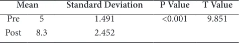
Table 1. Comparison between pre test and post test values of Glasgow coma scale in experimental group (within group comparison of GCS in experimental group)
Mean Standard Deviation P Value T Value
Pre 5 1.491 <0.001 9.851
The two-tailed P value is <0.001, considered extremely significant. t = 9.851 with 9 degrees of freedom.
Table 2. Comparison between pre test and post test values of Glasgow coma scale in control group (within group comparison of GCS in control group)
Mean Standard
Devi-ation P Value T Value
Pre 4.9 1.524 0.0039 3.857
Post 5.8 2.150
The two-tailed P value is 0.0039, considered very
significant. t = 3.857 with 9 degrees of freedom.
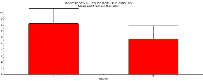
Table 3. Comparison between Post tests values of both groups (between groups comparisons of GCS score)
Mean Standard
Deviation P Value T Value
Post E 8.3 2.452 0.0261 2.424
Post C 5.8 2.150
The two-tailed P value is 0.0261, considered significant. t = 2.424 with 18 degrees of freedom.
Graph 1. Diagrammatic representation of Glasgow Coma Scale score before and after treatment in experimental group.
Graph 2. Diagrammatic representation of Glasgow Coma Scale score before and after treatment in control group.
4. Results
The statistical analysis concluded that the comparison between Pre-test and Post-test values of experiment group considered extremely significant with P value <0.001 and comparison between Pre-test and Post-test values of control group considered very significant with P value 0.0039. Comparison between Post-test values of both the groups considered significant with P value 0.0261.
5. Discussion
Severe brain injury is a cause for high morbidity and mortality rates. Individuals who sustain severe acquired brain injury, experience prolonged disorders of consciousness. So the purpose of this study is to find out the effectiveness of right median nerve stimulation on the level of consciousness in subjects with traumatic brain injury.
The results of this study have concluded that experimental group shows significant changes (P = 0.0261) when level of consciousness was measured by Glasgow coma scale. Although there is significance in experimental group in between the groups, significant difference noticed within the groups also (P < 0.0001, 0.0039). Post test values in experimental group have revealed that younger age group subjects have responded in much better way and there is increase in post test values of level of consciousness.
Experimental group showed improved level of consciousness this might be due to, median nerve stimulation brings numerous afferent inputs to the Ascending Reticular Activating System (ARAS) via the spino-reticular component of the median nerve synapsing with the neurons of the Ascending Reticular Activating System21. Median nerve stimulation will lead to activation
of the entire central nervous system. It is proposed that this peripheral stimulus reaches the Ascending Reticular Activating System, which further connects with the intra laminar nuclei of the thalamus and then cortical layers are stimulated. Improvement of level of consciousness, whether in persons in acute coma, or those in a chronic vegetative or minimally conscious state, is driven by the electrically induced elevation of dopamine and norepinephrine22,23. Increase in cerebral blood flow, is
another important factor in neuro-stimulation for re-awakening24
.
Unconscious patients have inhibition of Levator
palpebrae muscle which elevates the upper eyelid. Median nerve stimulation will modulate the levator palpebrae muscle activity. Existence of electrophysiological relationship between nucleus of the levator palpebrae muscle and ascending reticular activating system in brainstem is also evident25.
The right median nerve not only stimulate the brain stem and cerebrum to increase awareness but also better pattern of speech and abilities to calculate have been observed after right median nerve electrical stimulation26,27. As Broca’s speech area is in the left
fronto-temporal region, this area has been shown to become more active in positron emission tomography when a subject moves his or her hand28.
In this study it has found that after median nerve stimulation there is increase in scores of Glasgow coma scale, improvement in speech, which helped in faster recovery of the subjects. So, right median nerve stimulation can be included as treatment in clinical practice to improve consciousness.
6. Conclusion
Right median nerve stimulation have positive role in improving the levels of consciousness in subjects with traumatic brain injury and there by promotes faster recovery. Even though there is a need to study the mechanism behind the improvement of consciousness with right median nerve stimulation, this treatment option is evident in the recovery of comatose patients after traumatic brain injury.
7. References
1. Menon D, Schwab K, Wright D, Maas A. Position statement: Definition of traumatic brain injury. Archives of Physical Medicine and Rehabilitation. 2010; 91(11):1637–40. 2. Gururaj G. Epidemiology of traumatic brain injuries:
Indi-an scenario. Neurological Research. 2002; 24(1):24–8. 3. Agrawal A, Galwankar S, Kapil V, Coronado V, Basavaraju
S, McGuire L et al. Epidemiology and clinical characteris-tics of traumatic brain injuries in a rural setting in Maha-rashtra, India. Int J Crit Illn Inj Sci. 2012; 2(3):167.
4. Iranmanesh F. Outcome of head trauma. Indian J Pediatr. 2009; 76(9):929–31.
injury: A review. Epilepsia. 2003; 44:2–10. 7. Editorial Board. Endocrinology. 2015; 156(5):2C.
8. Liu C. Combined therapies. Neurosurgery. 2008; 63(4):N12. 9. Gill P, Gill T, Kamath A, Whisnant B. Readability assess-ment of concussion and traumatic brain injury publications by Centers for Disease Control and Prevention. Interna-tional Journal of General Medicine. 2012; 923.
10. Laureys S, Owen A, Schiff N. Brain function in coma, veg-etative state, and related disorders. The Lancet Neurology. 2004; 3(9):537–46.
11. Gultekin R, Huang S, Clavisi O, Pattuwage L, König T, Gru-en R. Pharmacological intervGru-entions in traumatic brain in-jury: Can we rely on systematic reviews for evidence? Inju-ry. 2016; 47(3):516–24.
12. Madder H. Treatment interventions for severe traumatic brain injury: Limited evidence, choice limitations. Journal of Medical Ethics. 2012; 38(11):662–3.
13. Lombardi F, Taricco M, De Tanti A, Telaro E, Liberati A. Sensory stimulation of brain-injured individuals in coma or vegetative state: results of a Cochrane systematic review. Clin Rehabil. 2002; 16(5):464–72.
14. Mandeep Kumar P. Effectiveness of early intervention of coma arousal therapy in traumatic head injury patients. International Journal of Head and Neck Surgery. 2012; 3:137–42.
15. Tolle P, Reimer M. Do we need stimulation programs as a part of nursing care for patients in “persistent vegetative state”? A conceptual analysis. Axon. 2003; 25(2).
16. Lombard L, Zafonte R. Agitation after traumatic brain inju-ry. American Journal of Physical Medicine and Rehabilita-tion. 2005; 84(10):797–812.
17. Sung H, Chang A, Abbey J. The effects of preferred music on agitation of older people with dementia in Taiwan. Int J Geriat Psychiatry. 2006; 21(10):999–1000.
18. Bradt J, Magee WL, Dileo C, Wheeler BL, McGill way E. Music therapy for acquired brain injury. Cochrane Data-base of Systematic Reviews. 2010.
19. Arumugam G, Brammatha, Shivananda V, Jose N, Sashidar. The effect of right side median nerve stimulation along with
multi sensory coma stimulation program on level of con-sciousness and neurobehavioral function among diffuse ax-onal injury patients - An experimental study. Internatiax-onal Journal of Physiotherapy and Research. 2013; 1(3):83–7. 20. Tadrisi SD, Bahari N, Ebadi A, Madani SJ. Validity and
re-liability of coma scale (Four Score) in adult patient hospi-talized in critical care units. Iran J Crit Care Nurs. 2012; 5(2):95–100.
21. Kaur H, Gupta D, Sharma V. Right median nerve stimula-tion for improving consciousness: A case series. IJNT. 2015; 12(2):144–8.
22. Hayashi N. Prevention of vegetation after severe head trau-ma and stroke by combination therapy of cerebral hypo-thermia and activation of immune, dopaminergic nervous system. Society for Treatment of Coma. 1997; 6:133–47. 23. Moriya T, et al. Usefulness of median nerve stimulation in
patients with severe traumatic brain injury determined on the basis of changes in cerebrospinal fluid, dopamine. Soci-ety for Treatment of Coma. 2000; 9:159–61.
24. Liu JT, Wang CH, Chou IC, Sun SS, Koa CH, Cooper E. Regaining consciousness for prolonged comatose patients with right median nerve stimulation. Acta Neurotic Suppl. 2003; 87:11–4.
25. Uysal H, Kizilay F, Selcuk B, Ersoz M, Akyuz M. A silent period of levatorpalpebrae activity induced by median nerve stimulation. Journal of Neurological Sciences. 2010; 27:28–34.
26. Cooper J, Jane J, Alves W, Cooper E. Right median nerve electrical stimulation to hasten awakening from coma. Brain Injury. 1999; 13(4):261–7.
27. Cooper E, Scherder E, Cooper J. Electrical treatment of reduced consciousness: Experience with coma and Alz-heimer’s disease. Neuropsychological Rehabilitation. 2005; 15(3-4):389–405.