ASSOCIATIONS BETWEEN GENETIC POLYMORPHISMS IN DNA BYPASS POLYMERASES AND BASE EXCISION REPAIR GENES WITH THE RISK OF
BREAST CANCER
Leila Family
A dissertation submitted to the faculty of the University of North Carolina at Chapel Hill in partial fulfillment of the requirements for the degree of Doctor of Philosophy in the
Department of Epidemiology.
Chapel Hill 2014
Approved by:
Andrew F. Olshan
Jeanette T. Bensen
Melissa A. Troester
Michael C. Wu
ii ©2014 Leila Family
iii
ABSTRACT
LEILA FAMILY: Associations between genetic polymorphisms in DNA bypass polymerases and base excision repair genes with the risk of breast cancer
(Under the direction of Andrew F. Olshan)
Mutations in BRCA1, a DNA repair gene, have been associated with a lifetime increased
risk of breast cancer (1). Therefore, researchers hypothesized there may be other DNA repair
genes associated with breast cancer risk. However thus far, studies of common low-penetrant
DNA repair SNPs have not yielded consistent results. In this proposed study, we hypothesized
one or more of the following mechanisms may explain the lack of main SNP effects: combined
SNP effects, modification by race or breast cancer subtype, and functional redundancy. To
evaluate these hypotheses, we used genotype data from the Carolina Breast Cancer Study (1,972
cases and 1,776 controls) to investigate race-specific, subtype-specific, and combined SNP
associations using unconditional logistic regression in two DNA damage pathways, base excision
repair (BER) and translesion synthesis (TLS). For BER, we evaluated the association between
31 single-nucleotide polymorphisms (SNPs) in 15 genes and breast cancer risk. SKAT, a
pathway-based analytic method, was used to evaluate the combined SNP effects within the BER
pathway. Among Whites, our results showed a significant positive association for NEIL2
rs1534862 and a significant inverse association for PCNA rs17352. Among African Americans,
we found a suggestive positive association for UNG rs3219275 and an inverse association for
NEIL2 rs8191613. Tumor subtype analysis showed that NEIL2 rs1534862 was associated with
luminal and HER2+/ER- subtypes. SKAT analysis showed no significant combined effects
single-iv
nucleotide polymorphisms (SNPs) in 7 bypass polymerase genes and breast cancer risk. We
found similar increased odds ratios for breast cancer with three POLQ SNPs (rs487848,
rs532411, rs3218634), which were also in high LD in both races. Furthermore, analysis by
specific tumor subtypes showed all three SNPs were associated with increased risk of luminal
breast cancer. These significant findings need to be replicated independently in other studies.
Overall, our results did not indicate associations with breast cancer, which may concur with the
theory that our cells possess an intricate system of functionally redundant DNA repair
v
To Dr. Robert C. Millikan
Who always believed in me
“The thing always happens that you really believe in,
and the belief in a thing makes it happen.”
vi
ACKNOWLEDGEMENTS
First, I would like to thank God for giving the strength and endurance to continue and
finish my dissertation work. Also, I would like to thank my dissertation committee members, Dr.
Andrew Olshan, Dr. Jeannette Bensen, Dr. Melissa Troester, Dr. Michael Wu, and Dr. Carey
Anders, for providing me with their expertise and valuable feedback. I would like to extend a
special thanks to my chair, Dr. Andrew Olshan, for his continuous guidance and support and for
navigating me through the dissertation process and dealing with unforeseen circumstances.
I would also like to thank everyone who has contributed their time and energy to the
Carolina Breast Cancer Study. Without their hard work and dedication, this work would not be
possible. I would especially like to thank Mary Beth Bell, CBCS Project Manager, for her strong
commitment to the study. Of course, I would also like to acknowledge the selfless contributions
of all the women that participated in the Carolina Breast Cancer Study.
I would also like to thank my vast circle of support, including the helpful staff in the
Epidemiology Department. I especially want to thank Nancy Colvin for her unwavering support
and dedication to my success. I would also like to thank my awesome friends and family. In
particular, I would like to give special thanks to Katie O’Brien and Lauren McCullough. These
ladies went out of their way to make sure I was successful, including volunteering countless
hours of their time to provide advice and feedback.
I would also like to take a moment to acknowledge and honor the work of Dr. Robert
Millikan and Dr. Keith Amos, whose impact in the field of breast cancer will be felt for years to
vii
Last, but certainly not least, I would like to thank my amazing parents, Gity and Siamak
Family, for their unwavering support and unconditional love throughout my whole life, and the
many selfless sacrifices they have made for me. I am truly blessed and honored to know such
wonderful and genuinely humble people.
I would also like to acknowledge the sources of financial support that enabled me to
complete this research: the University Cancer Research Fund of North Carolina, the National
Cancer Institute Specialized Program of Research Excellence (SPORE) in Breast Cancer (NIH/
NCI P50-CA58223) and NRSA Pre Doctoral Training Grant (2007-2009), University of North
viii
TABLE OF CONTENTS
LIST OF FIGURES ... xvii
LIST OF ABBREVIATIONS ... xviii
CHAPTER 1. REVIEW OF THE LITERATURE ... 1
1.1 Introduction ... 1
1.2 Definition of breast cancer ... 1
1.3 Epidemiology of breast cancer... 2
1.3.1 Breast cancer incidence ... 2
1.3.2 Breast cancer mortality ... 3
1.3.3 Non-genetic risk factors of breast cancer ... 3
1.3.3.1 Non-genetic risk factors of breast cancer by race ... 4
1.3.4 Genetic risk factors of breast cancer ... 5
1.3.4.1 Genetic risk factors of breast cancer by race ... 7
1.4 Heterogeneity of breast cancer... 7
1.4.1 Non-genetic risk factors of breast cancer by subtype ... 9
1.4.2 Genetic risk factors of breast cancer by subtype... 10
1.4.3 Summary of breast cancer risk factors ... 12
ix
1.5.1 Low-penetrant common DNA repair variation in breast cancer ... 13
1.6 DNA damage responses ... 13
1.6.1 Overview of DNA repair... 14
1.6.2 Overview of base excision repair (BER) ... 14
1.6.2.1 Base excision repair and breast cancer ... 16
1.6.2.1.1 UNG ... 16
1.6.2.1.2 SMUG1 ... 16
1.6.2.1.3 MBD ... 17
1.6.2.1.4 MPG ... 17
1.6.2.1.5 MYH/MUYTH ... 17
1.6.2.1.6 TDG ... 18
1.6.2.1.7 OGG1 ... 18
1.6.2.1.8 NEIL1 ... 19
1.6.2.1.9 NEIL2 ... 19
1.6.2.1.10 APE1 ... 19
1.6.2.1.11 POLB ... 20
1.6.2.1.12 XRCC1 ... 20
1.6.2.1.13 LIG3 ... 22
1.6.2.1.14 FEN1 ... 22
x
1.6.2.1.16 PCNA ... 23
1.6.2.1.17 RFC1 ... 23
1.6.2.2 Critique and Summary of BER literature... 23
1.6.3 Overview of DNA tolerance ... 26
1.6.4 Overview of translesion synthesis (TLS) ... 27
1.6.4.1 DNA bypass polymerases and cancer ... 29
1.6.4.1.1 POLH ... 30
1.6.4.1.2 POLI ... 31
1.6.4.1.3 REV1 ... 31
1.6.4.1.4 POLQ ... 31
1.6.4.1.5 REV3L ... 32
1.6.4.1.6 POLL ... 32
1.6.4.2 Critique and summary of bypass polymerase literature ... 33
1.6 Conclusions ... 33
CHAPTER 2. METHODS... 46
2.1 Specific Aims ... 46
2.2 Study population: Carolina Breast Cancer Study (CBCS) ... 49
2.2.1 Case ascertainment ... 49
2.2.2 Control ascertainment ... 50
2.2.3 Randomized recruitment ... 50
xi
2.2.5 Baseline study interview ... 52
2.3 Exposure assessment ... 54
2.3.1 CBCS SNP selection ... 54
2.3.2 Genotyping analysis ... 55
2.3.3 Genotyping quality control ... 56
2.4 Outcome Assessment ... 57
2.4.1 Ascertainment of intrinsic subtype markers... 57
2.4.2 IHC for in situ cases ... 59
2.5 Covariate Assessment ... 59
2.5.1 Traditional Confounding ... 59
2.5.2 Confounding by ancestry (population stratification) ... 60
2.6 Statistical Analysis ... 63
2.6.1 Assessment of Hardy-Weinberg Equilibrium ... 63
2.6.2 Genetic Model Specification ... 63
2.6.3 Race-specific effects ... 64
2.6.4 Correction for multiple testing ... 66
2.6.5 Combined within-pathway effects ... 67
2.7 Power calculations ... 70
2.8 Limitations ... 70
xii
2.8.2 Outcome (phenotype) misclassification ... 71
2.8.3 Covariate misclassification ... 72
2.8.4 Selection bias ... 73
2.8.5 Missing data ... 73
2.9 Strengths of the study... 74
2.10 Public health significance ... 76
CHAPTER 3. SINGLE NUCLEOTIDE POLYMORPHISMS IN BASE EXCISION REPAIR PATHWAY GENES AND ASSOCIATION WITH BREAST CANCER AND BREAST CANCER SUBTYPES AMONG AFRICAN AMERICANS AND WHITES ... 94
3.1 Introduction ... 94
3.2 Materials and Methods ... 95
3.2.1 Study population ... 95
3.2.2 Baseline Study Visit ... 96
3.2.3 SNP selection and genotyping ... 96
3.2.4 IHC analysis and subtype ascertainment ... 97
3.2.5 Statistical analysis ... 98
3.2.6 Subtype analyses ... 99
3.2.7 Correction for multiple testing ... 99
3.2.8 Pathway-based analysis ... 100
xiii
3.3.1 Genotype associations by race ... 101
3.3.2 Genotype associations by subtype ... 101
3.3.3 Pathway-based analysis ... 102
3.4 Discussion ... 102
CHAPTER 4. SINGLE NUCLEOTIDE POLYMORPHISMS IN DNA BYPASS POLYMERASE GENES AND ASSOCIATION WITH BREAST CANCER AND BREAST CANCER SUBTYPES AMONG AFRICAN AMERICANS AND WHITES ... 122
4.1 Introduction ... 122
4.2 Materials and Methods ... 124
4.2.1 Study population ... 124
4.2.2 Baseline Study Visit ... 125
4.2.3 SNP selection ... 125
4.2.4 Genotyping methods and quality control ... 125
4.2.5 IHC analysis and subtype ascertainment ... 126
4.2.6 Statistical analysis ... 127
4.2.7 Subtype analyses ... 128
4.2.8 Correction for multiple testing ... 128
4.2.9 Pathway-based analysis ... 129
4.3 Results ... 129
xiv
4.3.2 Genotype associations by subtype ... 130
4.3.3 Pathway-based analysis ... 130
4.4 Discussion ... 131
4.5 Conclusions ... 134
CHAPTER 5. DISCUSSION ... 145
5.1 Summary of Results ... 145
5.2 Strengths and Limitations ... 149
5.2.1 Study design ... 149
5.2.2 Genotyping methods ... 150
5.2.3 Tumor Subtyping ... 150
5.2.4 SKAT analysis ... 151
5.2.5 Power issues ... 151
5.3 Public health significance ... 152
5.4 Future research ... 153
5.5 Conclusion ... 154
xv
LIST OF TABLES
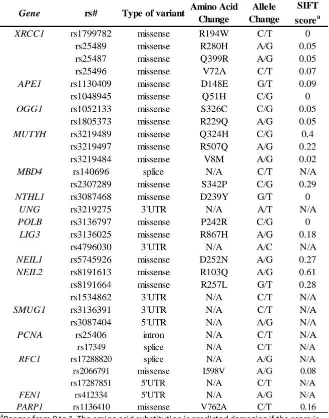
Table 1. Functions of BER genes ... 36
Table 2. DNA Glycosylases ... 37
Table 3. Associations between BER genes and breast cancer risk ... 38
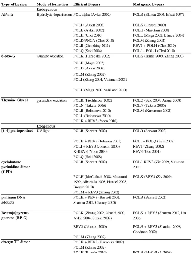
Table 4. Efficient and Mutagenic Bypass of DNA Lesions... 40
Table 5. CBCS Sampling Probabilities ... 78
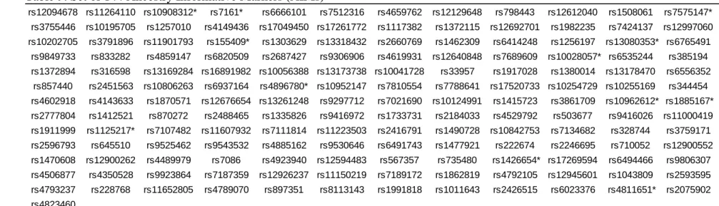
Table 6. Base Excision Repair SNPs ... 79
Table 7. Bypass polymerase SNPs... 81
Table 8. Subtype distribution by race ... 83
Table 9. Set of 144 Ancestry Informative Markers (AIMs) ... 87
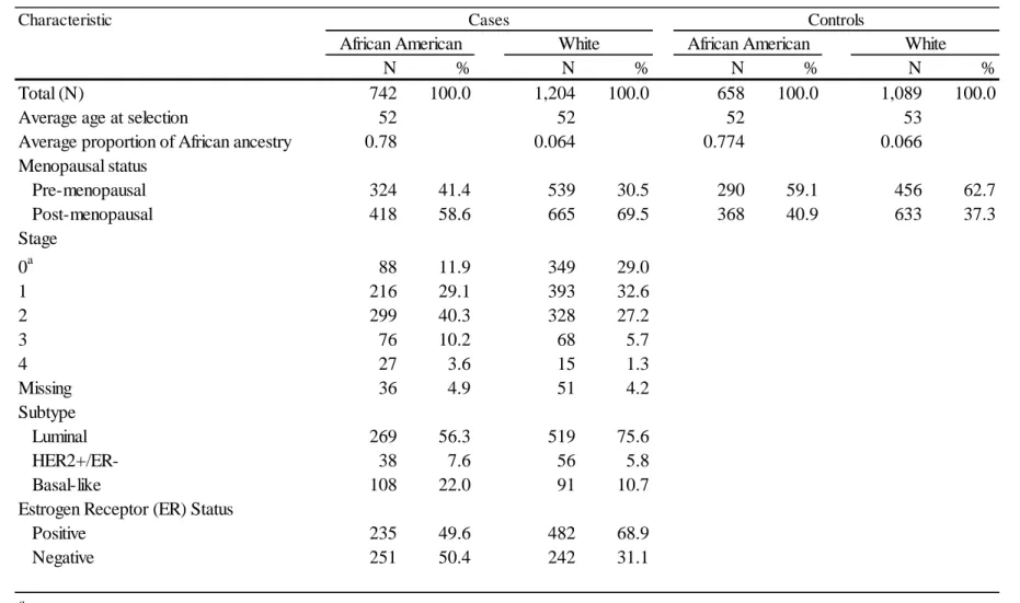
Table 10. Characteristics of CBCS participants with genotyping data ... 108
Table 11. List of successfully genotyped BER SNP in HWE ... 109
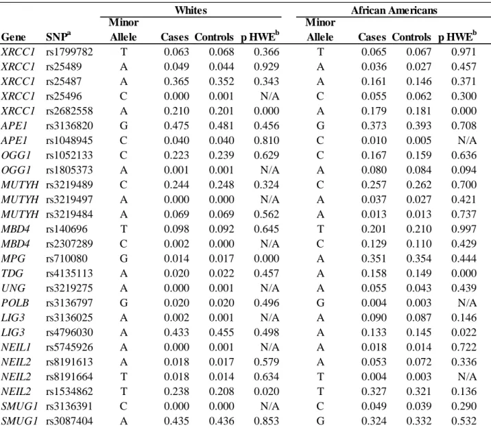
Table 12. Minor Allele Frequencies (MAFs) of BER variants stratified by race and case status ... 110
Table 13. Association of BER variants with breast cancer stratified by race ... 112
Table 14. Association of BER variants with breast cancer stratified by subtype ... 116
Table 15. Assocation of BER variants with breast cancer stratified by estrogen receptor (ER) status... 118
Table 16 SKAT analysis ... 120
Table 17. Linkage Disequilibrium by race ... 121
Table 18. Characteristics of CBCS participants ... 135
Table 19. List of successfully genotyped TLS variants ... 136
Table 20. Minor Alleles Frequencies of bypass polymerase SNPs stratified by race ... 137
xvi
Table 22. Association of bypass polymerase variants with breast cancer
stratified by subtype ... 140
Table 23. Association of bypass polymerase variant with breast cancer
stratified by ER status ... 141
Table 24. SKAT analysis of bypass polymerase SNP sets ... 142
xvii
LIST OF FIGURES
Figure 1. Breast anatomy ... 41
Figure 2. Breast cancer incidence and mortality by race and age ... 42
Figure 3. DNA Damage Responses ... 43
Figure 4. Sources of DNA Damage and associated lesion and repair pathway genes ... 44
Figure 5. Short-Patch vs. Long-Patch BER ... 45
Figure 6. Carolina Breast Cancer Study Area (Phase 1 and 2) ... 77
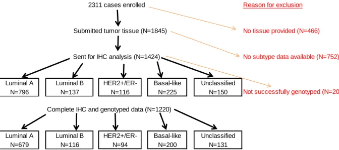
Figure 7. Enrolled cases with genotyped data ... 82
Figure 8. Enrolled cases with complete IHC and genotyped data ... 84
Figure 9. Directed Acyclic Graph (DAG)... 85
Figure 10. Confounding by ancestry ... 86
Figure 11. Classification schema for tumor subtypes ... 88
Figure 12. Power curves for African Americans ... 90
Figure 13. Power curves for Whites ... 90
Figure 14. Power curves for Luminal vs controls ... 91
Figure 15. Power curves for Basal-like vs controls ... 92
xviii
LIST OF ABBREVIATIONS
AA African-American
ACS American Cancer Society
ADP Adenosine diphosphate
AIMs Ancestry informative markers
AP Apurinic/Apyrimindinic site
APE1 AP endonuclease 1
ATM Ataxia telangiectasia mutated homolog
ATP Adenosine triphosphate
ATR Ataxia telangiectasia and Rad3 related
BER Base excision repair
BMI Body mass index
BRCA1 Breast Cancer Gene 1
BRCA2 Breast Cancer Gene 2
BRIP1 BRCA1 interacting protein C-terminal helicase 1
CBCS Carolina Breast Cancer Study
CEU
HapMap population of individuals of northern and western European ancestry living in Utah
CGEMS Cancer Genetic Markers of Susceptibility
CGHFBC Collaborative Group on Hormonal Factors in Breast Cancer
CHEK2 Checkpoint kinase 2
CI Confidence interval
xix
CIS Carcinoma in situ
CK 5/6 Cytokeratin 5/6
CLR Confidence Limit Ratio
DAG Directed acyclic graph
DCIS Ductal carcinoma in situ
Df Degrees of freedom
DNA Deoxyribonucleic acid
dNMP Deo-nucleotide monophosphate
dNTP Deo-nucleotide triphosphate
DRC DNA repair capacity
DSB Double strand break
EGFR Epidermal growth factor receptor
ER Estrogen receptor
FEN1 Flap-structure-specific endonuclease 1
GWAS Genome-wide association study
HER2 Human Epidermal Growth Factor Response 2
HR Homologous recombination
HRT
HWE
Hormone replacement therapy
Hardy Weinberg Equilibrium
IHC Immunohistochemistry
IR Ionizing radiation
LCIS Lobular carcinoma in situ
xx
LIG3 Ligase III, DNA, ATP-dependent
LOH
MAF
Loss of heterozygosity
Minor allele frequency
MDB4 Methyl-CpG binding domain protein 4
MMR Mismatch repair
MPG N-methylpurine-DNA-glycosylase
MYH A/G specific adenine DNA glycosylase
NAACCR North American Association of Central Cancer Registries
NC North Carolina
NCBI National Center for Biotechnology Information
NCI National Cancer Institute
NEIL1 Nei endonuclease VIII-like 1
NEIL2 Nei endonuclease VIII-like 2
NHEJ Non-homologous end-joining
NHS Nurses' Health Study
OC Oral contraceptive
OGG1 8-oxoguanine DNA glycosylase
OR Odds ratio
PAH Polycyclic aromatic hydrocardon
PALB2 Partner and localizer of BRCA2
PARP1 Poly(ADP-ribose) polymerase 1
PARP3 Poly(ADP-ribose) polymerase family, member 3
xxi
POLB DNA polymerase beta
POLH DNA polymerase eta
POLI DNA polymerase iota
POLL DNA polymerase lambda
PR Progesterone receptor
PTEN Phosphatase and tensin homolog
R2 Pairwise correlation coefficient
RFC1 Replication factor C (activator 1)
SCCOOP Squamous cell carcinomas of the oral cavity and oropharynx
SEER Surveillance, Epidemiology and End Results Program
SMUG1 Single-strand-selective monofunctional uracil-DNA glycosylase 1
SNP Single nucleotide polymorphism
SSB Single strand break
TDG Thymine-DNA glycosylase
TLS Translesion synthesis
TP53 Tumor protein p53
UNC University of North Carolina at Chapel Hill
UNG Uracil-DNA glycosylase
US United States
UTR Untranslated region
UV Ultraviolet radiation
WEB Western New York Exposures and Breast Cancer
xxii
XRCC1 X-ray repair complementing defective repair in Chinese hamster cells 1
1
CHAPTER 1. REVIEW OF THE LITERATURE 1.1 Introduction
In the past few decades, there have been significant strides in breast cancer research in
both etiology and treatment. Breast cancer is one of the most investigated types of cancer in the
US, garnering a large proportion of both private and public cancer research funding as well as
media attention. Consequently, the results spawned from these research efforts have afforded
many women new treatment and prevention options improving overall survival from breast
cancer, with an estimated 3 million survivors in the United States (2).
1.2 Definition of breast cancer
The term “breast cancer” refers to a malignant tumor that has developed from cells in the
breast. There are two main types of breast cancer: ductal carcinoma and lobular carcinoma.
Most breast cancers are categorized as ductal carcinomas; that is they originate from the ducts,
the passages that transfer milk from the lobule to the nipple. Lobular carcinoma originates from
the lobules, the milk-producing glands. A less frequent type of breast cancer can begin in the
stromal tissues, which include the fatty and fibrous connective tissues of the breast (2) (Figure 1)
Breast cancer can be further classified as invasive or noninvasive. Invasive cancer has
spread from the milk duct or lobule to other tissues in the breast. Noninvasive or in situ cancers
are confined within the ducts or lobules and named accordingly, ductal carcinoma in situ (DCIS)
2
accounted for about 83% of in situ cases diagnosed during 2004-2008. Lobular carcinoma in situ
(LCIS) is a marker for an increased risk of invasive cancer in the same or both breasts (2).
1.3 Epidemiology of breast cancer 1.3.1 Breast cancer incidence
Breast cancer is the most commonly diagnosed (non-skin) cancer in women in the United
States, representing 29% of all female cancer cases (3). According to the American Cancer
Society (ACS) 2013 Cancer Statistics Report, there will be an estimated 232,340 new cases of
invasive and 64,640 new cases of carcinoma in situ this year (3). In North Carolina, there will be
an estimated 7,090 new cases of female breast cancer in 2013 (3). In 2012, the age-adjusted
incidence rate for breast cancer in the United States was 124.3 per 100,000 women per year.
These rates are based on cases diagnosed during 2005-2009 from 18 SEER geographic areas (4).
At a population level, women have a 12% risk (1 in 8) of developing breast cancer in the course
of their lifetime.
After increasing for more than two decades, female breast cancer incidence rates began to
slowly decrease in 2000, and then dropped abruptly by about 7% from 2002 to 2003. This large
decrease was thought to be due to the decline in use of hormone replacement therapy (HRT) after
menopause that occurred after the Women's Health Initiative ended their clinical trial early. The
preliminary data showed that the risks of HRT may outweigh their benefits. The study linked the
use of hormone therapy to an increased risk of breast cancer and heart disease (5). In the past
couple of years, breast cancer incidence rates have stabilized (3).
Age-adjusted breast cancer incidence rates (per 100,000) also vary by race. White women
are at the highest risk of breast cancer (122.3) followed by African Americans (116.1). Asians
3
Carolina, the incidence rates (per 100,000) are slightly higher for Whites and moderately higher
for African-Americans compared to the national average (124.5 and 122.3 respectively) (3).
According to data from 18 SEER geographic regions, while incidence is similar for
premenopausal White and African-African women, after menopause the incidence rates diverge,
and postmenopausal White women have substantially higher incidence compared to
postmenopausal African-American women (4) (Figure 2).
1.3.2 Breast cancer mortality
Although breast cancer mortality has declined by 30% in the past 25 years, it is still the
second leading cause of cancer mortality in the United States after lung cancer for women
(2).The ACS estimates 39,620 deaths due to breast cancer in 2013. Breast cancer accounts for
about 3% of all-cause mortality and 14% of all cancer deaths in the US (2). At the state level,
there will be an estimated 1,260 deaths from breast cancer in North Carolina in the year 2013.
Death rates from breast cancer have been declining in the past few decades especially in
premenopausal women. These decreases are believed to be the result of earlier detection through
screening and increased awareness, as well as improved treatment.
There are differences in survival by both age and race. Although the mortality gap has
lessened over the past several years, African-American women have higher mortality rates from
breast cancer compared to White women, especially for younger women, despite the fact that
Whites have a higher incidence (4) (Figure 2). Understanding this survival paradox is the first
step in helping to improve breast cancer survival among younger African American women.
1.3.3 Non-genetic risk factors of breast cancer
In the past two decades, there have been a multitude of epidemiological studies
4
Many of these individual studies yielded inconsistent results due to small sample size. The
Collaborative Group on Hormonal Factors in Breast Cancer (CGHFBC) was established to
aggregate data from multiple studies (10,000s of cases and controls) for a number of putative risk
factors such as menarche and menopause, abortion, breastfeeding, alcohol and tobacco, and
family history (6-10). Results from the Collaborative Group studies have provided conclusive
evidence for several hormone-related factors such as nulliparity, older age at first birth, younger
age at menarche and older age at menopause, long-term use of HRT being associated with
increased risk of breast cancer (11-14). Lifestyle factors such as moderate alcohol use and
postmenopausal weight gain have also been established to be positively associated with risk
while physical activity has been associated with an inverse association (9, 11-19).The strongest
risk factors are older age and personal family history; the latter which is correlated with genetic
factors.
1.3.3.1 Non-genetic risk factors of breast cancer by race
Results from past breast cancer research studies may not provide a fully representative
story. Most of this research relied on data collected from postmenopausal White women. In the
past, it was difficult to examine risk factors in other racial/ethnic groups, since many studies did
have enough power to evaluate breast cancer by race. Recent studies consisting of larger, more
diverse cohorts of women have allowed researchers to re-evaluate these “well-established” risk
factors by race (20, 21). These studies have identified several risk factors that may differ by race.
For example, the effects of body mass index (BMI) may vary by race. While higher BMI in
post-menopausal White women may increase breast cancer risk, in African-American women of any
age BMI may act as a protective factor (22). While increasing parity and early age at first birth
5
African-American women. Multi-parity was associated with increased risk of breast cancer
among younger African-American women (for 3 or 4 pregnancies: OR =1.5, 95% (CI): 0.9, 2.6;
for 5 or more pregnancies: OR = 1.4, 95% CI: 0.6, 3.1), but not among younger White women
with the same number of pregnancies (20).
1.3.4 Genetic risk factors of breast cancer
Family history, one of the strongest risk factors for breast cancer, is linked to inherited
genetic susceptibility. About 20 - 30% of women with breast cancer have a family history of the
disease. Having one first-degree relative (i.e. mother, sister) may increase risk by two-fold while
having two first-degree relative may increase risk by as much as three-fold (10, 23, 24). In the
CGHFBC cohort, risk ratios for breast cancer increased significantly with increasing numbers of
affected first-degree relatives compared with women who had no affected relatives (p<0.0001)
(10). However a recent study reported no significant differences by the number of affected
first-degree or second-first-degree family history (24). Overall, only 2.5% of breast cancer cases were
found to be attributable to a positive family history (23).
In 1990, BRCA1 was identified as one of the first breast cancer susceptibility genes
followed by the discovery of BRCA2 (1, 25). BRCA1 and BRCA2 genes are frequently mutated
in familial breast and ovarian cancers. Women who carry these mutations have a lifetime
increased risk of developing breast cancer (10). The average cumulative risks of breast cancer
among BRCA1 and BRCA 2 carriers by age 70 are 65% (95% CI: 44-78%) and 45% (95% CI:
31-56%) respectively (26). However, BRCA1 and BRCA2 mutations are estimated to account for
only 5-10% of all breast cancers and 15-20% of familial cases (27, 28). Several moderate
penetrant genes such as ATM, CHEK2, PTEN, and TP53 that predispose patients to genetic
6
consistently associated with higher risk of breast cancer (28, 29). Recent studies have identified
BRIP1 and PALB2 as two novel breast cancer susceptibility markers involved in DNA repair (30,
31). Many of these genes are involved in the regulation of DNA repair and checkpoint signaling
(28).
It has been proposed that there are other common low-penetrant genes that may modify
breast cancer risk in BRCA1/2 carriers. Antoniou and others established the Consortium of
Investigators on Modifiers of BRCA1/2 (CIMBA), a large research collaborative between 40
study centers in 22 countries to investigate potential modifiers in this high risk subgroup. Results
from these studies demonstrated that common variants in LSP1 and ZNF365 as well as several
susceptibility loci, 2q35, 8q24, 12p11, 12q24, 9p21, 9q31.2, were associated with breast cancer
in BRCA1 and/or BRCA2 carriers (32-40)
With the completion of the Human Genome Project in 2003 (41), researchers were
enabled to use this cache of comprehensive genome-wide data to evaluate associations that were
not possible with candidate gene studies. Large collaboration efforts such as Cancer Genetic
Markers of Susceptibility (CGEMS) and Breast Cancer Association Consortium (BCAC)
increased sample size and hence power in genome-wide association studies (GWAS). As a
result, since the advent of GWAS, several breast cancer susceptibility markers from genetic
association studies have been replicated and several novel susceptibility markers have been
identified (42-47). While GWAS have enhanced our current understanding of breast cancer
susceptibility genes and loci, these known genetic factors still only account for about 28% of the
7
1.3.4.1 Genetic risk factors of breast cancer by race
In addition, genetic risk factors may vary by race. Compared to White women with breast
cancer, African American cases are less likely to have a BRCA1 mutation (48). Furthermore,
recent results from GWAS suggest that susceptibility loci may differ by race. Initially, many
GWAS loci such as TERT-CLPM1L were identified in populations of European descent (33, 47,
49). However, Zheng 2012 failed to replicate this positive association in women of African
descent (50). In addition, GWAS studies have shown modification by race for various breast
cancer susceptibility loci (51).
1.4 Heterogeneity of breast cancer
Research from the past decade has shown that breast cancer is a complex and
heterogenous disease involving multiple pathways and a combination of genetic and non-genetic
risk factors. There is evidence of heterogeneity by both hormone receptor status and more
recently by intrinsic tumor subtype. Historically, breast cancer tumors are classified based on
their hormone receptor status (i.e. estrogen receptor (ER) and progesterone receptor (PR)) and
HER2 status mainly to guide clinical treatment options (52). Endocrine therapies such as
aromatase inhibitors were used for hormone receptor positive (ER+ and PR+) tumors, while
therapeutics such as Herceptin were used for tumors overexpressing HER2 (52). Tumors that
were negative for all three of these markers were classified as triple-negative and were not
candidates for endocrine therapy targeted treatments. Several studies show risk profile
differences between hormone-positive vs. hormone-negative tumors (53, 54). A Carolina Breast
Cancer Study (CBCS) report showed that several hormone-related factors were associated with
stronger increased risks for ER+PR+ than for ER-PR- breast cancer; including early age at
8
among postmenopausal women and high waist to hip ratio (WHR) among premenopausal
women. Conversely, family history and medical radiation exposure were associated with
ER-/PR- tumors (53). Prospective data from the Nurses’ Health Study confirmed significant
differences by ER/PR status for age, menopausal status, postmenopausal BMI, adverse effect of
first pregnancy, and past use of postmenopausal hormones (54). Additionally, risk factors
profiles may vary by HER2/neu status. A CBCS report showed that early age at menarche,
higher WHR, and family history of breast or ovarian cancer were associated with increased odds
ratios (ORs) for both HER2/neu+ and HER2/neu- breast cancers while breastfeeding for more
than a year was inversely associated (53).
Recent technological developments in microarray analysis have led to the molecular
subtyping of breast cancer tumors to further discern breast cancer heterogeneity. Perou et al. used
cDNA microarrays to measure the gene expression of more than 1700 genes. A hierarchical
clustering algorithm identified four different patterns of gene expression in in vitro human breast
cells and tumors: ER+/luminal, basal-like, HER2+/ER-, and normal (55, 56). In a second study
with more samples, Perou et al. further dichotomized luminal tumors into luminal A and luminal
B (56). In addition, Sorlie et al. was able to define these molecular types using a set of only 534
genes (57). Nielsen et al. used immunohistochemistry to categorize molecular profiles based on a
panel of four antibodies (ER, EGRF/HER1, HER2, and cytokeratin 5/6) and found that this
method was equivalent to the gene expression technique (58). Carey further updated IHC
subtype definitions to include PR status as well as dichotomize HER2 status into HER2+ or
HER2- (59). Therefore, breast cancer tumors were classified into 4 distinct molecular subtypes:
9
PR-, HER2+), basal-like (ER-, PR-, HER2-, CK 5/6+ and/or EGFR+) and an unclassified
category (59).
1.4.1 Non-genetic risk factors of breast cancer by subtype
More recently, molecular profiling of breast cancer tumors has enabled researchers to
evaluate risk factors based on these “intrinsic” subtypes. Several studies have observed
differences in the associations between breast cancer risk factors and subtypes of breast cancer.
Basal-like subtype may have a different risk factor profiles compared to the luminal A subtype
(22, 60-65). Compared to the luminal A subtype, basal-like cases were also more likely to have
younger age at menarche (62), younger age at first full-term pregnancy(22, 62, 66), higher parity
(22, 62, 66, 67) were less likely to breastfeed (22, 62, 64, 66, 67). While long term breastfeeding
(>6 months) was inversely associated with breast cancer across subtypes, the protective effect
was strongest for basal-like tumors (68). In addition to reproductive factors, obesity or elevated
BMI was also associated with increased risk of basal-like breast cancer and luminal B cancers
compared to luminal A cases, especially for premenopausal women (22, 62, 64, 66, 68). Many
studies also reported that family history may play a bigger role for women with basal-like tumors
compared to other subtypes, especially for premenopausal women (24, 61, 64, 69).
Reports from the CBCS have suggested heterogeneity among in situ tumors, Phillips et
al. showed that many risk factors for invasive and high grade in situ tumors were similar, but
differed from risk factors for low or medium grade in situ tumors in both strength and magnitude
of effects. For example, higher parity showed a strong inverse association with high grade DCIS
but had a weaker inverse association for low to medium grade DCIS. In addition, ten or more
10
inverse association for low to medium DCIS (70). In summary, there may be heterogeneity
within in situ tumors which needs to be further investigated.
In addition, basal-like tumors have poorer prognoses compared to luminal tumors (57, 59,
71). Basal-like tumors showed more aggressive features compared to Luminal A, including
higher mitotic index (P < 0.0001), higher grade (P < 0.0001), and a higher frequency of p53
mutations (P < 0.001)(57). In situ basal-like cancers also shared similar poor clinical outcomes
with invasive cases (72)
Several studies have shown that basal-like tumors occur at a higher incidence among
Americans compared to whites (71, 73, 74). In a study of Ghanaian women,
African-American and white women from the US, proportion of African ancestry was significantly
associated with triple-negative tumors. Ghanaians had the highest prevalence of triple-negative
tumors (82.2%), followed by African Americans (32.8%) and White Americans(10.2%) (75).
1.4.2 Genetic risk factors of breast cancer by subtype
Carriers of BRCA1 mutations are at higher risk of developing basal-like tumors (76-81).
It has been estimated that 80-90% of cancers in BRCA1 mutation carriers are of basal-like
subtype (81). Furthermore, the prevalence of BRCA1 mutation carriers in triple-negative tumors
was approximately 20% and 11%, respectively, in two studies (79, 80). Therefore, loss of
BRCA1 function may have an etiological role in the development of the basal-like phenotype.
Expression microarray analyses have also indicated similarities in gene expression between
BRCA1 cancers and sporadic basal-like cancers (82). BRCA1-mutated and basal-like tumors
share many similar characteristics including higher levels of genomic instability compared to
ER+ or luminal tumors. Compared to other subtypes, basal-like tumors have the highest levels of
11
alterations. At least three studies reported loss of 5q and gain of 10p in basal-like cancers
(83-85). Van Loo et al. showed that basal-like tumors were associated with low ploidy, high
frequency of loss of heterozygosity (LOH), and the highest frequency of copy number events
when compared to the other subtypes (86). There are also a higher number of chromosomal
rearrangements and aneuploidy increase in defective DNA repair genes. These results provide
evidence for both deficient DNA repair genes and basal-like breast cancer being associated with
genomic instability.
Recently identified breast cancer susceptibility loci in GWAS (CASP8, FGFR2, TNRC9,
MAP3K1, LSP1, 8q24, 2q35, 5p12, 16q12) may also vary by tumor characteristics such as
hormonal status or intrinsic subtype. Susceptibility loci in FGFR2, TNRC9, 8q24, 2q35, and
5p12, 9q13.2 had stronger associations for estrogen receptor-positive (ER+) disease than
estrogen receptor-negative (ER-) disease (51, 87-89). Two candidate loci in CASP8 (rs1045485,
rs17468277) and TGFB1 (rs1982073), were strongly associated with the risk of PR- tumors and
16q12 and 2q35 were associated with basal-like subtype (87).
A common variant at the TERT-CLPTM1L locus was also found to be associated with
estrogen receptor-negative breast cancer (90). Of note, this locus was not replicated in women
with African ancestry (91). In a recent population-based case-control study, Domagala et al.
found several CHEK2 mutationsassociated with different molecular subtypes of breast cancer.
Truncating mutations were associated with luminal B, and I157T CHEK2 mutation was
associated with luminal A. (92, 93). The GWAS discovery of a novel locus 19p13 was shown to
both modify risk of breast cancer in BRCA1 mutation carriers as well as in hormone receptor
negative and triple negative cases in the general population (32, 33, 43). In the CIMBA study,
12
breast cancer for both BRCA1/2 mutation carriers (32, 33). Results from the Breast Cancer
Association Consortium (BCAC) further showed that 19p13 was associated with triple negative
subtype (43). MERIT40 interacts with BRCA1 and plays a role in the repair of double-strand
break in the HR pathway. These results provide evidence that DNA repair may vary by breast
cancer tumor subtype.
1.4.3 Summary of breast cancer risk factors
Breast cancer is a multifactorial disease, which results from the combined effect of
genetic and non-genetic risk factors that can vary by both race and subtype. Linkage association
studies were the first to hint at a genetic component underlying familial breast cancer.
Furthermore, women with a first degree family history of breast cancer were found to be at
almost twice the risk as women without a family history (10). Therefore, a positive family
history of breast cancer may serve as a surrogate for shared genetic variation (94).
1.5 Variation in DNA repair capacity
The discovery of mutations in BRCA1 in the early 1990s offered insight into the genetic
etiology of familial breast cancer. Carriers of BRCA 1/2 mutations were found to have deficient
DNA repair capacity (DRC) compared to normal controls (95). Experimental studies showed that
the near complete loss of DNA repair capacity can lead to genetic instability and a high risk of
developing cancer (96). However the prevalence of BRCA1 mutations and mutations in other
moderate to high penetrant DNA repair genes such as BRCA2, ATM, CHEK2, PTEN, BRIP1, and
PALP2 are rare in the general population and only explain 15-20% of genetic susceptibility to
13
1.5.1 Low-penetrant common DNA repair variation in breast cancer
The polygenic model of cancer was proposed to explain the missing heritability (100,
101). Under this model, a combination of multiple low penetrance genes would contribute to
overall genetic risk. There is increasing evidence that mild reductions in DNA repair capacity,
assumed to be the consequence of common genetic variation, can also affect breast cancer
susceptibility. The extensive variation in the coding regions of DNA repair genes and the large
number of genes in each DNA pathway results in complex genotypes with potential to impact
cancer risk in the general population (94, 102). In our proposed study, we focused our
investigation on these variants in common, low-penetrant DNA repair variants to evaluate their
potential association with breast cancer.
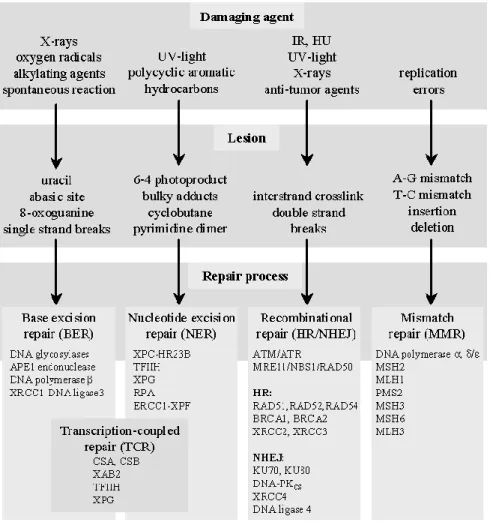
1.6 DNA damage responses
The combination of endogenous and exogenous sources of DNA damage can result in as
many as one million DNA lesions per cell per day (103). Endogenous sources include
replication errors and spontaneous reactions, while exogenous sources of DNA damage include
X-rays, oxygen radicals, alkylating agents, UV light, polycyclic aromatic hydrocarbons (PAHs),
IR, and anti-tumor agents. Unrepaired DNA damage can result in gene mutations such as point
mutations, deletions and insertions as well as chromosomal alterations such as chromosomal
rearrangements and aneuploidy. In order to maintain genomic stability, organisms have evolved
a complex series of damage repair responses to process DNA damage in a timely and efficient
manner including 1) apoptosis, 2) checkpoint signaling 3) DNA repair and 4) damage tolerance
(Figure 3). The proposed study will focus on the latter two mechanisms, specifically base
14
1.6.1 Overview of DNA repair
DNA repair mechanisms protect somatic cells from mutations in tumor suppressor genes
and oncogenes that can lead to cancer initiation and progression. Over the course of evolution,
cells have evolved several DNA repair pathways for repairing distinct types of DNA damage.
Specialized DNA repair pathways include direct reversal repair, mismatch repair (MMR), base
excision repair (BER), nucleotide excision repair (NER), and recombinational repair
(homologous recombination (HR) and non-homologous end-joining (NHEJ) (104). Figure 4
summarizes the source of DNA damage, the ensuing DNA lesion, and the DNA repair pathway
used to repair the lesion. Functional DNA repair plays an important role in tumor suppression.
There have been dozens of epidemiological studies examining common variation in multiple
DNA repair pathway. The focus of this study will be on single nucleotide polymorphisms (SNPs)
in the BER pathway.
1.6.2 Overview of base excision repair (BER)
Base excision repair (BER) is the fundamental pathway responsible for the repair of
damaged DNA bases induced by various sources of endogenous and exogenous damage. BER is
specialized to repair non-bulky DNA base lesions such as base adducts and abasic sites caused
by deamination, alkylation, or oxidation. The repair process consists of five enzymatic steps: 1)
cleavage of the sugar-phosphate chain, 2) excision of the abasic (AP) site, 3) removal of the
remaining sugar-phosphate chain, 4) DNA synthesis, and 5) ligation (105). Table 1 summarizes
BER genes and their functions in DNA repair.
To date, there are at least 11 known human DNA glycosylases. DNA glycosylases play
an important role in the initial recognition of a lesion and recruitment to the site of the damage
15
recognize more than one substrate (Table 2). DNA glycosylases initiate repair by releasing the
modified/damaged base out of the double helix and cleaving the N-glycosidic bond of the
damaged base, resulting in an apurinic/ apyrimidinic (AP) site. The location and type of the AP
site can also be determining factors on which glycosylase is recruited to the site (107, 108). If the
AP site was created by a glycosylase that does not possess AP lyase activity (UNG, SMUG1,
TDG, MPG, MDB4, MYH), or NTH1 and OGG1, repair of the AP site is APE1-dependent. A
newly discovered family of glycosylases (NEIL1, NEIL2, and NEIL3) was shown to efficiently
repair AP site independently from APE1 (107, 109, 110).
The repair of AP sites is crucial since they can interrupt normal DNA replication, and
become a threat to genomic integrity. APE1 or a member of the NEIL family converts the lesion
into a single-strand break (SSB). The SSB requires removal of the altered 3′-terminal groups
prior to ligation. After removal of obstructive termini, replacement of the excised nucleotide can
be completed either via short-patch where a single nucleotide is replaced or long-patch BER
where 2-10 new nucleotides are synthesized (111). Choice of pathway depends on several
different factors including the type of lesion, the cell cycle stage, and whether the cell is
terminally differentiated or actively dividing (112). The short-patch pathway requires a different
set of genes from the long-patch pathway (111, 113). The main distinction is whether the abasic
sugar is oxidized or reduced, which dictates if POLB is involved (short-patch) or not
(long-patch) (114).
Finally, the posttranslational modification of proteins is mediated by poly (ADP ribose)
polymerases (PARPs). Members of the PARP family (PARP1, PARP2, and PARP3) catalyze the
16
thereby recruiting DNA polymerases to the site of DNA synthesis to the 3’ end of primer,
promoting DNA synthesis (116, 117).
1.6.2.1 Base excision repair and breast cancer
It has been proposed that base excision repair may be involved in tumor suppression
(118, 119). Experimental studies have demonstrated that deletion of certain BER genes is
associated with an increased mutation rate in a variety of organisms, and hypothesize that this
loss could contribute to the development of cancer in humans (105). In addition, several dozen
case-control genetic association studies have been conducted (Table 3).The following section
summarizes the experimental and epidemiologic literature for the association between base
excision repair and breast cancer.
1.6.2.1.1 UNG
While no variants have been associated with breast cancer, two novel SNPs (UNG
Arg88Cys and UNG Gly143Arg) have been identified using mutational analysis in colorectal
cancer and glioblastoma cell lines, respectively (106, 120). In an in vivo study, knockout of the
UNG gene led to carcinogenesis in mice. Older (>18 months) UNG knockout mice developed B
cell lymphomas compared with only 1.3% of control animals (121).
1.6.2.1.2 SMUG1
In a 2011 Western New York Exposures and Breast Cancer (WEB) report (1,077 cases,
1,910 matched controls), two polymorphisms in the SMUG1 promoter region (rs2029166 and
17
95% CI: 1.1-1.5)(122). Another study examined the association between SMUG1 variants and
uracil blood concentration in 431 participants from the Boston Puerto Rican Health Study and
found a significant association with the SNPs studied. Increased level of uracil misincorporation
may induce mutagenic lesions and possibly lead to increased cancer risk (123).
1.6.2.1.3 MBD
Frameshift mutations in MBD4 have been associated with gastrointestinal cancers in two
Asian study populations (124, 125). The Glu346Lys polymorphism has been associated with
lung, esophageal, and gastrointestinal cancers (125-127). To our knowledge, there are no
experimental or epidemiologic studies investigating genetic variants of MBD4/MED1 with breast
cancer risk.
1.6.2.1.4 MPG
Based on the literature, there are no known experimental or epidemiologic studies
associating genetic variants of MPG with breast cancer risk. Three laboratory studies reported
altered expression of MPG in human gonad cells and astrocytic tumors (128-130).
1.6.2.1.5 MYH/MUYTH
Mutations in the MUYTH gene result in MAP (MUTYH-associated polyposis) a heritable
predisposition to colorectal tumors (131, 132). While a Dutch study initially reported increased
mutation frequency of several MUTYH SNPs among women of families with HBCC (Hereditary
Breast and Colon Cancer) (133), a validation study failed to replicate these results in a larger
case-control study (132). In a Chinese case-control study (545 cases, 545 controls), there were no
18
significantly associated with increased risk of early-onset breast cancer (<55 years old)
OR=1.51: 95% CI: 1.09-208) (134).
1.6.2.1.6 TDG
Polymorphisms G199S and V367M are the most common genetic polymorphisms in
human TDG. A recent Polish study revealed a possible association with these TDG
polymorphisms and lung cancer however these results may be biased due to small sample size
(135). Further studies are needed to fully understand the relationship between TDG and cancer.
1.6.2.1.7 OGG1
Functional lab evidence has suggested that rs1052133 (S326C) in OGG1 may be
associated with decreased DNA glycosylase activity in the repair of 8-oxoG, a mutagenic
byproduct of exposure to reactive oxygen (136). However, the results from epidemiological
studies have been less conclusive. At least six independent epidemiologic studies have evaluated
the association between the S326C polymorphism with breast cancer risk (137-142). Two reports
suggested an increased risk for S326C (137, 138) while two reports failed to find any significant
association (141, 143). An earlier review by Goode had identified S326C as being associated
with increased breast risk (144), however two recent meta-analyses were not able to replicate this
finding (145, 146). In a review of 14 functional studies and 19 epidemiological studies, Weiss et
al. found no significant association between the OGG1 polymorphism and breast cancer (146). A
recent case-control study in China (518 cases, 777 controls) showed two functional variations in
5'-UTR of OGG1 gene were significantly associated with the risk of breast cancer (OR=2.0 95%
19 1.6.2.1.8 NEIL1
In experimental studies, NEIL1 have been shown to interact with other BER genes
including POLB, LIG3, and PCNA (109, 148). In an in vitro study, NEIL1 downregulation
enhanced spontaneous mutation by three-fold in Chinese hamster and human cell lines (149). To
our knowledge, no epidemiologic studies have been conducted.
1.6.2.1.9 NEIL2
NEIL2 was shown to interact with POLB and LIG3 (109, 110, 150). Variant risk
genotypes in NEIL2 have been associated with increased risk in SCCOOP (head and neck
cancers) and lung cancers (151, 152). In an in vivo study, NEIL2 expression was significantly
reduced by over 50% in the presence of 2 SNPs (rs74800505 and rs8191518) which were in
significant LD (153). NEIL2 rs6982453 was associated with a significantly protective effect in
breast cancer in the Multiethnic Cohort Study (OR=0.86, 95% CI: 0.79-0.94, p<0.001) (154).
1.6.2.1.10 APE1
A number of functional polymorphisms have been identified in APE1 with the most
commonly studied SNP being APE1 Asp148Glu (155). This polymorphism has been associated
with risk of bladder, lung, prostate and gastric cancers (156-159). Overexpression of APE1 has
been linked to chemotherapy and radiation therapy resistance (160). However the epidemiologic
evidence for APE1 and risk of breast cancer is inconclusive. While two case-control studies
reported a borderline significant protective association for carriers of heterozygous variant
(Asp/Glu) in Thai and White American women respectively (138, 161), two other reports found
null associations for African American and White American women respectively (143, 161). A
20
genetic models (162). However, a recent lab study has linked deregulation of APE1 acetylation
to triple negative breast cancer (163).
1.6.2.1.11 POLB
DNA polymerase beta or POLB has been shown to be overexpressed in several cancers
(164-166). Seven germline mutations (P242R, E295K, G231D, K289M, E232K, T233I) have
been identified in POLB (167). In an in vitro study, Yamtich et al. 2012 found that expression of
these variant germline SNPs could be related to increased cancer susceptibility following
treatment with an alkylating agent (165). Gieseking also identified two POLB SNPs (E232K and
T233I) to be associated with lower fidelity when processing undamaged DNA, which may lead
to mutagenesis (168).Estimates of somatic mutations in Pol β range from 15% to 75% of tumors
in various types of cancer (169, 170). Functional analyses have implicated many of these variants
in cancer etiology and/or progression (170-173). An in vivo study showed that overexpression of
POLB variants in mouse cells resulted in cellular transformation. Furthermore, knockout of
POLB caused embryonic lethality. While there have been no positive associations between
POLB and breast cancer, there have been multiple reports suggesting POLB involvement in lung
and colorectal cancers in other epidemiological studies (174-176)
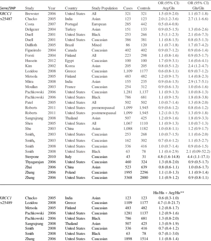
1.6.2.1.12 XRCC1
While the majority of studies did not find any significant associations (177-183), the
XRCC1 Arg399Gln polymorphism was associated with a protective effect in one report (184)
and an increased risk in seven other reports (105, 138, 139, 185-188). We suspect that these
significant positive findings were mostly false positives due to study design and low power
issues. Several of these studies had smaller sample sizes which may not have had adequate
21
CLR indicating imprecise estimates in several studies (138, 139, 186, 187, 189, 190). In addition,
results may be biased due to selection of controls (i.e. hospital-based controls (189) or cases (i.e.
cases selected for family history) (184). Alternatively, since the majority of significant findings
were from studies in Asian populations, there is the possibility of effect modification by Asian
race/ethnicity. At least four independent meta-analyses of XRCC1 Arg399Gln have provided
evidence for this theory (143, 191-193). Additionally, two U.S.-based population-based
case-controls studies found no overall associations, but showed subgroup effects for
African-American and postmenopausal women in the CBCS and WEB study, respectively (92, 137).
While no significant associations were observed in premenopausal women, postmenopausal
women with any Gln variant had increased risk of breast cancer (OR = 1.24; 95% CI: 1.01-1.51)
(137). Duell found a similar increased risk for African-Americans in the CBCS (OR=1.5 95%
CI: 1.1-2.3) (92).
Two reports found an increased risk for at least one variant of XRCC1 Arg194Trp (161,
189), while another report did not (92). In a meta-analysis of 11 studies including both White
and Asian populations, Zhang found no association between Arg194Trp and breast cancer risk
(143).
The majority of these studies reported no association with XRCC1 Arg280His with the
exception of one population-based case-control study of women from Cyprus. Loizidou et al.
found homozygous carriers of XRCC1 280His to have an increased risk of breast cancer
(OR=4.7; 95% CI: 1.0-21.7, P=0.03). Although this study contained 1,109 cases and 1,177
controls, a highly imprecise estimate was reported (194). The authors reported that this SNP
failed HWE (p<0.05) which may indicate genotyping error. Meta-analyses of XRCC1 Arg280His
22
and the XRCC1 Arg280His SNP, two other meta-analyses reported an overall increase risk of
cancer for the variant genotypes (His/His + Arg/His) compared with the wild-type homozygote
genotype (Arg/Arg) (191, 195).
1.6.2.1.13 LIG3
Knockout of LIG3 are embryonic lethal in mice (196). However, to our knowledge, there
are no known LIG3 SNPs that have been studied for association with cancer in the epidemiologic
study literature (114).
1.6.2.1.14 FEN1
FEN1 was significantly upregulated and aberrant expression was associated with
promoter hypomethylation in breast cancer cells in a gene expression study of 241 matched pairs
of cancer and normal tissues (197).
1.6.2.1.15 PARP1
PARP1 has been shown to inhibit DNA repair in both the short and long patch pathways
(198, 199). Conversely, cells deficient in PARP1 show increased rates of repair (198). Bieche
and colleagues reported overexpression of PARP1 and low genomic instability in a study of
breast cancer cells (200). In another study, inhibition of PARP1 was shown in tumors from
BRCA mutation carriers (201). However, a recent meta-analysis of 8 studies did not show an
association between PARP1 V762A and breast cancer (162).
In a lab-based study, PARP1-deficient cells were assessed for their capacity to repair AP
sites induced by uracil or 8-oxoguanine. For both DNA lesions, PARP1-deficient cells were
23
inefficient in the long-patch repair pathway. Inefficient BER occurred when both PARP1 and
POLB were absent (199).
In a subset of Nurses’ Health Study II cohort (NHS II), Han 2009 found PARP1
rs10915985 to be significantly associated with premenopausal breast cancer in the additive
model (OR=1.31, 95% CI: 1.04-1.64), however this SNP was not genotyped in CBCS (202).
1.6.2.1.16 PCNA
Several yeast models have associated PCNA mutations with cancer and genomic
instability (203). In addition, Ma and colleagues sequenced the coding region and adjacent
noncoding region of PCNA in 60 individuals and identified 9 sequence variants, including 7
SNPs which were located in introns involved in the control of PCNA gene expression. Results
from the analyses showed no associations with melanoma, breast cancer or lung cancer
compared with healthy controls (204).
1.6.2.1.17 RFC1
Experimental studies in have shown RFC1 to function in both DNA replication and
repair, specifically NER (116, 205). Replication factor C (RFC) is a five-subunit DNA
polymerase accessory protein that functions as a structure-specific, DNA-dependent
ATPase. RFC acts as a sensor in the DNA damage checkpoint pathway and plays a role in DNA
synthesis. To our knowledge, we are not aware of any epidemiologic studies examing RFC1
variants.
1.6.2.2 Critique and Summary of BER literature
Despite the strong associations of BRCA1 and BRCA2 and moderate penetrant genes such
24
penetrant BER genes for breast cancer have been underwhelming and attempts to understand the
contribution of low penetrant SNPs has been challenging. To date, there have been dozens of
population-based case-control genetic studies, including the Carolina Breast Cancer Study
(CBCS), that have investigated the association between common genetic variation in BER genes
(XRCC1, OGG1, APE1, NEIL1 and NEIL2) and breast cancer risk (92, 143, 182, 184, 189,
206-208). Most studies examined BER SNPs in the XRCC1, APE1, and OGG1 genes. While the
majority of studies of White women showed no significant associations with XRCC1 SNPs
(rs1799782, rs25489, and rs25487), several studies in non-White populations indicated potential
effect modification by race/ethnicity for rs25487, Arg399Gln.The evidence for OGG1
Ser326Cys and APE1 Asp148Glu polymorphism and breast cancer risk was null to weak (137,
138, 141-143). Additionally, findings from other BER SNPs studies have been inconclusive.
This failure to reveal significant associations between individual BER SNPs and breast
cancer is not surprising, given that carcinogenesis is a multistep, multi-genic process. Therefore
it is plausible that any one single genetic polymorphism would not have a dramatic effect on
cancer risk. Interaction between multiple common low-penetrant SNPs may be needed to
produce a significant effect. The polygenic model of cancer posits that although the risks
conferred by an individual locus are small, some risks may act multiplicatively or additively. In
this model, each variant is only one of the many genetic and environmental causal factors, each
of which are neither necessary nor sufficient to individually cause the disease. Therefore,
accumulation of mutations may be more important than a single SNP mutation (209).
Supporting evidence for the polygenic or multi-SNP effect in DNA repair is abundant.
Despite not finding main effects, many DNA repair studies have found significant multi-SNP
25
GWAS-validated breast cancer SNPs in a large European biobank-based study (3,584 cases,
5063 controls) and found a highly significant trend for increasing breast cancer risk with
increasing number of previously validated risk alleles (p-trend 5.6 x 10⁻²⁰) and for the maximum
versus the minimum number of risk alleles (OR=1.84, 95% CI: 1.59-2.14) (210).
Recent studies have used hierarchical modeling and other multi-SNP methods to evaluate
cancer risk at a gene or pathway level in various cancers (158, 211, 212). For breast cancer, two
reports from the Cancer Genetic Markers of Susceptibility (CGEMS) Project, a study nested
within the Nurses’ Health Study, evaluated the combined effects of low-penetrant SNPs in
multiple DNA repair pathways using Admixture Maximum Likelihood (AML) and Kernel
machine tests (202, 213). Han 2009 found several significant main effects for SNPs in PARP1,
NEIL2, APE1, and POLD for premenopausal women (p<0.05)(202), while a second report failed
to replicate any of this findings in postmenopausal women (213).
Another potential theory relates to the functional redundancy of genes to maintain
genomic stability. For example, in mouse models, knockouts of core BER proteins such as
XRCC1, POLB, APE1, and FEN1 all result in embryonic lethality (214-217). Furthermore, the
coding regions of PCNA and FEN1 are highly conserved (204). On the other hand, for DNA
glycosylases with multiple redundant pathways, there are no obvious phenotypes in nullizygous
mice lacking a single oxidative DNA glycosylase. Studies of double knockout mice have shown
they are prone to tumorigenesis. Chan et al. showed that targeted deletion of NTH and NEIL1
resulted in mice with a higher frequency of lung and liver tumors compared to single knockout
mice (218). In another experimental study, knockout of MYH or OGG1 individually showed very
26
formation (219). These studies suggest functional redundancy of DNA glycosylases and
highlight the integral role of BER genes to preserve genomic integrity.
1.6.3 Overview of DNA tolerance
The process of maintaining accurate DNA replication is essential to the genomic stability
of all cells. In the event that DNA damage should escape repair surveillance prior to initiation of
DNA replication, organisms have evolved a series of tolerance mechanisms for allowing
replication and cell division to process.
The first step in DNA replication involves the unwinding of DNA at the origin. The
replication fork is a structure that forms within the nucleus during DNA replication. It is created
by helicases, which break the hydrogen bonds holding the two DNA strands together. The
resulting structure has two branches, each one made up of a single strand of DNA. These two
strands serve as the template for the leading and lagging strands, which will be created as DNA
polymerases match complementary nucleotides to the templates. The leading strand is
synthesized continuously in the direction of replication fork, 5’ to 3’, while the lagging strand is
synthesized in small pieces (Okazaki fragments) backward from the overall direction of
replication (220, 221). Several DNA polymerases are involved in DNA replication. DNA
polymerase alpha initiates DNA synthesis on both the leading and lagging strands providing an
RNA primer and synthesizing approximately 20-30 bases of DNA. Pol epsilon (POLE) and pol
delta (POLD2) elongate these primers created by pol alpha (222). PCNA is the sliding clamp for
POLD1 and POLE (223). POLD1 and POLE also possess proofreading 3’-5’ exonuclease
activity that is important in preventing mutations.
DNA replicative polymerases, such as pol alpha, pol epsilon (POLE), and pol delta
27
efficient, with an estimated error rate of 1 in 10 billion base pairs (224). Despite this high
fidelity, a replication error may generate a one-sided double-strand break (DSB) or degrade to a
full DSB if it not repaired prior to initiation of DNA replication (225, 226). In order to resume
DNA replication at a stalled replication fork, two damage tolerance mechanisms have been
proposed; template switching in homologous recombination (HR) and translesion synthesis
(TLS) (227). Posttranslational modification of PCNA by ubiquitin may play a role in
determining which DNA repair tolerance mechanism to employ. Studies showed that the
mono-ubiquitylation of PCNA may activate translesion synthesis by damage-tolerant DNA
polymerases, while poly-ubiquitylation of PCNA may activate error-free pathway involving
template switching in HR (228-231). During template switching in HR, although normal
synthesis of DNA is blocked by a lesion on one of the template strands, synthesis on the
undamaged template strands can continue to a limited extent. The newly synthesized daughter
strand is used as the template, hence the term “template switching”. If template switching is
unsuccessful, translesion synthesis is activated to bypass the lesion (119, 222, 227).
1.6.4 Overview of translesion synthesis (TLS)
The focus of this study will be on the second DNA tolerance mechanism: translesion
synthesis (TLS). Translesion synthesis is conducted by a specialized type of DNA polymerases.
Aptly named, bypass polymerases do not directly repair the damage, but rather bypass or tolerate
the damage to prevent replication fork stalling. Unlike replicative polymerases, bypass
polymerases lack 3' to 5' exonuclease (proofreading) activity and are able resume replication
without an undamaged template (232, 233). However, this also contributes to their low fidelity