Immunogenicity of AGS-004 Dendritic Cell
Therapy in Patients Treated During Acute HIV Infection
Cynthia L. Gay,1,2Mark A. DeBenedette,3Irina Y. Tcherepanova,3Alicia Gamble,3 Whitney E. Lewis,3 Anna B. Cope,2,4JoAnn D. Kuruc,1,2 Kara S. McGee,6Mary F. Kearney,7John M. Coffin,8
Nancie M. Archin,1,2Charles B. Hicks,6Joseph J. Eron,1,2Charles A. Nicolette,3 and David M. Margolis1,2,5
Abstract
AGS-004 consists of matured autologous dendritic cells co-electroporated with
in vitro
transcribed RNA
encoding autologous HIV antigens. In an open-label, single arm sub-study of AGS-004-003, AGS-004 was
administered monthly to suppressed participants who started antiretroviral therapy (ART) during acute HIV
infection. HIV-1 specific T cell responses were measured by multicolor flow cytometry after 3–4 doses. The
frequency of resting CD4
+T-cell infection (RCI) was measured by quantitative viral outgrowth assay.
Parti-cipants demonstrating increased immune response postvaccination were eligible for analytic treatment
inter-ruption (ATI). AGS-004 induced a positive immune response defined as
‡2-fold increase from baseline in the
number of multifunctional HIV-1 specific CD28
+/CD45RA
-CD8
+effector/memory cytoxic T-lymphocytes
(CTLs) in all six participants. All participants underwent ATI with rebound viremia at a median of 29 days.
Immune correlates between time to viral rebound and the induction of effector CTLs were determined. Baseline
RCI was low in most participants (0.043–0.767 IUPM). One participant had a
>2-fold decrease (0.179–0.067
infectious units per million [IUPM]) in RCI at week 10. One participant with the lowest RCI had the longest
ATI. AGS-004 dendritic cell administration increased multifunctional HIV-specific CD28
+/CD45RA
-CD8
+memory T cell responses in all participants, but did not permit sustained ART interruption. However, greater
expansion of CD28
-/CCR7
-/CD45RA
-CD8
+effector T cell responses correlated with a longer time to viral
rebound. AGS-004 may be a useful tool to augment immune responses in the setting of latency reversal and
eradication strategies.
Keywords:
dendritic cell vaccine, HIV, acute HIV infection, HIV eradication, analytic treatment interruption
Introduction
G
iven a rapidly expanding need for antiretroviral therapy (ART) for HIV-infected individuals worldwide, innovative therapies that control or cure HIV-1 infection are needed. One approach is to strengthen and broadenHIV-specific CD8+ T cell responses, based on the temporal
as-sociation between the appearance of HIV-specific CD8+
responses and viral decline during acute HIV infection
(AHI).1,2One HIV therapeutic immunization strategy utilizes
monocyte-derived dendritic cells (MD-DCs) loadedex vivo
with RNA encoding HIV antigens, as MD-DCs are potent
antigen-presenting cells and induce primary and secondary
immune responses in CD4+and CD8+T cells. This approach
can induce and increase antigen-specific T cell responses in animal models and patients with chronic infections and
cancer.3Further, HIV-1 peptide-loaded DCs administered to
infected individuals can induce some degree of
HIV-specific immunogenicity.3–7
004-003 was a randomized controlled trial of AGS-004 on HIV RNA set point following analytic treatment in-terruption (ATI) in chronically HIV-infected (CHI)
partici-pants.4,8AGS-004 is an autologous DC therapy consisting of
matured patient-derived autologous DCs co-electroporated
1University of North Carolina HIV Cure Center, Chapel Hill, North Carolina.
Departments of2Medicine,4Epidemiology,5Microbiology and Immunology, University of North Carolina Chapel Hill, Chapel Hill, North Carolina.
3
Argos Therapeutics, Durham, North Carolina.
6
Duke University, Durham, North Carolina.
7
National Cancer Institute, Frederick, Maryland.
8
with in vitro transcribed RNA encoding HIV-1 Gag, Nef, Rev, and Vpr amplified from participants’ pre-ART plasma and RNA encoding human CD40 ligand (CD40 L).
Auto-logous DCs are maturedex vivoby sequential exposure to tumor
necrosis factor alpha (TNF-a), interferon gamma (IFN-c), and
PGE2before adding the CD40 L adaptive signal. CD40 L is
employed to maximize DC antigen presentation9and improve
immunopotency, by inducing secretion of interleukin (IL)-12, a necessary cytokine for CD8+T cell responses.10,11
In this sub-study, we evaluated AGS-004 in participants who started ART during AHI, as immune-enhancing strate-gies may be particularly suited to these individuals. ART
initiation during AHI can preserve HIV-1-specific CD4+and
CD8+T cell responses12and limit the size of the latent
res-ervoir.13–17 Further, most individuals are infected with a
single or limited number of founder viruses18,19 and early
treatment minimizes virus evolution, as few variants escape
from CD8+T cell pressure in the first weeks.18,20The
com-bination of preserved cellular responses and limited viral diversity in acutely treated individuals provides a unique population for vaccination studies.
Materials and Methods
This was an open-label, two-site, single arm sub-study of AGS-004 DC therapy in participants ages 18–59 years who initiated ART within 45 days of AHI, defined as a negative or indeterminate enzyme immunoassay or negative HIV RNA test within 45 days of detectable plasma HIV RNA.
Elig-ibility included HIV RNA levels <50 copies/ml (c/ml) on
ART for at least 6 months, CD4+ nadir ‡200 cells/mm3,
baseline CD4+count>400 cells/mm3, and a pre-ART frozen
plasma sample within 90 days of starting ART. AHI partic-ipants received five monthly doses of AGS-004 (weeks 0, 4, 8, 12, and 16) while on ART (Fig. 1), with each dose
con-sisting of 1.2·107DCs administered as three 0.2 ml
intra-dermal injections. Participants began ATI at*week 20 and
continued monthly AGS-004 dosing. In contrast to the parent
study in CHI participants,4 only AHI participants with a
positive CTL response postvaccination were eligible for
voluntary ATI. CD4+ counts and HIV RNA levels were
evaluated every 2 weeks following ATI. Restarting ART
could be triggered by CD4+ count <350 cell/mm3, >20%
decline in absolute CD4+count or CD4+ count percentage,
confirmed HIV RNA level‡10,000 c/ml, or participant
re-quest. Participants were followed through week 48 after the last AGS-004 dose or ART reinitiation (Fig. 1). The Uni-versity of North Carolina at Chapel Hill (UNC) and Duke University Institutional Review Boards approved the study. All participants provided written informed consent.
Autologous DCs used to produce AGS-004 have been
described.4 Monocytes are collected during leukapheresis
and differentiated in culture. Autologous pre-ART HIV RNA encoding Gag, Nef, Vpr, and Rev (GNVR) is amplified and co-electroporated with synthetic CD40 L RNA into mature
DCs.9A reduced quantity of Nef RNA is used to preserve DC
functionality by minimizing HLA downregulation.21 Vpr
RNA is truncated due to its ability to inhibit production of
IL-1222 and reduce activation of antigen-specific memory and
recall CD8+CTL responses.10,23,24
A second leukapheresis was collected before ATI for immune monitoring purposes. Autologous DCs were again co-electroporated with CD40 L RNA and GNVR, or with CD40 L RNA, and each individual antigen RNA or with CD40 L RNA and GFP RNA (DCs expressing green fluorescent protein (GFP) served as control DCs). This allows measurement of HIV-specific T cell responses in vitroto the combination RNA antigen payload (GNVR), to each individual HIV antigen (Gag, Nef, Vpr, or Rev) and to control DC for six separate DC targets. Peripheral blood mononuclear cells (PBMCs) were collected at baseline (immediately before the first AGS-004 dose), and 1 month after the third (week 12) or fourth dose (week 16), and 2 days after the fourth dose in one participant (51–102). Given a limited number of PBMCs collected, cultures were set up as individual wells and positive responses determined by
subtracting two times the total number of functional CD28+/
CD45RA- effector/memory CTL at baseline, from the total
number of functional CD28+/CD45RA- effector/memory
CTL measured in response toin vitrostimulation with DC
encoding each antigen.
To determine immune responses by multicolor flow
cy-tometry, PBMCs collected at baseline and after‡3
immu-nizations were cultured in vitrowith autologous DCs as
described.10,25 PBMCs were labeled with BrdU
(bromo-deoxyuridine) to track CTL proliferation and cocultured with autologous DC targets prepared as above. Autologous
DC targets forin vitro stimulation were either DCs
co-electroporated with CD40 L RNA and all antigens (GNVR) or with DCs co-electroporated with CD40 L and each in-dividual antigen. To determine background nonspecific
activation ofin vitro CTL cultures, PBMCs at each time
point were stimulated with CD40 L DCs co-electroporated with GFP RNA. GFP RNA was not included in the ad-ministered product. Cocultures were incubated at 37C for 6 days. On day 6, each culture was restimulated with the same autologous DCs used in the initial culture, anti-CD107a antibody was added and cultures were incubated at 37C for 5 h. Viable cells were identified by staining for viability using Live/Dead Fixable Dye (Invitrogen)
FIG. 1. Study schema. ART, antiretroviral therapy; ATI, analytic treatment interruption; BL, baseline leukapheresis; PS,
treatment-related adverse events in the study; none>grade 1, with mild injection site reactions the most common. No participant discontinued or interrupted study treatment due to adverse events.
Three participants (51–100, 51–102, and 54–100) received AGS-004 DC therapy with all four RNAs (GNVR). Two participants (54–101 and 54–102) received AGS-004 with three RNAs, Gag, Nef, and Rev (GNR) as Vpr was not re-covered in the pre-ART sample; one participant (54–104) received AGS-004 with three RNAs, Gag, Vpr, and Rev (GVR) as Nef was not recovered in the pre-ART sample.
Previous work demonstrated that in vitro priming with
postmatured DC co-electroporated with CD40 L RNA and HIV RNA encoding specific target antigens expanded
multifunctional antigen-specific CTL, exhibiting a CD28+/
CD45RA-effector/memory phenotype.4,11,28Similar DC
therapy administered to participants with metastatic renal cell carcinoma showed a significant correlation between the
ex-pansion of functional CD28+/CD45RA- effector/memory
CTL and clinical outcomes.25 Therefore, our primary
im-mune end point focused on the expansion of functional
HIV-1-specific CD28+/CD45RA-effector/memory CTLs.
After in vitro stimulation, the number of CD8+ T cells
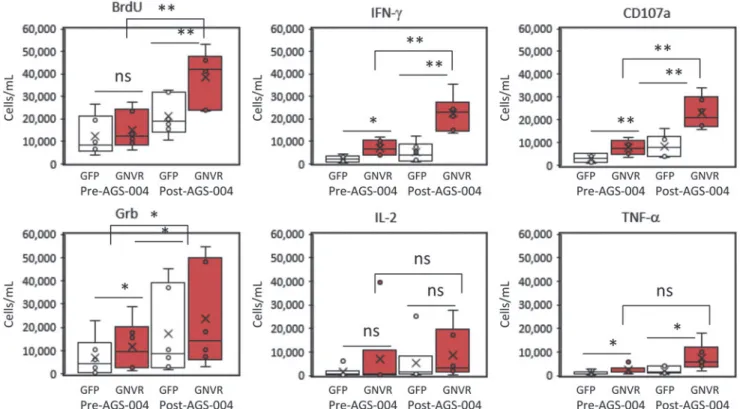
expressing each functional marker was calculated at baseline (W0, pre-AGS-004) and after AGS-004 administration (W12 or W16) for each participant (Fig. 3). Statistically significant
increases in the number of CD8+T cells proliferating (BrdU)
and expressing IFN-c, CD107a and Grb were detected to the total GNVR total RNA payload after AGS-004
admin-istration in all six participants. The number of CD8+T cells
expressing each functional marker (red dots) for a represen-tative participant is shown (Fig. 4A). Post-AGS-004, the
number of proliferating CTLs increased afterin vitro
resti-mulation from 14,011 to 32,875 cells/ml. Increases in the numbers of cells/ml were seen for the lytic markers CD107a (6,467–20,478) and GrB (15,971–27,793) and for the
cyto-kines IFN-c(6,718–20,557) and TNF-a (517–806). For this
representative participant, IL-2 decreased from 7,307 cells/ ml at baseline to 4,285 cells/ml post-AGS-004. Moreover, the
majority of functional CTLs resided in the CD28+/CD5RA
-effector/memory CTL subset (Fig. 4A red dots). Given in-creases in the number of functional CTL present in the
CD28+/CD45RA- CTL population, we determined
func-tional marker expression after AGS-004 administration as a
quantitative value (cells/ml) for CTLs in the CD28+/
CD45RA-effector/memory CTL subset. Determining
chan-ges in the number of cells/ml with functional activity in the
CD28+/CD45RA- effector/memory CTL subset from
base-line to post-treatment enables a direct comparison between measurements performed within a single participant and be-tween participants. Six of six participants had greater than a
two-fold increase in the total number of CD28+/CD45RA
-effector/memory CTLs expressing any functional marker after post-AGS-004 (W12 or W16), and in five participants the increases over baseline (W0) were statistically significant (Fig. 4 B–F). While the increase in response for participant 054–104 (Fig. 4G) was not statistically significant, it was greater than two-fold over baseline (from 30,613 cells/ml at baseline to 95,709 cells/ml at week 12). This contrasted to responses measured to GFP where only two participants, 054–101 and 054–102 (Fig. 4E, F, respectively) had
statis-tically significant increases in the total number of CD28+/
followedby surface staining with specific antibodiesfor
CD28,CCR7,CD27,CD45RA,CD3,andCD8expression.
Cellswerefixed,permeabilized,andDNasetreatedto
de-tectBrdU(BDbiosciences).Intracellularstainingfor
IFN-c, TNF-a,IL-2,GrB,andBrdUwasperformed.Cellswere
transferred to BD TruCountTubes foranalysis on a BD
LSRIIcytometer,with400,000–600,000 events collected
persample.
ViableCD3+CD8+TcellsweredividedintoeitherCD28
receptor-positive orCD28 receptor-negativesubsets.
Bool-eangatingwasusedtodefinethecentralmemory(CM)
ef-fectormemory(EM)andeffector(E)CTLsubsetsdefinedby
the expression of CD45RA, CCR7, and CD27 within the
CD28 receptor-positive and CD28 receptor-negative CTL
subsets(Fig.2A).WithintheCM,EM,andECTLsubsets,
positive staining for cytokines (IFN-c, TNF-a, and IL-2),
cytolyticmarkers(GranzymeB[GrB],CD107a,and
prolif-eration (BrdU) (Fig. 2B) identified functional marker
ex-pression.Increasesineffectormarkerexpressionarereported
as the absolute number of cells/ml determined using
Tru-CountTubes(BDbiosciences)duringsampleacquisitionby
flowcytometry.Thenumberofcells/mlwithinagivengateis
calculatedasfollows: (numberofcellulareventscollected/
numberofTrucountbeadscollected)·(Trucountbead
con-centration)/collectedvolume[lL])·1,000.
As in the parent study (AGS-004-003),4 a positive
re-sponse to AGS-004 was defined as >2-fold increase over
baselineinthenumber ofCD28+/CD45RA-
effector/mem-oryCTLs(cells/ml)exhibitingatleastoneeffectorfunction
defined by the expression of IFN-c, TNF-a, IL-2, GrB,
CD107a,or proliferationto the total HIV antigenpayload
(GNVR)present.Statisticallysignificantdifferencesbetween
CD28+/CD45RA-effector/memoryCTLresponsesat
base-lineandafterAGS-004administrationweredetermined
us-ingatwo-tailedStudent’st-TEST.Correlatesbetweentime
to viral rebound and change in the number of effector/
memoryand effector multifunctional CTLsafter AGS-004
administrationweredeterminedbynonparametricbivariate
Spearman’sRhostatisticalanalysis.
PlasmaHIVRNAassayswereperformedbyasingle-copy
assay (SCA), with two measurements at baseline and at
weeks10and16.AnadditionalSCAwasperformedatweek
28 if HIV RNA remained undetectable during ATI. The
frequencyofrestingCD4+Tcellinfection(RCI)was
mea-suredbyquantitativeviraloutgrowthassay(QVOA)as
de-scribed26,27 at baseline and pre-AGS-004 dosing and after
threedosesonART(week10).Correlationbetweentimeto
viralreboundduringATIandRCIandCTLfoldincreasewas
assessed by Spearman correlation. In participant (51–102)
withprolongedaviremiafollowingrebound duringtheATI
period,antiretroviral drugtestingwasperformedatMcGill
UniversityandtheUNCSchoolofPharmacy.
Results
BetweenJune 2012and January 2013, six male
partici-pantsenrolledwithamedianageof35years(range26–56;
Table1).Atenrollment,themedianCD4+Tcellcountwas
618cells/mm3 (range397–937),andthemediantime from
AHIdiagnosistoARTinitiationwas15days(range9–20).
Themediantimefromparticipant ARTstartdateto
CD45RA-effector/memory CTLs expressing any functional marker after AGS-004 administration.
We then examined the multifunctionality of the response, defined as an increase in at least two functional markers after post-AGS-004. Five of six participants had statistically sig-nificant increases in multiple functional markers with par-ticipants 54–100 (Fig. 4J) and 54–102 (Fig. 4L) having increases in all six functional markers. Participants 51–100 (Fig. 4H) and 51–102 (Fig. 4I) had statistically significant increases in all functional markers excluding IL-2, and par-ticipant 54–101 (Fig. 4K) had statistically significant in-creases in the number of cells/ml expressing markers for BrdU, CD107a, IFN-c, and TNF-a. Participant 54–104 (Fig. 4M) had increases in functional markers over baseline; however, the only statistically significant change was de-tected for CTLs that produced IL-2.
The increased numbers of CD28+/CD45RA- effector/
memory CTL (cells/ml) determined from pre- to post-AGS-004 treatment for each functional marker was plotted versus time to viral rebound and showed an inverse correlate be-tween time to rebound and the magnitude of proliferating
CTL (q= -0.811p= <.05 or CTL expressing Grb (q= -0.811
p= < .05) (Fig. 5A). If the CD28+/CD45RA- subset was further sub-gated to include only CTL expressing CCR7, to define CM CTL, additional inverse correlates were seen with
CTL expressing IFN-c(q= -0.927p= <.007), and CD107a
(q= -0.927 p= < .007), BrdU (q= -0.927 p= < .007), and GrB (q= -0.927p= <.007) (Fig. 5B). This prompted analysis of other CTL subsets defined by the absence of the CD28 receptor and CCR7, to see whether other functional subsets correlated with time to viral rebound. Interestingly, the in-crease in the number of functional CTL representative as
effector CTL defined as CD28-/CCR7-/CD45RA-positively
correlated with time to viral rebound (Table 1) for numbers of
CTL that are BrdU positive (q=0.811 p= < .05) express
CD107a, (q=0.927p= <.007), IL-2 (q=0.840p= <.036), or
TNF-a (q=0.927p= <.007) (Fig. 5C). These data suggest
participants with the longest time to viral rebound accumu-lated increased numbers of effector CTL in peripheral blood with a concurrent decrease in numbers of CM CTL in the periphery.
Evaluation of response to individual RNA antigens after in vitrostimulation revealed an increase in total numbers of
CD28+/CD45RA- effector/memory CTLs expressing any
functional marker for each participant to each individual HIV
antigen (Fig. 6). All six participants had ‡2-fold increased
responses over baseline to Gag (range 310–11,323 cells/ml) (Fig. 6A), and responses were multifunctional. The percent-age of each functional marker within the total response to Gag is represented for each participant (Fig. 6B). While all participants contained multifunctional CTLs, there was no clear pattern of functional marker expression across partici-pants. In some cases the CTL population response to Gag contained CTLs that proliferated (shown in blue), or
pro-duced cytokines IFN-c(purple) or TNF-a (orange), or IL-2
(light blue). Lytic activity was detected with GrB expression in three participants (green) and CD107a (red) for all par-ticipants.
The multifunctional responses to Nef (Fig. 6C) ranged from 2,380 to 37,834 cells/ml. Proliferating CTLs were ob-served in three participants and two of these participants
contained all six functional markers. Similarly, the
Table 1. Demographic and Clinical Characteristics of Acute Hiv Infection Participants Who Received AGS -004 Dendritic Cell Therapy Participant ID Age (years) Race/ethnicity Gender
Baseline CD4 count
composition of responses were multifunctional with 051–100 containing CTL expressing CD107a, IFN-c, and IL-2, par-ticipant 051–102 contained proliferating CTL and all six functional markers, 054–100 contained proliferating CTL and CD107a and GrB. 054–101 contained proliferating CTL and CD107a, IFN-c, and TNF-a. 054–102 contained CTL expressing all six functional markers (Fig. 6D).
Four participants tested for response to Vpr revealed re-sponses over baseline ranging from 8 to 67,160 cells/ml and were multifunctional for two participants (051–102 and 054– 104), expressing all six functional markers. Responses for
participants 051–100 and 054–100 were below 100 cells/ml and more mono-functional with CTL expressing only IL-2,
(051–100) or IFN-cand TNF-a(054–100) (Fig. 6F).
Responses to Rev over baseline ranged from 2,950 to 76,429 cells/ml (Fig. 6G); all participants had CTLs that expressed two functional markers. For participant 51–100, the response was dominated by CTL expressing IFN-c, GrB, and TNF-a, and for 051–102, the response contained prolif-erating CTL and CD107a, GrB, and IFN-c. 051–100 con-tained CTL expressing IL-2 and CD107a, 054–101 concon-tained proliferating CTL and IFN-c, CD017a, and TNF-a. Both
FIG. 3. CD8+ T cell responses in participants receiving AGS-004. Boxand whisker plots show the distribution of the
number of CD8+T cells (cells/ml) proliferating (BrdU), expressing IFN-c, CD107a, Grb, IL-2, or TNF-adetermined before
or post AGS-004 administration for participants. Statistically significant differences between the GFP background response (open barandwhisker plots) and the total antigen payload (GNVR)-specific response (red barandwhisker plots) are shown. Statistically significant differences between the total antigen payload (GNVR) responses determined pre- and post-AGS-004 administration are shown. *p<.05; **p<.01. GNVR, Gag, Nef, Vpr, and Rev; NS, not statistically significant.
FIG. 4. Multifunctional immune responses to the total antigen RNA payload in participants treated with AGS-004. CD8 T
cell responses at baseline (W0) or after AGS-004 administration (W12 or W16) were measured afterin vitrostimulation
with AGS-004 autologous DCs and functional marker expression was detected by multicolor flow cytometry.(A)Dotplot
overlays from one representative participant before AGS-004 administration (W0) and after AGS-004 administration (W12)
show the distribution of cellular events representing each functional marker (red dots) BrdU, CD107a, Grb, IFN-c, IL-2, and
TNF-aoverlaid on the total CD8+T cell population (blue dots) gated on the CD28 (y-axis) and CD45RA (x-axis) plots. The
numbers are representative of cells/ml expressing each functional marker (red dots).(B–G)The total number of CD28+/
CD45RA-CTL (cells/ml, Y-axis) expressing any functional marker after stimulation with DCs encoding GFP or the final
product HIV payload (GNVR, GNR, or GVR) was calculated at baseline (W0) and post-AGS-004 (W12 or W16). Total
numbers of cells/ml were determined by adding the numbers of cells/ml measured for each individual functional marker.(B)
51–100 to GNVR,(C)51–102 to GNVR,(D)54–100 to GNVR,(E)54–101 to GNR,(F)054–102 GNR, and(G)054–104
to GVR. Statistically significant increases in the total number of cells/ml expressing functional markers over baseline are shown. NS, not significant. The contribution of each functional marker (cells/ml, y-axis) within the total response at baseline
(W0) or post-AGS-004 (W12 or W16) for each participant is shown,(H)51–100 to GNVR,(I)51–102 to GNVR,( J)54–
100 to GNVR,(K)54–101 to GNR,(L)054–102 GNR, and(M)054–104 to GVR. BrdU , CD107a , Grb , IFN-c ,
IL-2 , and TNF-a .Starred(*) markers indicate statistically significant increased CTL responses from pre- to
post-AGS-004 administration. GNR, Gag, Nef, and Rev; GVR, Gag, Vpr and Rev.
054–102 and 054–104 contained CTL expressing all six functional markers (Fig. 6H).
After confirming increases in the number of functional
CD28+/CD45RA- memory CTL responses post-treatment,
all participants underwent ATI. Figure 7 shows the time
course of CD4+T cell counts and changes in viral load for
participants. Participants rebounded to ‡40 copies/ml at a
median of 29 days (range 13–55), with a median ATI of 66 days (range 33–280; Table 1). Three participants restarted
ART due to confirmed HIV RNA>10,000 c/ml, two restarted
due to a>20% decline in CD4+count, and one restarted after
meeting both CD4+and viral load criteria. The median
ob-served peak HIV RNA level following ATI was 21,650 c/ml (range 3,290–287,000), and no symptoms consistent with acute retroviral syndrome were observed. All participants rapidly suppressed viremia after restarting pre-entry ART regimens (Fig. 7); however, two had confirmed viremia during follow-up with self-reported nonadherence to re-sumed ART (54–101, 54–104).
In one participant (051–102), initial viral rebound to >10,000 c/ml was followed by repeat viral load of 3,579 c/ml and continued ATI. Due to prolonged aviremia (280 days) following initial viral rebound in this participant, therapeutic drug testing was performed for efavirenz, emtricitabine, and tenofovir on stored samples obtained during ATI. Efavirenz was not detected eight days following ATI and before viral rebound, but detected from three subsequent stored samples from separate time points over 3 months during the ATI; tenofovir and emtricitabine were detected in the one sample tested. This participant denied restarting ART, and had a second viral rebound of 12,200 c/ml at day 280.
The frequency of RCI at pre-entry and pre-AGS-004 dosing was low in most participants, ranging 0.043–0.767
infected resting CD4+cells per million (IUPM; Table 1), and
in comparison with participants treated during CHI.29Time
to viral rebound was inversely correlated with RCI (q= -0.84; p=.04). The only participant (054–100) with a statistically significant decrease in RCI (2.5-fold from 0.179
IUPM at baseline to 0.067 IUPM at week 10)30 also had
increases in the total number of functional CD28+/CD45RA
-CTLs (Fig. 4D), and increases in CD28-/CCR7-/CD45RA
-CTLs after AGS-004 dosing, but did not have the longest ATI. Increases were multifunctional in nature with CTLs positive for all six functional markers (Fig. 4J). Participant 054–100 had increases in the number of CTL responding to Gag (Fig. 6A, B), Nef (Fig. 6C, D), and Rev (Fig. 6G, H) and the longest time to rebound (55 days, Table 1) with an ATI duration of 90 days (Fig. 7C) in the absence of an HLA allele
associated with HIV control.31Despite an increase in both
effector/memory and effector CTL responses, this participant had the highest observed peak HIV RNA level off ART
(287,000 c/ml), but suppressed to <50 c/ml 4 weeks after
reinitiating ART.
The participant (54–101, Fig. 7D) with the lowest baseline RCI (0.043 IUPM) rebounded at 49 days, but underwent ATI
for 151 days, reinitiating ART due to decline in CD4+count
percentage (44.1%–33.8%). The peak observed HIV RNA level during this participant’s ATI was 3,290 c/ml followed by rapid suppression after restarting ART, but developed subsequent viremia with reported nonadherence to ART. This participant showed an increase in the total number of functional CTL after AGS-004 administration (Fig. 4E) and
FIG. 5. Increased numbers of effector CTL within the
CD28-/CCR7-/CD45RA- CTL subset correlates with longer
time to viral rebound. CD8+T cell responses at baseline (W0)
or after AGS-004 administration (W12 or W16) were
mea-sured afterin vitrostimulation with AGS-004 autologous DCs
and functional marker expression was detected by multicolor
flow cytometry.(A)Absolute numbers of CTL (cells/mL) for
each effector marker (BrdU, CD107a:, Grb,, IFN-cX,
IL-2 B, and TNF-a 6) were calculated by subtracting the
number of CTL determined at baseline (W0) from the number of CTL determined after AGS-004 administration (W12 or W16). Absolute numbers of CTL with effector marker
ex-pression for the CD28+/CD45RA- CTL subset (A), the
CD28+CCR7+CD27-CD45RA- CTL subset (B), and the
CD28-CCR7-CD45RA- CTL subset (C) were plotted versus
in the CD28-CCR7-CD45RA-effector CTL subset with a multifunctional response to the total GNR HIV antigen payload and to the individual antigens Gag (Fig. 6A), Nef (Fig. 6C) and Rev (Fig. 6G).
HIV RNA measurement by SCA was detectable above 1 c/ ml in only one participant at one time point after dosing, preventing assessment of the impact of AGS-004 on low-level
viremia. No association of virologic or T cell response was observed with HLA type (data not shown).
Discussion
AGS-004 DC therapy administered to individuals who started ART during AHI led to statistically significant
FIG. 6. Multifunctional
im-mune responses to the indi-vidual HIV antigens present in
the administered AGS-004
payload. The total number of
CD28+/CD45RA-CTL (cells/
ml, X-axis) expressing the any functional marker afterin vitro stimulation with DCs encod-ing the sencod-ingle HIV antigens are expressed as the values (cells/ml) greater than twofold
over baseline.(A) Participant
responses to GAG, (C)
par-ticipant responses to Nef,(E)
participant responses to Vpr,
and (G)participant responses
to Rev. The percent contribu-tion of each individual func-tional marker to the total response is represented by pie
charts for participant
re-sponses to(B)GAG,(D)Nef,
(F) Vpr, and (H) Rev. Pie
charts of representative re-sponses to BrdU , CD107a ,
Grb , IFN-c , IL-2 ,
TNF-a are shown
increases in HIV-specific CD28+/CD45RA- effector/mem-ory CTL responses to the total antigen payload in five of six participants. Further, increased responses could be detected to single HIV antigens present in the payload. All six par-ticipants had an increased response to Gag and Rev, five participants had increased responses to Nef, and two partic-ipants to Vpr. The induced responses to the total antigen payload or the individual antigens displayed a
multifunc-tional phenotype characterized by CD28+/CD45RA-
effec-tor/memory CTL that proliferated, produced cytokines such
as IFN-c, IL-2, and TNF-a and exhibited markers of lytic
activity, a critical function necessary to kill virally infected
cells. Further, the expansion of multifunctional CD28-/
CCR7-/CD45RA- effector CTLs correlated with a longer
time to viral rebound during ATI.
We observed a significant association between reservoir size measured by QVOA and time to rebound, supporting the hypothesis that a replication competent reservoir in resting CD4 cells fuels HIV rebound. Of note, the one participant with a significant decline in RCI had the longest time to rebound. Given concerns that even brief periods of viral re-bound would be unacceptable among individuals treated during AHI, conservative criteria for reinitiating ART re-sulted in variability in the reason for reinitiation, and limited our ability to determine correlations. Due to the small sample size, comparison of immune responses and time to viremia
with CHI participants who received AGS-004 or other acutely treated patients is limited.
We hypothesized that time to viral rebound in acutely treated participants might be substantially delayed as re-ported in other cohorts treated early following
acquisi-tion.12,32,33 However, despite expansion of multifunctional
CD28+/CD45RA- effector/memory CTL responses,
ex-tended viral control was not observed. A lack of delay in viral rebound has been recently reported in a large cohort of
par-ticipants treated during AHI.34 In fact, an inverse correlate
with time to viral rebound was seen with expansion of both effector/memory and central/memory CTLs. Unexpectedly, a positive correlation between longer time to viral rebound and expansion of multifunctional effector CTL defined by the lack of CD28, CCR7, and CD45RA was observed in our study. Taken together, these data suggest that a conversion of
AGS-004 induced CD28+/CCR7+/CD45RA-
central/mem-ory CTL toward multifunctional effector CTL may contrib-ute to immunological control of viral rebound. This hypothesis is supported by the observation that participants with the longest time to viral rebound during ATI shifted from central/memory multifunctional CTLs toward a more differentiated effector CTL phenotype that maintained mul-tifunctionality. The ability to convert expanded CM CTL to effector CTL may be essential to an effective antiviral im-mune response.
FIG. 7. Measurement of viral load and CD4 T cell counts in participants receiving AGS-004. Plasma HIV RNA levels
(red lines) and CD4 cell counts (blue lines) are shown for participants treated with AGS-004. Theblue shadedareas indicate
the period of analytic treatment interruption (ATI) across study visits andoverlying arrowsindicate the duration of ATI
(days). AGS-004 administrations are denoted byblack arrows. Participant 51–102 is not shown given drug monitoring
Disclosure Statement
C.G. has received research support from Bristol Myers Squibb, Gilead Sciences, Abbott, and Janssen (formerly Ti-botec Therapeutics). C.H. has received grant support and/or consulting/honoraria from BMS, GSK, Merck, Tibotec Therapeutics, Gilead, Myriad Pharmaceuticals, and Pfizer. D.M. has received research support from Bristol Myers Squibb, Gilead Sciences, and Janssen, has consulted for Merck, and holds common stock in Gilead Sciences. J.E. receives research support from ViiV Healthcare and is a consultant to Bristol Myers Squibb, Merck, Gilead, Janssen, and ViiV Healthcare. M.D., I.T., E.V., A.G., W.L., and C.N. are employees of Argos Therapeutics, Inc., and M.D., E.V., I.T., and C.N have been granted stock options in the com-pany. A.C., J.K., K.M., and M.M. have no competing finan-cial interests.
Funding Sources: This project has been funded in whole
or in part by Federal funds from the National Institute of Allergy and Infectious Diseases, National Institutes of Health, Department of Health and Human Services, under contract no. N01-AI-60019, AI50410 to the UNC CFAR, and in part by NIH U19-AI096113 to D.M.M. Single-copy assays were funded by intramural NIH funding.
Prior presentation of interim data by:Gay C, Archin N,
Tcherepanova I, Villiard E, Hicks C, Kearney M, Coffin J, DeBenedette, M, Eron J, Nicolette C, Margolis DM. Im-munogenicity of AGS-004 Dendritic Cell Therapy in Patients Treated during Acute HIV Infection. 21st Conference on Retroviruses and Opportunistic Infections 2014, Boston, MA. March 2–6, Abstract 344.
References
1. Borrow P, Lewicki H, Hahn BH, Shaw GM, Oldstone MB: Virus-specific CD8+ cytotoxic T-lymphocyte activity as-sociated with control of viremia in primary human immu-nodeficiency virus type 1 infection. J Virol 1994;68:6103– 6110.
2. Koup RA, Safrit JT, Cao Y,et al.: Temporal association of cellular immune responses with the initial control of vire-mia in primary human immunodeficiency virus type 1 syndrome. J Virol 1994;68:4650–4655.
3. Garcia F, Routy JP: Challenges in dendritic cells-based therapeutic vaccination in HIV-1 infection Workshop in dendritic cell-based vaccine clinical trials in HIV-1. Vac-cine 2011;29:6454–6463.
4. Jacobson JM, Routy JP, Welles S, et al.: Dendritic cell immunotherapy for 1 infection using autologous HIV-1 RNA: A randomized, double-blind, placebo-controlled clinical trial. J Acquir Immune Defic Syndr 2016;72:31–38. 5. Lu W, Arraes LC, Ferreira WT, Andrieu JM: Therapeutic dendritic-cell vaccine for chronic HIV-1 infection. Nat Med 2004;10:1359–1365.
6. Garcia F, Lejeune M, Climent N, et al.: Therapeutic immu-nization with dendritic cells loaded with heat-inactivated au-tologous HIV-1 in patients with chronic HIV-1 infection. J Infect Dis 2005;191:1680–1685.
7. Garcia F, Climent N, Guardo AC,et al.: A dendritic cell-based vaccine elicits T cell responses associated with control of HIV-1 replication. Sci Transl Med 2013;5: 166ra2.
8. Debenedette M, Tcherepanova I, Gamble AH,et al.:Immune Function and Viral Load Post AGS-004 Administration to
These correlations must be confirmed prospectively in
future clinicaltrials. Lackof acontrol group prevents
de-terminingwhetherHIV-specificCTLresponses inducedby
AGS-004 contributed to viremic control. The lackof
pro-longedviralcontroldespiteenhancedHIV-specificCD28+/
CD45RA- effector/memoryCTL responses maybe due to
insufficient frequencies of induced responses, lack of
dif-ferentiationofcentral/memoryCTLtoeffectorCTL,lackof
responsestocriticalHIV-1antigentargetsnotincorporatedin
AGS-004,orthepresenceofpreexistingescapevariants.Itis
possible that early after AGS-004 treatment, CD8+ T cell
activationinduces CTLs, identified as effector/memory by
theexpressionofCD28andCCR7.Expansionofthispoolof
CTLsleadstoadifferentiationofeffectorCTL,whichlack
CD28andCCR7.Therefore,whileAGS-004administration
inducesexpansionof effector/memoryandcentral/memory
CTL, it may require further differentiation of these CTL
subsetsintoamoreeffectorlikephenotypeshownto
corre-latewithcontrolofviralrebound.
Our findings highlight challenges of studies in patients
treatedduringAHI.Thepreservationofimmunefunctionin
AHIparticipantssuggeststheyarethepopulationmostlikely
torespondtoimmuneenhancinginterventionsin
proof-of-concepteradicationstudies,supportedbytheobservationthat
multifunctional CTL responses could be recalled in vitro
after administration of AGS-004 in all participants.
Con-sistentwith otherfindings,17 AHI participantsinourstudy
had <1 c/ml measured by SCA, precluding our ability to
assess whether immunization impacted low level viremia.
Further,allparticipantsdemonstratedlowfrequencyofRCI
atbaselinecomparedtoCHIpatients17 onsuppressive
ther-apy,35 limiting the ability toassess depletion of RCI. We
werealsoprecludedfromcomparingviralsetpointpre-and
postvaccinationas participants initiatedART before
estab-lishingasetpointduringAHI.Therefore,thevirologic
im-pactofAGS-004inacutelytreatedparticipantswaslimitedto
evaluatingtime-to-viral-reboundduringATI.
OurfindingsmayinformtheuseofATIinHIVeradication
studies.Inastudyofsixparticipantswithdurablysuppressed
HIV,oneparticipantsurreptitiouslyresumedARTduringthe
ATI,highlightinganeedtoincorporatethemeasurementof
ART levelsinany ATIstudy.As twopreviously adherent
participants became nonadherent after restarting ART, our
findingssuggestnonadherencefollowingATIshouldbe
ad-dressedasariskforparticipants.36
AlthoughimmuneresponsesinducedbyAGS-004didnot
prevent rebound viremia off ART as anapproach to
func-tionalcure,thefindingthatAGS-004inducedresponsesinall
participantssuggestsAGS-004 mightenhanceclearanceof
virus-expressing cells in the setting of latency reversal in
ART-suppressed individuals. Given ex vivo data suggest
augmentationofHIV-specificimmunitywillbe criticalfor
HIV eradication, including one which reactivates latent
HIV,37 ourfindingsindicateAGS-004shouldbeconsidered
infuturecombinederadicationstrategies.
Acknowledgments
Weearnestly thank EstherVilliard,Dain Melendez, Ken
Wood,AmandaCrooks,DeborahMcMullen,andNilu
Goo-netillekefortheirinvaluableworkonthisstudy.Weare
Chronic HIV Subjects Undergoing STI. 2014 Conference on Retroviruses and Opportunistic Infections. The CROI Foundation, Boston, MA, 2014.
9. Routy JP, Boulassel MR, Yassine-Diab B, et al.: Im-munologic activity and safety of autologous HIV RNA-electroporated dendritic cells in HIV-1 infected patients receiving antiretroviral therapy. Clin Immunol 2010;134: 140–147.
10. DeBenedette MA, Calderhead DM, Ketteringham H,et al.: Priming of a novel subset of CD28+ rapidly expanding high-avidity effector memory CTL by post maturation electroporation-CD40 L dendritic cells is IL-12 dependent. J Immunol 2008;181:5296–5305.
11. Calderhead DM, DeBenedette MA, Ketteringham H,et al.: Cytokine maturation followed by CD40 L mRNA electro-poration results in a clinically relevant dendritic cell product capable of inducing a potent proinflammatory CTL response. J Immunother 2008;31:731–741.
12. Rosenberg ES, Altfeld M, Poon SH,et al.: Immune control of HIV-1 after early treatment of acute infection. Nature 2000;407:523–526.
13. Gianella S, von Wyl V, Fischer M,et al.:Impact of Early ART on Proviral HIV-1 DNA and Plasma Viremia in Acutely Infected patients. 17th Conference on Retroviruses and Opportunistic Infections. The CROI Foundation, San Francisco, CA, 2010.
14. Schmid A, Gianella S, von Wyl V, et al.: Profound de-pletion of HIV-1 transcription in patients initiating anti-retroviral therapy during acute infection. PLoS One 2010; 5:e13310.
15. Archin NM, Cheema M, Sackman R,et al.: Correlation of peak and duration of viremia and resting CD4+T-Cell in-fection in acute HIV inin-fection. 17th Conference on Retro-viruses and Opportunistic Infections, Abstract 464; 2010; San Francisco, CA.
16. Hocqueloux L, Prazuck T, Avettand-Fenoel V,et al.: Long-term immunovirologic control following antiretroviral therapy interruption in patients treated at the time of pri-mary HIV-1 infection. AIDS 2010;24:1598–1601. 17. Archin NM, Vaidya NK, Kuruc JD,et al.: Immediate
an-tiviral therapy appears to restrict resting CD4+cell HIV-1 infection without accelerating the decay of latent infection. Proc Natl Acad Sci U S A 2012;109:9523–9528.
18. Goonetilleke N, Liu MK, Salazar-Gonzalez JF,et al.: The first T cell response to transmitted/founder virus contributes to the control of acute viremia in HIV-1 infection. J Exp Med 2009;206:1253–1272.
19. Keele BF, Giorgi EE, Salazar-Gonzalez JF,et al.: Identi-fication and characterization of transmitted and early founder virus envelopes in primary HIV-1 infection. Proc Natl Acad Sci U S A 2008;105:7552–7557.
20. McMichael AJ, Borrow P, Tomaras GD, Goonetilleke N, Haynes BF: The immune response during acute HIV-1 infection: Clues for vaccine development. Nat Rev Im-munol 2010;10:11–23.
21. Quaranta MG, Mattioli B, Giordani L, Viora M: The im-munoregulatory effects of HIV-1 Nef on dendritic cells and the pathogenesis of AIDS. FASEB J 2006;20:2198–2208. 22. Tcherepanova I, Starr A, Lackford B, et al.: The
immu-nosuppressive properties of the HIV Vpr protein are linked to a single highly conserved residue, R90. PLoS One 2009;4:e5853.
23. Majumder B, Janket ML, Schafer EA,et al.: Human im-munodeficiency virus type 1 Vpr impairs dendritic cell
maturation and T-cell activation: Implications for viral immune escape. J Virol 2005;79:7990–8003.
24. Muthumani K, Desai BM, Hwang DS,et al.: HIV-1 Vpr and anti-inflammatory activity. DNA Cell Biol 2004;23: 239–247.
25. Amin A, Dudek AZ, Logan TF,et al.: Survival with AGS-003, an autologous dendritic cell-based immunotherapy, in combination with sunitinib in unfavorable risk patients with advanced renal cell carcinoma (RCC): Phase 2 study re-sults. J Immunother Cancer 2015;3:14.
26. Archin NM, Eron JJ, Palmer S,et al.: Valproic acid without intensified antiviral therapy has limited impact on persistent HIV infection of resting CD4+ T cells. AIDS 2008;22: 1131–1135.
27. Archin NM, Cheema M, Parker D, et al.: Antiretroviral intensification and valproic acid lack sustained effect on residual HIV-1 viremia or resting CD4+ cell infection. PLoS One 2010;5:e9390.
28. DeBenedette MA, Calderhead DM, Tcherepanova IY, Ni-colette CA, Healey DG. Potency of mature CD40 L RNA electroporated dendritic cells correlates with IL-12 secre-tion by tracking multifuncsecre-tional CD8(+)/CD28(+) cytotoxic T-cell responses in vitro. J Immunother 2011;34:45–57. 29. Eriksson S, Graf EH, Dahl V,et al.: Comparative analysis
of measures of viral reservoirs in HIV-1 eradication studies. PLoS Pathog 2013;9:e1003174.
30. Crooks AM, Bateson R, Cope AB,et al.: Precise quanti-tation of the latent HIV-1 reservoir: Implications for erad-ication strategies. J Infect Dis 2015;212:1361–1365. 31. Goulder PJ, Walker BD: HIV and HLA class I: an evolving
relationship. Immunity 2012;37:426–440.
32. Kaufmann DE, Lichterfeld M, Altfeld M, et al.: Limited durability of viral control following treated acute HIV in-fection. PLoS Med 2004;1:e36.
33. Saez-Cirion A, Bacchus C, Hocqueloux L, et al.: Post-treatment HIV-1 controllers with a long-term virological remission after the interruption of early initiated anti-retroviral therapy ANRS VISCONTI Study. PLoS Pathog 2013;9:e1003211.
34. Colby D, Chomont N, Kroon E,et al.: HIV RNA rebound postinterruption in persons suppressed in Fiebig I Acute HIV, Abstract #124. Conference on Retroviruses and Op-portunistic Infections; February 13–16, 2017; Seattle, WA. 35. Ananworanich J, Schuetz A, Vandergeeten C,et al.: Impact of multi-targeted antiretroviral treatment on gut T cell de-pletion and HIV reservoir seeding during acute HIV in-fection. PLoS One 2012;7:e33948.
36. Henderson GE. The Ethics of HIV ‘‘Cure’’ research: What can we learn from consent forms? AIDS Res Hum Retro-viruses 2014;31:56–63.
37. Shan L, Deng K, Shroff NS,et al.: Stimulation of HIV-1-specific cytolytic T lymphocytes facilitates elimination of latent viral reservoir after virus reactivation. Immunity 2012; 36:491–501.
Address correspondence to: Cynthia L. Gay, MD University of North Carolina 130 Mason Farm Road, CB #7030 Suite 2112 Bioinformatics Building Chapel Hill, NC 27599-7030