5
III
March 2017
Technology (IJRASET)
©IJRASET: All Rights are Reserved
1191
A Novel Method to Resolve Artifact Errors on
Retinal Images
A. Senthil Kumar1, S. Raja2, Aiswarya. M3, Gayathri. R4, Gomathi S5, Lavanya M6
1,2
Assistant Professor, 3,4,5,6UG Students , Dept. of ECE Sri Shakthi Institute of Engg and Technology, Coimbatore, TN, India.
Abstract: The retina (fundus) has a very specific diagnostic role regarding human health. The eye is a window into the body responsible for sensing information in the visible light domain, thus, it is also suitable to make clinical diagnoses in a non-invasive manner. The retina is a layered structure with several layers of neurons interconnected by synapses. By-products are produced while absorbing light. It leads to the formation of fundus. If the fundus is identified at early stages the patient can be easily overcome from vision loss. Suppose if a person is affected due to fundus and if the light reflections overlaps on fundus it is very difficult to diagnose. Fundus image can be identified by using ophthalmoscopy or using fundus photography. In this fundus spot is categorized through extraction of features from retinal fundus images. This tries to address the problem of removing unwanted reflection layer from the retinal image .These reflections may occur due to semi-reflective glass mediums. This type of problem falls under the category of blind source separation. An image processing technique is proposed for the detection of artifacts on retinal images due to light reflections. It can be eliminated using mat lab. The major advantage of this process is to detect fundus spot completely without any unwanted blur.
Keywords: RPE cells, fundus photography, macular degeneration, lipso-fusion.
I. INTRODUCTION
[image:2.612.202.414.440.575.2]An image is an array or a matrix of square pixels (picture elements) Arranged in columns and rows. An image (from Latin: imago) is an artifact, for example a two dimensional picture, that has a similar appearance to some subject usually a physical or a person. It is also defined as a two dimensional array having a set of pixel represented in a matrix form [1]. The figure 1.1 shows the representation of an image.
Figure 1 An fundus image-an array or a matrix of pixels arranged in columns and rows
The interior lining of the eyeball, including the retina (the light sensitive screen), optic disc (the head of the nerve to the eye),and the macula(the small spot in the retina where vision is keenest).
Technology (IJRASET)
©IJRASET: All Rights are Reserved
1192
cones or S-cone, M-cones, and L-cones for short. The light levels where both are operational called metopic.
A fundus camera or retinal camera is a specialized low power microscope with an attached camera designed to photograph the interior surface of the eye, including the retina, retinal vasculature, optic disc, macula, and posterior pole (i.e. the fundus).Fundus photography is used to inspect anomalies associated to diseases that affect the eye and to monitor the progression of the disease. It is vital for disease processes such as macular degeneration, retinal neoplasms, choroid disturbances and diabetic retinopathy. While capturing the fundus photograph there is a possibility of arising artefact errors due to eye movement, camera position and reflection of the light.
II. IMAGE PROCESSING
Image processing in any form of signal processing foe which the input is an image, such as a photograph or video frame; the output of image processing may be either an image or a set of characteristics or parameters related to the image. Most image-processing techniques involve treating the image as a dimensional signal and applying standard signal-image-processing techniques to it, Image processing usually refers to digital image processing, but optical and analog image processing are also possible. The acquisition of images (producing the input image in the first place) is referred to as an imaging..
An image is digitized to convert it to a form which can be stored in a computer’s memory or on some form of storage media such as a hard disk or CD-ROM.This digitization procedure can be done by a scanner, or by a video camera connected to a frame grabber board in a computer.
Image compression is familiar to most people. It involves reducing the amount of memory needed to store a digital image. Image processing consists of a wide variety of techniques and mathematical tools to process an input image. An image is processed as soon as we start extracting data from it. The data of interest in object recognition systems are those related to the object under investigation. An image usually goes through some enhancement steps, in order to improve the extractability of interesting data and subside other data. Extensive research has been carried out in the area of image processing over the late 30 years. Image processing has a wide area of applications. Some of the important areas of applications are, medicine, military and automation. Image processing has been defined as a wide variety of techniques that includes coding, filtering, enhancement restoration registration, and analysis. In many applications, such as the recognition are not separate disiplines.Pattern recognition has been defined as a process of extracting features and classifying objects. In every three-dimensional (3-D) object recognition system there are units for image processing and there are others for pattern recognition.
A. Main Tasks of Image Processing
1) Image formation(Reconstruction)
2) Correcting sensors, signal calibration,standardization,image restoration
3) Measuring quantitative data and signal detection and parameter estimation
4) Interactive image processin
5) Automated signal and image analysis and understanding
6) Image data compression for achiving and transmission
7) Image data mining
B. Applications of Digital Image Processing
1) Image Acquired by Aerial and Satellites are used in: a) GPS and Tracking of earth resources
b) Weather forecasting and environmental assessment
c) Astronomical observations
d) Marine acquisition and Seismic imaging
2) Image Transmission and Storage Applications are:
a) Television Broadcasting and Teleconferencing
b) Transmission of image in facsimile
Technology (IJRASET)
©IJRASET: All Rights are Reserved
1193
b) Gamma rays imaging for nuclear medicine
III. IMAGE SEGMENTATION
Image segmentation is the process of partitioning a digital image into disjoined, meaningful regions. The meaningful regions may represent objects in an image of three-dimensional scene, regions corresponding to industrial, residential, agriculture, or natural terrain in an aerial recognizance application, and so on. A region is a connected set of pixels and the objects are considered, or they can be four-connected, if only laterally adjacent pixels are also considered to be connected. Image segmentation is an efficient and natural process for humans. A human eye (or rather, mind) sees not a complex scene, but rather a collection of objects. In contrast, image segmentation is not an easy task in digital image processing, and it may become a serious problem if the number of objects is large or unknown or if the boundaries between objects are not clear .Three of the most commonly used techniques for digital image segmentation are the grey-level thresholding technique, gradient-based segmentation technique, and the region growing technique.
IV. LITERATURE SURVEY
P.echevarria T.miller J.o’meara (2004) proposed a segmentation of blood vessel in retinal imaged allows early diagnosis of disease; automating this process Provides several benefits including minimizing subjectivity and eliminating a Painstaking, tedious task. Previous approaches, while satisfactory in some cases, still leave room for improvement, especially in abnormal retinal images. Our method utilizes a tracking based algorithms based on level sets and fasting marching methods and also the concept of level sets to remove noise, enhance the image, and track the edges of the vessels. Initially, to improve the contrast in the image, filter is implemented. Then with a level set method to enhance the image and remove noise. After this, the starting points are followed and finally finish with a tracking method featuring an interface which propagates through the blood vessels. The level set and fast marching methods have been researched extensively for use on medical images from brain imaged, it is not fully automats, and cohesive study on a large number of normal and abnormal has never been conducted .thus far, each stage has been partially implemented .After these process the reimplementation of the chaudhiri Gaussian matched filter is carried out. The level set image enhancement algorithms is applied to both our filtered images and to the publicly available pre-filtered. In attempting to locate starting points for the tracking algorithms, thus experimented with a number of methods including thresholding skeletonizing and mat lab filters. But currently in the process of fleshing out the final stage of propagating the interface o yield the final segmentation. There are Number of alternative to generate starting points for the tracking algorithms as it is difficulty to use interface algorithms Lili Xu, Shaquan Lou (2010) proposed a novel method to segment retinal blood vessels to overcome the variation in contrast of large and thin vessels. This method uses adaptive local thresholding to produce a binary image then extract large connected components as large vessels, The residual fragments in the binary image including some thin vessel segments (or pixels),are classified by support vector machine (SVM).The tracking growth is the thin vessel segments to form the whole vascular network. In this paper .Lili Xu distinguished large vessels by adaptive local thresholding for their good contrast. Then identify some thin vessel segment with bad contrast by SVM, which can be lengthened by tracking, this proposed method can avoid heavy cooptation and manual intervention.
Due to the acquisition process, retinal images often have a variation gray level contrast .In general, large vessel display good contrast while the thin ones show bad contrast. Thereby pixels attached to large and thin vessel show the different gray level and geometrical correlation with the nearby pixels. Thus extract large and thin vessels separately. The proposed method is made us of four fundamentals parts,(1)preprocessing, which involves background normalization image linearization and large vessel extraction,(2) feature extraction of fragments, which are the residual parts of binary retinal image with large vessels excluded,(3) classification of fragments ,support vector machine is used to distinguish thin vessel from all the fragments,(4) thin vessel growth, based on tracking method .The retinal blood that papers split into two parts ,due to the contast,large and thin vessels.
V. PROPOSED METHOD
Technology (IJRASET)
[image:5.612.233.413.78.303.2]©IJRASET: All Rights are Reserved
1194
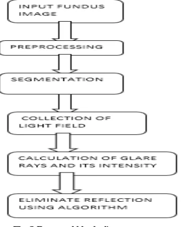
Fig 2 Proposed block diagram
The fundus of the eye is the interior surface of the eye opposite the lens and includes the retina, optical disc, macula, fovea and posterior poles. The color of the fundus varies both between and within species. In one study of PRIMATES the retina is blue, green, yellow, and red; only the human fundus (from a lightly pigmented blood person) is red. The major differences noted among the “higher “primate species were size and regularity of the border of muscular area, size and shape of the optic disc, appearing ‘texturing’ of retina and pigmentation of retina.
An RGB image is referred to as a true color image pixel ,which defines red, green, and blue color components for each individual pixel.RGB images do not a palette .Color of each pixel the is determined by the combination of the red, green and blue intensities stored in each color plane at the pixel’s location. Graphics file formats store RGB images as 24-bit images, where there, green and blue components are 8 bits.RGB is a device-dependent color model; different detect or reproduce a given RGB value differently, since the color elements (such as phosphorous or dyes) and their responses to the R, G and B levels vary from manufacturer to manufacturer, or even in the same device over time. Thus an RGB value does not define the same color across device without some kind of color management.
[image:5.612.160.449.506.710.2]VI. RESULTS AND DISCUSSIONS
Technology (IJRASET)
©IJRASET: All Rights are Reserved
1195
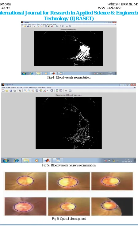
Fig 4 : Blood vessels segmentation
[image:6.612.82.532.7.743.2]Fig 5 : Blood vessels neurons segmentation
Technology (IJRASET)
©IJRASET: All Rights are Reserved
1196
. VII. CONCLUSION
This paper introduces a new approach and fast method for optic disc segmentation in fundus image. The proposed method is tested on three different publicly available database, DiaretDb1, Messidor, Optomed. For the Optomed database, the optic disc was correctly detected with 98% accuracy. The average speed of the proposed algorithm is founded to be 0.8 seconds which is highest speed when compared to other algorithms. The experimental results show assuring performance, and the proposed method appears to be robust to different image conditions. The algorithm attains higher accuracy by the statistical approach in detecting Optic disc in the fundus image. This helps in faster segmentation of optic disc in fundus images.
REFERENCES
[1] Jiang Liu and Wing Kee Wong”Origa-light: an online retinal fundus image database for glaucoma analysis and research”.
[2] T.Y. Wong, “Prediction of Diseases via Ocular Imaging: The Singapore Retinal Archival and Analysis Imaging Network”, Inaugural Ocular Imaging Symposium, June 2008.
[3] Fundus auto fluorescence applications in retinal imaging, Andrea Gabai1, Daniele Veritti1, 2, Paolo Lanzetta1, [4] Fundus imaging with a nasal endoscope, P Mahesh Shanmugam, Rajesh Ramanjulu, Divyansh Mishra kc.,
[5] Multimodal imaging in multifocal pattern dystrophy simulating fundus Flavimaculatus, Rupak Roy, Saurabh Kumar, Dhileesh P Chandrasekharan1, Avirupa Ghose1, Preeti Sharma .,
[6] Fundus autofluorescence imaging of retinal dystrophies, Camiel J.F. Boon, B. Jeroen Klevering, Jan E.E. Keunen, Carel B. Hoyng, Thomas Theelen, Vision Reseach 48 (2008) 2569–2577
[7] Change of retinal pigment epithelial atrophy after anti-vascular endothelial growth factor treatment in exudative age-related macular degeneration, Moosang Kim*, Eung Suk Kim1,*, Kyung Hoon Seo1, Seung-Young Yu1, Hyung-Woo Kwak1
[8] Z. Zhang, B.H. Lee, J. Liu, D.W.K. Wong, N.M. TAN, J.H. Lim, F.S.Ying, W.M. Huang, H. Li, “Optic Disc Region of Interest Localizationin Fundus Image for Glaucoma Detection in ARGALI”, In Proc. ofthe 5th International Conference on Industrial Electronics & Applications (2010)