JOURNAL OFCLINICALMICROBIOLOGY, June 2008, p. 2083–2087 Vol. 46, No. 6 0095-1137/08/$08.00⫹0 doi:10.1128/JCM.00611-08
Copyright © 2008, American Society for Microbiology. All Rights Reserved.
Rapid Differentiation of Methicillin-Susceptible
Staphylococcus aureus
from Methicillin-Resistant
S. aureus
and MIC Determinations by
Isothermal Microcalorimetry
䌤
Ueli von Ah, Dieter Wirz, and A. U. Daniels*
Laboratory for Orthopaedic Biomechanics, Clinical Morphology & Biomedical Engineering, University of Basel Faculty of Medicine, Basel, Switzerland
Received 31 March 2008/Accepted 10 April 2008
In this study, the use of isothermal microcalorimetry (IMC) for differentiation between methicillin-resistantStaphylococcus aureus(MRSA) and methicillin-susceptibleS. aureus(MSSA) and MIC determi-nation was evaluated. It was possible to differentiate between MRSA and MSSA within 4 h, whereas the standard method required 24 h. The MICs of cefoxitin were successfully determined for MRSA and MSSA by using IMC.
Methicillin-resistant Staphylococcus aureus (MRSA) in-fections continue to be associated with significant adverse outcomes and increased health care costs (7). One of the reasons for this is the time required for detection, which is still at least 48 h by conventional methods (4, 11). A faster MRSA detection method would allow faster interventions and rapid implementation of targeted infection control mea-sures (3, 6, 9).
Isothermal microcalorimetry (IMC) is a universal tool for real-time evaluation of rate processes in small (e.g., 3- to 20-ml) ampoules, including processes involving cultured cells. Mi-crocalorimetry has long been used to study the metabolism of cultured cells. James reviewed work in cellular microcalorim-etry in 1987 (8) and reported a 1918 paper by Hill as the earliest employing microcalorimetry to study bacteria. Recent studies suggest that IMC may have clinical diagnostic poten-tial. We have reported the potential use of IMC to detect Staphylococcus aureusor other microorganisms in blood prod-ucts, i.e., platelet concentrates (12).
In this study, we evaluated the use of IMC to differentiate between MRSA and methicillin-susceptibleS. aureus(MSSA) and to determine the MICs of cefoxitin for MRSA and MSSA. The strains used in this study, S. aureus ATCC 43300 (MRSA) andS. aureusATCC 25923 (MSSA), were both cul-tivated overnight at 37°C in brain heart infusion (BHI) broth. They were then adjusted using phosphate-buffered saline to an optical density at 600 nm (OD600) of 0.01 and used to inoculate the samples at 0.01% (vol/vol).
The IMC instrument used was a TAM III thermostat (TA Instruments, New Castle, DE) equipped with 48 microcalorim-eters. The detection limit is⬃0.27W, which corresponds to approximately 135,000 CFU/ampoule (8). Samples were 4-ml sealed glass ampoules containing 3 ml BHI and the bacterial inoculum, leaving a headspace of 1 ml air. After equilibration
of the microcalorimeters themselves for at least 45 min at 37°C, the samples were lowered into the equilibration position. After 15 min, the samples were further lowered into the measuring position, and after 45 min (when the heat flow signal was stable), the actual measurement began. This time was taken as time zero.
The calorimetric differentiation between MRSA and MSSA was performed according to the protocol described by the British Society for Antimicrobial Chemotherapy (BSAC) (1), modified in that we used BHI as the growth medium and 4 mg liter⫺1oxacillin instead of 2 mg liter⫺1as the breakpoint
con-centration. For comparison, BHI agar plates and BHI broth cultures supplemented with either cefoxitin or oxacillin were prepared and kept in an incubator or water bath. In these control samples, the MSSA strain was completely inhibited for 24 h of incubation at 37°C whereas the MRSA strain showed visible growth after only 18 h. For 24 h of incubation, oxacillin inhibited the growth of the MRSA strain much more than cefoxitin did (final OD600, 0.35 and 0.76, respectively).
The calorimetric measurements showed the same results. For MSSA, only the culture without any antibiotics showed a measurable heat flow signal, and detection took only 60 min (Fig. 1A). The MRSA strain grew in every sample, but the increase in inhibition with increased antibiotic could be seen in the heat flow curves (Fig. 1B). It took 240 min for a detectable heat flow signal in the sample with oxacillin but only 100 min in the sample with cefoxitin. However, both antibiotics had metabolic inhibitory effects on the MRSA, which can be seen in the reduced maximum heat flow signal (Fig. 1B).
The determination of the MIC for cefoxitin was performed in cation-adjusted Mueller-Hinton broth, using a modified pro-tocol for the broth dilution test described by the CLSI (for-merly NCCLS) (2). Instead of 35°C, the samples were incu-bated at 37°C. Microcalorimetry was performed as described above. A set of ampoules was additionally incubated in a water bath, and the OD600was determined after 24 h of incubation.
The tested concentrations ranged from 0 to 64 mg liter⫺1
cefoxitin, as recommended by the BSAC (1). After 24 h of
* Corresponding author. Mailing address: LOB, c/o Biozentrum/ Pharmazentrum, Klingelbergstr. 50-70, 4056 Basel, Switzerland. Phone: 41 61 265 94 64. Fax: 41 61 265 95 74. E-mail: [email protected].
䌤Published ahead of print on 16 April 2008.
2083
on May 16, 2020 by guest
http://jcm.asm.org/
incubation, the MIC for the MSSA strain was determined as 4 mg liter⫺1and that for the MRSA strain as 32 mg liter⫺1, using
OD600measurements as well as heat flow measurements (Fig.
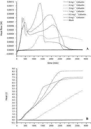
2 and 3). Figure 2A shows that almost all concentrations below the MIC started to grow immediately, except the sample with 2 mg liter⫺1cefoxitin, which exhibited a detectable heat flow
signal only after 500 min. Otherwise, after 250 min of growth the shapes of the heat flow curves started changing, indicating a change in the metabolic activity of the MSSA strain related to the cefoxitin concentration.
The integration of the heat flow rate curves up to a given timexgives the heat produced up to timex. The plot of the heat produced (rather than the heat flow rate) against the
time of the MIC determination results in the heat curves shown in Fig. 2B, which are analogous to cumulative colony or cell counts (data not shown). Although the sample with 2 mg liter⫺1cefoxitin is delayed compared to the other
sam-ples with detectable growth of MSSA, the slope of the curve is almost parallel to the others. It is, however, not yet fully understood why the samples with 0.5 and 1 mg liter⫺1
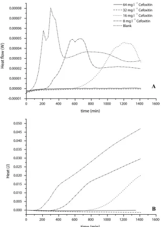
pro-duce more heat than the sample without antibiotic. Similar to the results for MSSA, the MIC of cefoxitin for the MRSA strain was confirmed as 32 mg liter⫺1by using
IMC. However, the shape and especially the delay in detec-tion time were different from those for MSSA. With increas-ing cefoxitin concentration, the delay in detection of the
FIG. 1. Heat flow rate curves for calorimetric measurements of MRSA-MSSA differentiation by use of the antibiotic cefoxitin or oxacillin. (A)S. aureusATCC 25923 (MSSA) in BHI supplemented with the given concentrations of antibiotics; (B)S. aureusATCC 43300 (MRSA) in BHI supplemented with the given concentrations of antibiotics. Blank, uninoculated BHI broth. Curves are averages for three experiments.
2084 NOTES J. CLIN. MICROBIOL.
on May 16, 2020 by guest
http://jcm.asm.org/
heat flow signal increased (Fig. 3A). Additionally, the max-imum heat flow was reduced compared to that in the sample with no added cefoxitin. Growth in the sample with the highest concentration still allowing growth of MRSA (16 mg liter⫺1) could be detected only after approximately 600 min.
In contrast to that for MSSA, the heat produced decreased with increasing cefoxitin concentration (Fig. 3B). This dif-ference might be related to the resistance mechanism of the MRSA strain.
In this study, we presented a potential calorimetric method for decreasing the time for differentiation of MRSA from MSSA. Although one can argue that the overall time for detection is 3 h (1 h for equilibration and 2 h until signal detection), the method is still much faster than traditional
culture methods. Also, there are calorimetric techniques which could greatly reduce the equilibration time. Taking into account that S. aureus ATCC 43300 is a low-level-resistance strain (5, 10), high-level-low-level-resistance strains may be detectable even earlier. This hypothesis and the direct de-tection of MRSA are currently being evaluated in our lab-oratory.
This study also showed that calorimetry works for the MIC determination of cefoxitin for the tested strains. Because cal-orimetry provides continuous real-time data, it was even pos-sible to see effects of cefoxitin concentration on the growth behavior of the strains. However, the time reduction for the evaluation was relatively small, 10 h compared to 24 h with the traditional broth dilution method. Nevertheless, IMC is a
suit-FIG. 2. MIC determination of cefoxitin forS. aureusATCC 25923 (MSSA) in the range of 0.0 mg liter⫺1to 8 mg liter⫺1. (A) Heat flow curve; (B) Heat curve. Blank, Mueller-Hinton plus 2% NaCl alone. Curves are averages for three experiments.
VOL. 46, 2008 NOTES 2085
on May 16, 2020 by guest
http://jcm.asm.org/
[image:3.585.135.449.68.513.2]able method for MIC determination of cefoxitin for MRSA or MSSA.
This work was supported mainly by grant no. 301 from the Velux Foundation, Zurich, Switzerland. We have also received support for microorganism and other cultured cell microcalorimetry from the De-partment of Orthopedic Surgery, University of Basel Faculty of Med-icine. Our laboratory receives general support from the Hardy & Otto Frey-Zu¨nd Foundation, Basel, Switzerland.
We thank A. Trampuz of the Infectiology Department, University of Basel Hospital, for his collaboration in some initial experiments, which one of us (D. Wirz) proposed and which led to the work described here. Finally, we are extremely grateful to J. Meyer and his team (Institute for Oral Microbiology and Preventive Dentistry, University of Basel Dental Clinics) for generously giving us access to their level II biosafety laboratory and equipment for our preparation of
microcalo-rimetry specimens and performance of standard microbiological eval-uations.
REFERENCES
1.Brown, D. F. J., D. I. Edwards, P. M. Hawkey, D. Morrison, G. L. Ridgway, K. J. Towner, and M. W. D. Wren.2005. Guidelines for the laboratory diagnosis and susceptibility testing of methicillin-resistantStaphylococcus aureus(MRSA). J. Antimicrob. Chemother.56:1000–1018.
2.Clinical Laboratory and Standards Institute.2006. Methods for dilution antimicrobial susceptibility tests for bacteria that grow aerobically: ap-proved standard—7th Edition. Clinical and Laboratory Standards Insti-tute, Wayne, PA.
3.de San, N., O. Denis, M.-F. Gasasira, R. De Mendonca, C. Nonhoff, and M. J. Struelens.2007. Controlled evaluation of the IDI-MRSA assay for detection of colonization by methicillin-resistantStaphylococcus aureusin diverse mucocutaneous specimens. J. Clin. Microbiol.45:1098–1101. 4.Fang, H., and G. Hedin.2006. Use of cefoxitin-based selective broth for
[image:4.585.135.451.66.511.2]improved detection of methicillin-resistantStaphylococcus aureus. J. Clin. Microbiol.44:592–594.
FIG. 3. MIC determination of cefoxitin forS. aureusATCC 43300 (MRSA) in the range of 8 mg liter⫺1to 64 mg liter⫺1. (A) Heat flow curve; (B) Heat curve. Blank, Mueller-Hinton plus 2% NaCl alone. Curves are averages for three experiments.
2086 NOTES J. CLIN. MICROBIOL.
on May 16, 2020 by guest
http://jcm.asm.org/
5.Felten, A., B. Grandry, P. H. Lagrange, and I. Casin.2002. Evaluation of three techniques for detection of low-level methicillin-resistant Staphylococ-cus aureus(MRSA): a disk diffusion method with cefoxitin and moxalactam, the Vitek 2 system, and the MRSA-screen latex agglutination test. J. Clin. Microbiol.40:2766–2771.
6.Gurran, C., M. G. Holliday, J. D. Perry, M. Ford, S. Morgan, and K. E. Orr.
2002. A novel selective medium for the detection of methicillin-resistant
Staphylococcus aureus enabling result reporting in under 24 h. J. Hosp. Infect.52:148–151.
7.Harbarth, S., C. Masuet-Aumatell, J. Schrenzel, P. Francois, C. Akakpo, G. Renzi, J. Pugin, B. Ricou, and D. Pittet.2006. Evaluation of rapid screening and pre-emptive contact isolation for detecting and controlling methicillin-resistant Staphylococcus aureusin critical care: an interventional cohort study. Crit. Care.10:R25.
8.James, A. M.1987. Calorimetry past, present and future.InA. M. James
(ed.), Thermal and energetic studies of cellular biological systems. IOP Publishing Ltd., Bristol, United Kingdom.
9.Johnson, G., M. Millar, S. Matthews, M. Skyrme, P. Marsh, E. Barringer, S. O’Hara, and M. Wilks.2006. Evaluation of BacLite rapid MRSA, a rapid culture based screening test for the detection of ciprofloxacin and methicillin resistantS. aureus(MRSA) from screening swabs. BMC Microbiol.6:83. 10.Swedish Reference Group for Antibiotics.8 June 2006, posting date.S.
aureusATCC 29213: target MIC ranges. http://www.srga.org/refstam/sa29213 .htm.
11.Tai, C. C., A. A. Nirvani, A. Holmes, and S. P. F. Hughes.2004. Methi-cillin-resistantStaphylococcus aureusin orthopaedic surgery. Int. Orthop.
28:32–35.
12.Trampuz, A., S. Salzmann, J. Antheaume, and A. U. Daniels.2007. Micro-calorimetry: a novel method for detection of microbial contamination in platelet products. Transfusion47:1643–1650.
VOL. 46, 2008 NOTES 2087