JOURNAL OFCLINICALMICROBIOLOGY, Mar. 2002, p. 943–950 Vol. 40, No. 3 0095-1137/02/$04.00⫹0 DOI: 10.1128/JCM.40.3.943–950.2002
Copyright © 2002, American Society for Microbiology. All Rights Reserved.
Specificities and Functions of the
recA
and
pps1
Intein Genes
of
Mycobacterium tuberculosis
and Application
for Diagnosis of Tuberculosis
Isabelle Saves,
1* Lee-Ann Lewis,
2Fabrice Westrelin,
1Robin Warren,
2Mamadou Daffé,
1and Jean-Michel Masson
1,3Institut de Pharmacologie et Biologie Structurale (UMR5089), CNRS/Université Paul Sabatier Toulouse III,1and Institut National
des Sciences Appliquées, Complexe Scientifique de Rangueil,3F-31077 Toulouse Cedex, France, and Department of
Medical Biochemistry, University of Stellenbosch Medical School, Tygerberg 7505, South Africa2
Received 23 July 2001/Returned for modification 28 October 2001/Accepted 30 December 2001
The worldwide recrudescence of tuberculosis and the widespread appearance of antibiotic resistance have strengthened the need for rapid and specific diagnostic tools. The prevailing microbiological identification of Mycobacterium tuberculosis, the causative agent of tuberculosis, which implies the use of in vitro cultures and
acid-fast staining microscopy, is time-consuming. Detection of M. tuberculosis directly in clinical samples
through PCR amplification of mycobacterium-specific genes, designed to shorten diagnostic delay, demon-strated reliability and high sensitivity. However, the quality of the diagnosis depends on the specificity of the
target sequence forM. tuberculosiscomplex strains. In the present study, we demonstrated the specificity ofrecA
andpps1inteins for this complex and thus the feasibility of using intein-coding sequences as a new target for
PCR diagnosis. Indeed, therecAandpps1genes of 36 clinical isolates ofM. tuberculosisand 10 field strains of
M. boviswere found to be interrupted by an intein sequence at the RecA-a and Pps1-b sites, respectively, while a large number of nontuberculous mycobacterial species failed to demonstrate these insertions. Besides, the
MtuPps1, which was cloned and expressed inEscherichia coli, was shown to possess an endonuclease activity.
The intein cleaves the 40-bp sequence spanning the intein insertion site Pps1-b in the inteinlesspps1gene. In
addition to the PCR amplification ofrecAandpps1intein genes as a tool for diagnosis, the specific
endonu-clease activity could represent a new molecular approach to identifyM. tuberculosis.
Tuberculosis remains a major public health problem in de-veloping countries. Despite the decline in incidence observed in the 1980s, resurgence has recently occurred. Furthermore, the recrudescence of infections withMycobacterium tuberculo-sisand the emergence of multidrug-resistant strains represent a serious public health threat in developed countries, where the incidence of tuberculosis is related to immunodeficient patients, homeless people, and the advancing age of the pop-ulation (World Health Organization web site at http://www .who.int/health-topics/tb.htm).
This worldwide resurgence of tuberculosis has strengthened the need for rapid and specific diagnostic tools to control its spread. Delay in diagnosis results, first, in late initiation of antitubercular therapy and, second, in prolonged transmission of the infection.
Until recently, the diagnosis of tuberculosis was based on clinical features and microbiological assays, radiological exam-inations, immunological tests, microscopy identification, or in vitro cultures (37). Acid-fast staining microscopy of specimens combined with isolation and culture of the bacilli remains the “gold standard” method to specifically identify mycobacteria. Because of the low growth rate ofM. tuberculosis, this method is time-consuming, and the diagnosis can take up to 8 weeks. More recently, molecular approaches have been designed to
ensure early detection ofM. tuberculosisin clinical samples. These methods include DNA amplification by strand displace-ment amplification (35) or by PCR ofM. tuberculosis-specific genes. The use of adequate oligonucleotide pairs to amplify target sequences such as rRNA 16S, IS6110, mtp40, or the
senX3-regX3intergenic region has demonstrated the reliability
and high sensitivity of PCR diagnosis (3). Moreover, technical adaptations of PCR protocols (13, 25, 40) have made it possi-ble to detect mycobacteria directly from clinical samples, such as sputum, blood, urine, and cerebrospinal or pleural fluid. These nucleic acid-based methods have been found to be more sensitive than conventional methods and were able to detect even small numbers ofM. tuberculosiscells (1, 12, 18). How-ever, the choice of the most favorable target sequence is con-troversial, since results from different clinical studies evaluat-ing the various amplification assays were divergent concernevaluat-ing their specificity and sensitivity (11, 17, 20, 31, 39; S. H. Gillespie, T. D. McHugh, and L. E. Newport, Letter, J. Clin. Microbiol.35:799–801, 1997). To optimize specificity and sen-sitivity, the target sequence should be present in all strains of
the M. tuberculosiscomplex, with a high degree of sequence
conservation between strains to avoid false-negative reactions, and should be absent in all other mycobacterium species, thereby eliminating the chance of false-positive results.
In a previous work undertaken to clarify the role of RecA inteins in mycobacteria (32), we showed that the insertion site and the sequence of therecAintein fromM. tuberculosis(8) are unique among mycobacteria. Similarly, theM. tuberculosis pps1
gene (6) possesses an insertion sequence with no homolog
* Corresponding author. Mailing address: Institut de Pharmacologie et Biologie Structurale (UMR5089), CNRS/Université Paul Sabatier Toulouse III, 205 Route de Narbonne, F-31077 Toulouse Cedex, France. Phone: (33) 561 175 471. Fax: (33) 561 175 994. E-mail: saves @ipbs.fr.
943
on May 15, 2020 by guest
http://jcm.asm.org/
known to date in other mycobacterial species (Inbase, the New England Biolabs intein database, at http://www.neb.com/neb /inteins/). The species specificities of both of these invading sequences make them potential targets for PCR amplification. Moreover, since inteins are potential endonucleases (Inteins-Protein Introns web site at http://bioinfo.weizmann.ac.il/⬃pietro /inteins/), the search for their specific functions could lead to a new molecular approach to diagnose tuberculosis.
In the present study, we show first that both intein coding sequences disruptingrecAandpps1genes fromM. tuberculosis
are specific to theM. tuberculosiscomplex and can thus effec-tively be used as DNA target sequences in a PCR-based diag-nosis. In addition, we demonstrated that, unlike the RecA intein, the Pps1 intein MtuPps1 exhibits an endonuclease ac-tivity, which could provide a new tool to unequivocally detect and identify members of theM. tuberculosiscomplex.
MATERIALS AND METHODS
Mycobacterial strains, growth conditions, and genomic DNA isolation. Thirty-three nontuberculous mycobacterial species (listed in Fig. 1) were used in this
study. They correspond to the strains previously described (32), except that only one strain each ofMycobacterium avium(IP140310013),Mycobacterium aurum (ATCC 23366),Mycobacterium chitae(IP141160001), andMycobacterium duvalii (IP141180002) was examined and that one strain of Mycobacterium xenopi (IP14035004) was added to the test. The strains were from the Reference Centre for Mycobacteria (Institut Pasteur, Paris) and were kindly provided by V. Vin-cent Lévy-Frébault. All of the strains were identified by conventional biochem-ical tests (38) and chemotaxonomic criteria, such as mycolic acid and species-specific glycolipid analyses (7). Strains were grown at their optimal temperature of growth (38) on Löwenstein-Jensen medium for a few days to several weeks, depending on the growth rate of the mycobacterial species. Genomic DNA was isolated from the 33 mycobacterial strains by the glass bead disruption method as described by Saves and collaborators (32). In addition, the cosmid B2235, which was used to amplify therecAgene fromMycobacterium leprae, was a gift from K. Eiglmeier (Institut Pasteur, Paris, France).
[image:2.587.63.520.70.452.2]M. tuberculosisisolates were collected from patients attending health care clinics in two suburbs of Cape Town, South Africa.M. bovisisolates were a gift from A. Michel (Tuberculosis Laboratory, Onderstepoort Veterinary Institute, Onderstepoort, South Africa). Cultures were grown on Löwenstein-Jensen slants until confluent growth was achieved. The bacilli were then inactivated by being heated to 80°C for 1 h. Three milliliters of extraction buffer (50 mM Tris-HCl [pH 8], 25 mM EDTA, 5% sodium glutamate) was added to each culture, and the cells were gently scraped to release them from the Löwenstein-Jensen medium. This cell suspension was then transferred to a 50-ml tube containing 20 g of glass
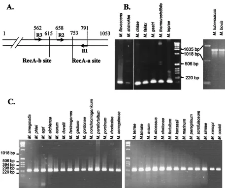
FIG. 1. Specificity of the MtuRecA intein. (A) Location of intein insertion sites in mycobacterialrecAgenes. Arrows represent the oligonu-cleotides used to amplify gene fragments. The positions of the RecA-a and RecA-b sites and of oligonuoligonu-cleotides are indicated as the number of base pairs from the translation start site in theM. tuberculosisH37Rv gene. (B) PCR amplification with R1 and R2 oligonucleotides from genomic DNA of nine species of mycobacteria. (C) PCR amplification with R1 and R3 oligonucleotides from genomic DNA of 27 species of mycobacteria.
on May 15, 2020 by guest
http://jcm.asm.org/
balls (each 0.5 mm in diameter). This process was repeated, and the cell sus-pensions were pooled. The cells were then dispersed by vigorous mixing. Ly-sozyme (25 mg) together with RNase A (25g) was added, and the cell suspen-sion was incubated at 37°C for 2 h. Proteinase K buffer (0.65 ml of 500 mM Tris-HCl [pH 7.5], 50 mM CaCl2) and proteinase K (150g) were added, and the suspension was incubated at 45°C for 16 h. Proteins were removed by phenol-chloroform-isoamyl alcohol (25:24:1) extraction, and the aqueous phase was reextracted with an equal volume of chloroform-isoamyl alcohol (24:1). The DNA was precipitated with isopropanol, washed with 70% ethanol, and dried. Finally, DNA was resuspended in TE buffer (10 mM Tris-HCl [pH 8], 1 mM EDTA).
Genomic DNA from the MT103 strain ofM. tuberculosiswas obtained from Institut Pasteur (Paris, France).
PCR assays.The primers R3 (5⬘ACCACGGCGATCTTCATCAACCAGCT 3⬘), R2 (5⬘GCGTCGGTGCGCATGGACGTGCG 3⬘), and R1 (5⬘AGGATG TCGAACTCGGCCAGCTTGAA 3⬘) correspond to theM. tuberculosis recA gene sequence or its complementary sequence at positions 562 to 587, 658 to 680, and 765 to 791, respectively, according to theM. tuberculosisH37Rv gene se-quence. The primers P2 (5⬘CATCCGCAACACCTACGACCGG 3⬘), P3 (5⬘
GAACATGGGCCZGTTCGAGCGGACG 3⬘), and P1 (5⬘GTCGTTGTTCGA CCAGTTCTGGATGGT 3⬘) correspond to theM. tuberculosis pps1gene se-quence or its complementary sese-quence at positions 357 to 378, 687 to 711, and 844 to 870, respectively, according to theM. tuberculosisH37Rv gene sequence. PCR amplifications were performed withTaqDNA polymerase in a mixture containing 10 mM Tris-HCl [pH 8.3], 1.5 mM MgCl2, 50 mM KCl, and 0.2 mM deoxynucleoside triphosphates (dNTPs), with 5l of the genomic DNA prepa-ration as a DNA template. Several PCR assays were performed under different reaction conditions with regard to oligonucleotide concentrations and hybridiza-tion temperature to ensure amplificahybridiza-tion specificity.
Cloning of MtuRecA and MtuPps1 coding sequences.The entire coding se-quences of MtuRecA and MtuPps1 inteins were amplified by PCR from genomic DNA of the MT103 strain of M. tuberculosis with oligonucleotide pairs MtuRecA-ATG (5⬘ ATGTGCCTCGCAGAGGGGACT 3⬘) andMtuRecA-3⬘
(5⬘GTTGTGCACGACAACCCCTTC 3⬘) andMtuPps1-ATG (5⬘ATGTGCCT GCCCGCCGGC 3⬘) andMtuPps1-3⬘(5⬘GTTGTGCACGGCGAACCCGT 3⬘), respectively. For this purpose, 1l (200 ng) of genomic DNA was incubated with TaqDNA polymerase in a mixture containing 10 mM Tris-HCl (pH 8.3), 1.5 mM MgCl2, 50 mM KCl, 0.2 mM dNTPs, and 10 pmol of each oligonucleotide in a 50-l mix, which was incubated as follows: 10 min at 92°C; 29 times for 1 min at 92°C, 1 min at 55°C, and 1.5 min at 72°C; and, finally, 5 min at 72°C.
These sequences were inserted in the pCR T7/CT-TOPO plasmid (Invitro-gen). The resulting constructs allow the expression of both inteins fused to C-terminal V5 and six-His tags under the regulation of T7 promoter and T7 RNA polymerase inEscherichia coli.
Production and extraction of MtuPps1 and MtuRecA inteins.E. coliBL21 (De3)(pLysS) bacteria were transformed with the two expression vectors. Bac-teria were grown at 30°C in Luria broth culture medium supplemented with 100
g of ampicillin (Sigma) and 30g of chloramphenicol per ml until they reached the exponential phase. Thus, the induction of MtuRecA or MtuPps1 expression with 1 mM IPTG (isopropyl--D-thiogalactopyranoside) (Sigma) was performed
for 4 h at 37°C or for 6 h at 30°C, respectively. Cells were lysed in 20 mM sodium phosphate (pH 7.5) by six cycles of freezing and thawing. The lysate was centri-fuged at 10,000⫻gfor 45 min to separate the soluble intein from insoluble proteins. The soluble fraction was dialyzed against a mixture containing 10 mM Tris-HCl (pH 7.5), 0.1 mM EDTA, 1 mM dithiothreitol, 200g of bovine serum albumin per ml, and 50 mM NaCl. A protein extract from nontransformed BL21 (De3)(pLysS) was collected under similar culture and extraction conditions and used as a negative control in endonuclease assays.
Protein extracts were quantified by the Bradford’s method (2). Samples con-taining 0.5 to 5 mg of proteins per ml were denatured at 95°C for 2 min in a sodium dodecyl sulfate (SDS)–urea–-mercaptoethanol buffer and analyzed by SDS-polyacrylamide gel electrophoresis (PAGE) (homogenous, 10% polyacryl-amide) according to the method of Laemmli (21). Separated proteins were electrically transferred to nitrocellulose membrane (BA45; Schleicher and Schuell) in a semidry apparatus (4). Membranes were incubated for 30 min in Tris-buffered saline (TBS; 10 mM Tris-HCl [pH 8], 137 mM NaCl) containing 1% Tween 20 and 10% nonfat dry milk for 1 h with anti-V5 antibodies (Invitro-gen), diluted 1:5,000 in TBS containing 1% Tween 20 and 1% nonfat dry milk, and for 1 h with a peroxidase-labeled antimouse immunoglobulin G conjugate diluted 1:10,000 in the same buffer. A detection reaction with chemiluminescent substrate was performed with the ECL enhanced chemiluminescence detection kit according to the manufacturer’s instructions (Amersham-Pharmacia Biotech).
Endonuclease assays.To assay MtuRecA and MtuPps1 for endonuclease activity, the 40-bp DNA sequences spanning their homing site were inserted
betweenXbaI andHindIII restriction sites of plasmid pUC19. Partially comple-mentary oligonucleotide pairsMtuRecA-XbaI (5⬘CTAGATCAAGGTCGTCA AGAACAAGTGTTCGCCCCCCTTCAAGCA 3⬘) andMtuRecA-HindIII (5⬘
AGCTTGCTTGAAGGGGGGCGAACACTTGTTCTTGACGACCTTGAT 3⬘) andMtuPps1-XbaI (5⬘CTAGAACGTGCACTACGTAGAGGGTCGCACC GCACCGATCTACAAAA 3⬘) andMtuPps1-HindIII (5⬘AGCTTTTGTAGAT CGGTGCGGTGCAGCCCTCTACGTAGTGCACGTT 3⬘) were annealed by boiling a mix of 1 nmol of each oligonucleotide in a mixture of 10 mM Tris-HCl (pH 7.5) and 50 mM NaCl for 5 min and slow cooling to room temperature. The annealed oligonucleotides were then inserted in pUC19 overdigested byHindIII andXbaI (New England Biolabs). The resulting constructs, substrates 1 and 2, were sequenced. They were linearized byScaI and extracted from a 1% agarose gel in TBE (90 mM Tris-borate, 2 mM EDTA) buffer, and the linear substrates were diluted in water to a concentration of 100 ng/l for cleavage assays.
Endonuclease activity assays were performed in a final volume of 10l, in various reaction buffers, and at temperatures ranging from 15 to 50°C. Moreover, the quantity of proteins added to the reaction mix varied from 25 ng to 5g. The reaction mixtures were analyzed on a 1% agarose gel in TBE buffer.
Nucleotide sequence accession numbers. The GenBank/EMBL accession numbers for the RecA and Pps1 inteins are X58485 and AL021184, respectively.
RESULTS
The MtuRecA intein sequence is specific for theM.
tubercu-losiscomplex.The 1,320-bp sequence coding for RecA intein is inserted at the RecA-a site, immediately downstream of the 753rd nucleotide of theM. tuberculosis recAgene, while all of the seven other RecA intein sequences identified inM. chitae,
Mycobacterium fallax,M. flavescens,M. gastri,M. leprae,
Myco-bacterium shimodei, and Mycobacterium thermoresistibile are
inserted at the RecA-b site (32), 138 nucleotides upstream (Fig. 1A). PCR oligonucleotides were designed according to the conservedrecAgene sequences betweenM. tuberculosis,M.
leprae, andMycobacterium smegmatis sequences. R1 and R2
primers were positioned on each side of the RecA-a site in order to amplify a 134-bp fragment spanning this intein inser-tion site (Fig. 1A). Hence, amplificainser-tion reacinser-tions with primers R1 and R2 produced a 1,454-bp fragment in the presence ofM.
tuberculosisandM. bovisDNA, while the amplified fragment is
only 134 bp long in the presence of DNA from different my-cobacterial species outside of the complex (Fig. 1B).
In a parallel experiment, amplification with primers R1 and R3 generated a 230-bp fragment spanning both RecA-a and RecA-b sites, allowing the confirmation of the absence of in-tein in 27 other mycobacterial species (Fig. 1C).
In order to check whether the R1-R2 primer pair can be used for tuberculosis screening, we performed an amplification reaction using genomic DNA preparations from 36 different
M. tuberculosis strains isolated from patients with confirmed
tuberculosis (36) and from 10 strains ofM. bovis. The 1,454-bp specific fragment was generated with all of theM. tuberculosis
orM. bovisstrains (not shown), implying that all strains ofM.
tuberculosis and M. bovis tested possess the intein in the
RecA-a site of therecAgene in their genomes.
The MtuPps1 intein gene is specific for theM. tuberculosis
complex. To date, three intein insertion sites have been
de-scribed in thepps1gene (33) (Inbase, the New England Biolabs intein database): Pps1-a, Pps1-b, and Pps1-c are the respective intein insertion sites ofM. leprae, M. tuberculosis, and
Myco-bacterium gastriinteins (Fig. 2A). To control for the specificity
of theM. tuberculosisintein amplification, we searched for the presence of intein sequences in 35 different mycobacterial spe-cies by PCR amplification of a 524-bp sequence spanning the
VOL. 40, 2002 MYCOBACTERIAL INTEINS AND TUBERCULOSIS DIAGNOSIS 945
on May 15, 2020 by guest
http://jcm.asm.org/
three insertion sites. P1 and P2 primers were designed accord-ing to the conservedpps1gene sequences inM. tuberculosisand
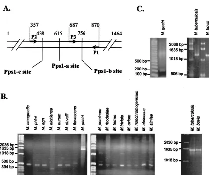
M. leprae. As shown in Fig. 2B, intein was absent from 15 of our
mycobacterial species, based on the amplification of a frag-ment of approximately 500 bp, while the presence of an intein
inM. gastri,M. tuberculosis, andM. boviswas demonstrated by
the amplification of larger fragments. In eightMycobacterium
species, namelyM. chitae,M. fallax,M. farcinogenes,M.
gordo-nae,M. parafortuitum,M. senegalense,M. thermoresistibile, and
M. shimoidei, no amplified DNA was obtained, whatever the
amplification conditions. Additionally, nonspecific amplified fragments were obtained from genomic DNA from the nine remaining species (M. gadium, M. chelonae,M. fortuitum, M.
kansasii,M. marinum,M. peregrinum,M. scrofulaceum,M.
xe-nopi, andM. cookii) under extremely permissive conditions of PCR, while no amplification was obtained under more strin-gent PCR conditions. That may be due to the lack of apps1
gene or to the strong divergence of the sequence of thepps1
gene in these 17 mycobacterial species.
A third primer, named P3 (Fig. 2A), was paired with primer P1 to specifically detect the intein from the M. tuberculosis
complex. As expected, a 1,260-bp fragment was obtained from
M. tuberculosisandM. bovisgenomic DNA amplification, while
a 183-bp fragment was amplified with all other mycobacterial strains and in particular withM. gastri(Fig. 2C).
Both oligonucleotide pairs (P1-P2 and P1-P3) were used to amplify genomic DNA from 36M. tuberculosisand 10M. bovis
strains (not shown). Interestingly, the identification of approx-imately 1,600- and 1,300-bp fragments, respectively, in the 46 amplification reactions confirmed the presence of the pps1
intein in all of theM. tuberculosisandM. bovisstrains tested.
Combined detection ofrecAandpps1inteins by PCR.In an
additional PCR assay, we tried to simultaneously amplifyrecA
andpps1genes in a one-tube reaction for a combined detection of both M. tuberculosis-specific inteins. pps1 and recA gene fragments were amplified fromM. tuberculosisH37v genomic DNA by using P1 and P3 and R1 and R2 oligonucleotide pairs, respectively, either in separate tubes or in the same reaction tube. As expected, the two specific fragments of 1,260 and 1,454 bp, resulting from the amplification ofpps1 and recA, respectively, were obtained when both oligonucleotide pairs were present in the reaction mix (Fig. 3).
MtuPps1 intein is a specific endonuclease.Both MtuRecA
and MtuPps1 coding sequences were cloned in the pCR
T7/CT-FIG. 2. Specificity of MtuPps1 intein. (A) Location of intein insertion sites in mycobacterialpps1genes. Arrows represent the oligonucleotides used to amplify gene fragments. The positions of Pps1-a, Pps1-b, and Pps1-c sites and of oligonucleotides are indicated as number of base pairs from the translation start site in theM. tuberculosisH37Rv gene. (B) PCR amplification with P1 and P2 oligonucleotides from genomic DNA of 18 species of mycobacteria. (C) PCR amplification with P1 and P3 oligonucleotides from genomic DNA of three species of mycobacteria.
on May 15, 2020 by guest
http://jcm.asm.org/
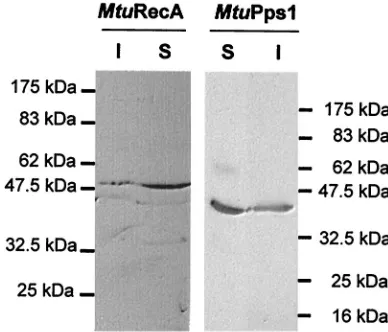
[image:4.587.79.502.67.422.2]TOPO cloning vector in fusion with the DNA sequence en-coding the C-terminal V5 and six-His epitopes. These con-structs allowed the expression of the inteins inE. colistrain BL21(De3)(pLysS). Induction of exponential cultures with 1 mM IPTG resulted in the production of soluble inteins as determined by Western blot analysis with anti-V5 antibodies (Fig. 4).
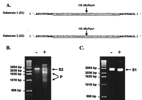
Few inteins are known to possess an endonuclease activity that recognizes and cuts a DNA sequence spanning the junc-tion site created when the intein coding region is deleted from the host gene (homing site). We thus constructed substrate 1 (S1) for MtuRecA and substrate 2 (S2) for MtuPps1 by cloning synthetic 40-bp homing sites in pUC19 plasmid (Fig. 5A). The
ScaI-linearized form of these DNA substrates was purified and used to assay the endonuclease activity of each intein, under various experimental conditions with regard to buffer pH and composition, to mono- and bivalent ions used as cofactors, and to temperature. Linear substrate S2 (2,730 bp) was cleaved in
two specific products (940 and 1,790 bp) in the presence of MtuPps1 extract in a 10 mM Tris-HCl (pH 8) buffer containing 10 mM MgCl2and 25 mM KCl at 37°C (Fig. 5B), while
sub-strate S1 could not be cleaved in the presence of MtuRecA, independent of the reaction conditions (Fig. 5C). Hence, MtuPps1 exhibits a specific endonuclease activity, which was named PI-MtuI according to the current nomenclature.
DISCUSSION
One of the limitations in the control of tuberculosis is the lack of highly sensitive and specific assays to rapidly identifyM.
tuberculosis, the causative agent of tuberculosis. While the
con-ventional methods (i.e., microscopy and cultural identification) still represent the most reliable way to diagnose the infection, these time-consuming methods do not always allow for the early detection and subsequent initiation of therapy. Indeed, a few weeks delay can account for increased morbidity and pro-longed transmission of the disease.
In order to reduce the delay in diagnosis, several methods have been developed. Semiautomated liquid culture systems, such as the BACTEC radiometric method or MB/Bact and MGIT systems, are able to detect mycobacterial slow growers within 10 to 14 days. While several of these growth detection methods have demonstrated good performance comparable to standard methods (23, 26), the necessity for a heavy and ex-pensive apparatus limits their use in poor countries. Moreover, additional tests may be necessary to finally identify the myco-bacterial species. Immunologic or enzymatic assays and phage systems (5, 30) were also proposed, but are generally nonuni-formly applicable or lack specificity and/or sensitivity.
One of the most important technical advances in diagnosis of tuberculosis is the development of molecular methods. The detection, through PCR amplification, of a DNA sequence specifically present in the genomes of members of the M.
tuberculosiscomplex has the potential to overcome the
sensi-tivity and specificity limitations of other diagnostic methods. However, while protocols used to amplify DNA from myco-bacteria present in a wide range of samples have been greatly optimized, the choice of the target sequence remains contro-versial. Various PCR assays have been suggested; these include amplification of sequences encoding mycobacterial antigens, insertion sequences, rRNA, and intergenic regions.
Among the proposed targets, the 16S rRNA gene is the sequence amplified by commercial kits: i.e., the Roche AMPLICOR MTB (Roche Diagnostic Systems, Somerville, N.J.) and the Gen-Probe AMTB (Gen-Probe, Inc., San Diego, Calif.). Although the performance of each of these tests is excellent, their specificity cannot be optimal, since the target gene is present in all mycobacterial species. Hence, the dis-crimination between mycobacteria necessitates hybridization with specific DNA probes (16, 41). Indeed, the use of these tests was initially restricted to acid-fast bacillus-positive spec-imens (1).
The most popular target sequence is the insertion sequence IS6110, also known as IS986 or IS987 (27). Even if some studies demonstrated excellent diagnostic results (15, 17), the specificity of IS6110-based assays is controversial (11; Gillespie et al., Letter). The multicopy IS6110sequence, identified in the majority ofM. tuberculosisstrains, accounts for the high
[image:5.587.95.233.72.240.2]sen-FIG. 3. PCR amplification with the P1-P3 oligonucleotide pair (lane 1), the R1-R2 oligonucleotide pair (lane 3), or both oligonucle-otide pairs (lane 2) from genomic DNA ofM. tuberculosisH37Rv.
FIG. 4. Expression of MtuPps1 and MtuRecA inteins in E. coli. Tagged inteins were detected by Western blotting of the SDS-PAGE (10% polyacrylamide) gels used to separate proteins from soluble (S) and insoluble (I) fractions of MtuPps1 and MtuRecA extracts and immunologic detection by using anti-V5 antibody.
VOL. 40, 2002 MYCOBACTERIAL INTEINS AND TUBERCULOSIS DIAGNOSIS 947
on May 15, 2020 by guest
http://jcm.asm.org/
[image:5.587.64.258.514.680.2]sitivity of the amplification procedure; however, strains with few or no copies of this insertion sequence have also been isolated (42). Moreover, since this sequence is a member of the IS3family, the most widespread group of insertion sequences in bacteria, homologous sequences have been found in nontu-berculous mycobacteria (20, 28). Hence, even if the use of various primer sets to amplify different segments of the se-quence may enhance the specificity of the test, the existence of strains lacking this sequence seriously compromises the use of IS6110-based PCR for tuberculosis diagnosis.
Little is known about the diagnosis based on the amplifica-tion of the MPB64- and Hsp65-encoding genes, but these pro-cedures have shown poor sensitivity and require an additional oligonucleotide hybridization step for the detection of the am-plified fragments. Therefore, the specificity remains rather un-certain (3, 22). Themtp40gene detection has been thoroughly studied. A comparative study showed thatmtp40is more reli-able than the IS6110-based amplification test (19). However, Weil et al. (39) demonstrated that this gene is not present in all strains ofM. tuberculosis.Moreover, since this gene is absent in the other species of theM. tuberculosiscomplex (9), the diag-nosis could fail to recognize many cases of tuberculosis. Hence,
it should instead be used to discriminate betweenM.
tubercu-losisand other species of theM. tuberculosiscomplex (10).
More recently, an interesting alternative was found in the amplification of the senX3-regX3 intergenic region (24, 34), since this sequence is distributed in all strains of theM.
tuber-culosiscomplex. However, one should note that, first, this
se-quence is also found in M. leprae genome, and, second, the length and the sequence of this intergenic region are variable, making interpretation of PCR results quite ambiguous.
In the present study, we demonstrated the feasibility of using intein coding sequences as a target for PCR diagnosis, since
theM. tuberculosis recAandpps1intein sequences are specific
for members of the M. tuberculosis complex (Fig. 1 and 2). Indeed, only the recAgenes of M. tuberculosisand M. bovis
were found to be interrupted by an intein sequence at the RecA-a site, while a total of 34 different mycobacterial species have failed to demonstrate this insertion. Likewise, the MtuPps1 intein sequence embedded at the Pps1-b site of the
M. tuberculosis pps1gene was absent from 16 other
[image:6.587.62.517.64.384.2]mycobac-terial species and was not detected in 17 other species. More-over, all of theM. tuberculosisandM. bovisstrains tested in this study harbor both intein sequences, suggesting a widespread
FIG. 5. Endonuclease assays. (A) DNA substrates for MtuPps1 and MtuRecA. The sequences of MtuPps1 and MtuRecA homing sites (HS) inserted in pUC19 (the sequence of which appears in boldface) are underlined. (B) Cleavage assay for MtuPps1. One hundred nanograms of linearized substrate 2 was incubated with either 2.5g of the crude extract of MtuPps1 (⫹) or 2.5g of a crude extract of nontransformedE. coli BL21(De3)(pLysS) (⫺), for 1 h at 37°C, in 10 mM Tris-HCl (pH 8) buffer containing 10 mM MgCl2and 25 mM KCl. Substrate (S2; 2,730 bp) and
products (P; 940 and 1,790 bp) were separated on a 1% agarose gel in TBE buffer. (C) Cleavage assay for MtuRecA. One hundred nanograms of linearized substrate 1 was incubated with either 2.5g of the crude extract of MtuRecA (⫹) or 2.5g of a crude extract of nontransformed E. coliBL21(De3)(pLysS) (⫺), for 1 h at 37°C, in 10 mM Tris-HCl (pH 8) buffer containing 10 mM MgCl2and 25 mM KCl. The reaction mixture
was analyzed on a 1% agarose gel in TBE buffer.
on May 15, 2020 by guest
http://jcm.asm.org/
distribution of these invading sequences among the members of theM. tuberculosiscomplex.
The primer pairs were designed in order to amplify a frag-ment of the host gene, spanning the intein insertion site, rather than the intein sequence itself. Hence, a DNA fragment has to be amplified even in absence of an intein coding sequence, so that it is possible to control the absence of inhibitory sub-stances in the PCRs. TherecAgene was efficiently amplified in all of the mycobacterial species and strains tested, and a 1,320-bp-longer fragment was obtained only in presence ofM.
tuber-culosisorM. bovisDNA (Fig. 1). These results indicated that
the recA gene is present in all 36 of the species, a result probably related to the essentiality of RecA recombinase in mycobacteria, as suggested by Frischkorn and collaborators and Papavinasasundaram and collaborators (14, 29). More-over, the amplification yields were rather high, suggesting that the sensitivity of the PCR diagnosis would be quite good, even if the target sequences are present in single copy in the M.
tuberculosisgenome.
In the case ofpps1, no amplification was obtained for some mycobacterial species. SincerecAamplification was performed from the same DNA preparations, these negative reactions were not attributed to the presence of any inhibitory sub-stances. The absence of amplified fragment could be explained by the absence of thepps1gene in these mycobacteria. How-ever, this hypothesis is unlikely, since inteins are generally present in essential host proteins, and a previous study sug-gested an important role of Pps1 in mycobacteria (33). Hence, a probable explanation may reside in sequence divergences of
pps1 genes between various mycobacterial species, hindering the hybridization of primers. Nevertheless, the detection of a 1,300-bp-long fragment in the presence ofM. tuberculosisand
M. bovisDNA, compared to the 200-bp fragment obtained in
presence of other mycobacterial species (Fig. 2), allowed the specific identification ofM. tuberculosisandM. bovis.
As formerly suggested by Brisson-Noël and collaborators (3), two amplification assays should be done in parallel to avoid misinterpretation of PCR results, without the need for an additional internal amplification control. Here, we propose the simultaneous detection of MtuRecA- and MtuPps1-specific sequences. The combination of amplifications of bothrecAand
pps1genes, in a multiprimer PCR (Fig. 3), can ensure the rapid and specific detection ofM. tuberculosis.
Moreover, the detection of the specific endonuclease activity of MtuPps1 intein could represent a new molecular approach to identifyM. tuberculosis. One can imagine a PCR amplifica-tion followed by in vitro translaamplifica-tion of the intein and a specific cleavage assay by using linear substrate S2 (Fig. 5). Effectively, the detection of the cleavage activity is even more specific than the sole PCR amplification of the intein sequence and, con-cerning specificity, should provide excellent results. However, the sensitivity of such a test is still to be determined. In vitro transduction assays have to be performed and optimized with this particular mycobacterial sequence in order to validate this new diagnostic approach.
In conclusion, the results described here highlight that the specificity of M. tuberculosis and M. bovis inteins found in RecA and Pps1 host proteins should be considered as an im-portant new molecular tool for diagnosis of tuberculosis.
ACKNOWLEDGMENTS
We thank A. Michel (Tuberculosis Laboratory, Onderstepoort Vet-erinary Institute, Onderstepoort, South Africa), V. Vincent Lévy-Frébault (Institut Pasteur, Paris, France), and K. Eiglmeier (Institut Pasteur) for providing the mycobacterial strains or genomic DNA; M. A. Lanéelle (IPBS/CNRS, Toulouse, France) for the cultivation of the mycobacterial strains; D. Zerbib (IPBS/CNRS) for helpful discus-sions; and N. Trinchero for technical assistance.
REFERENCES
1.Bergmann, J. S., and G. L. Woods.1996. Clinical evaluation of the Roche AMPLICOR PCRMycobacterium tuberculosistest for detection ofM. tuber-culosisin respiratory specimens. J. Clin. Microbiol.34:1083–1085. 2.Bradford, M. M.1976. A rapid and sensitive method for the quantitation of
microgram quantities of protein utilizing the principle of protein-dye bind-ing. Anal. Biochem.72:248–254.
3.Brisson-Noel, A., C. Aznar, C. Chureau, S. Nguyen, C. Pierre, M. Bartoli, R. Bonete, G. Pialoux, B. Gicquel, and G. Garrigue.1991. Diagnosis of tuber-culosis by DNA amplification in clinical practice evaluation. Lancet338:364– 366.
4.Burnette, W. N.1981. “Western blotting”: electrophoretic transfer of pro-teins from sodium dodecyl sulfate-polyacrylamide gels to unmodified nitro-cellulose and radiographic detection with antibody and radioiodinated pro-tein A. Anal. Biochem.112:195–203.
5.Chang, C. L., E. Y. Lee, H. C. Son, and S. K. Park.2000. Evaluating the usefulness of the ICT tuberculosis test kit for the diagnosis of tuberculosis. J. Clin. Pathol.53:715–717.
6.Cole, S. T., R. Brosch, J. Parkhill, T. Garnier, C. Churcher, D. Harris, S. V. Gordon, K. Eiglmeier, S. Gas, C. E. Barry III, F. Tekaia, K. Badcock, D. Basham, D. Brown, T. Chillingworth, R. Connor, R. Davies, K. Devlin, T. Feltwell, S. Gentles, N. Hamlin, S. Holroyd, T. Hornsby, K. Jagels, B. G. Barrell et al.1998.Deciphering the biology of Mycobacterium tuberculosis from the complete genome sequence. Nature393:537–544.
7.Daffé, M., and P. Draper.1998. The envelope layers of mycobacteria with reference to their pathogenicity. Adv. Microb. Physiol.39:131–203. 8.Davis, E. O., S. G. Sedgwick, and M. J. Colston.1991. Novel structure of the
recAlocus of Mycobacterium tuberculosis implies processing of the gene product. J. Bacteriol.173:5653–5662.
9.Del Portillo, P., L. A. Murillo, and M. E. Patarroyo.1991. Amplification of a species-specific DNA fragment ofMycobacterium tuberculosisand its pos-sible use in diagnosis. J. Clin. Microbiol.29:2163–2168.
10.Del Portillo, P., M. C. Thomas, E. Martinez, C. Marañón, B. Valladares, M. E. Patarroyo, and M. Carlos López.1996. Multiprimer PCR system for differential identification of mycobacteria in clinical samples. J. Clin. Micro-biol.34:324–328.
11.Doucet-Populaire, F., V. Lalande, E. Carpentier, A. Bourgoin, M. Dailloux, C. Bollet, A. Vachee, D. Moinard, J. Texier-Maugein, B Carbonelle, and J. Grasset.1996. A blind study of the polymerase chain reaction for the de-tection ofMycobacterium tuberculosisDNA. Tuber. Lung Dis.77:358–362. 12.Folgueira, L., R. Delgado, E. Palenque, J. M. Aguado, and A. R. Noriega.
1996. Rapid diagnosis ofMycobacterium tuberculosisbacteremia by PCR. J. Clin. Microbiol.34:512–515.
13.Folgueira, L., R. Delgado, E. Palenque, and A. R. Noriega.1993. Detection ofMycobacterium tuberculosisDNA in clinical samples by using a simple lysis method and polymerase chain reaction. J. Clin. Microbiol.31:1019–1021. 14.Frischkorn, K., P. Sander, M. Scholz, K. Teschner, T. Prammananan, and
E. C. Bottger.1998. Investigation of mycobacterial recA function: protein introns in the RecA of pathogenic mycobacteria do not affect competency for homologous recombination. Mol. Microbiol.29:1203–1214.
15.Githui, W. A., S. M. Wilson, and F. A. Drobniewski.1999. Specificity of IS 6110-based DNA fingerprinting and diagnostic techniques for Mycobacte-rium tuberculosiscomplex. J. Clin. Microbiol.37:1224–1226.
16.Glennon, M., T. Smith, M. Cormican, D. Noone, T. Barry, M. Maher, M. Dawson, J. J. Gilmartin, and F. Gannon.1994. The ribosomal intergenic spacer region: a target for the PCR based diagnosis of tuberculosis. Tuberc. Lung Dis.75:353–360.
17.Hellyer, T. J., L. E. DesJardin, M. K. Assaf, J. H. Bates, M. D. Cave, and K. D. Eisenach.1996. Specificity of IS6110-based amplification assays for Mycobacterium tuberculosiscomplex. J. Clin. Microbiol.34:2843–2846. 18.Hemal, A. K., N. P. Gupta, T. P. Rajeev, R. Kumar, L. Dar, and P. Seth.2000.
Polymerase chain reaction in clinically suspected genitourinary tuberculosis: comparison with intravenous urography, bladder biopsy, and urine acid fast bacilli culture. Urology56:570–574.
19.Herrera, E. A., and M. Segovia.1996. Evaluation ofmtp40genomic fragment amplification for specific detection ofMycobacterium tuberculosisin clinical specimens. J. Clin. Microbiol.34:1108–1113.
20.Kent, L., T. D. McHugh, O. Billington, J. W. Dale, and S. H. Gillespie.1995. Demonstration of homology between IS6110ofMycobacterium tuberculosis and DNAs of otherMycobacteriumspp. J. Clin. Microbiol.33:2290–2293.
VOL. 40, 2002 MYCOBACTERIAL INTEINS AND TUBERCULOSIS DIAGNOSIS 949
on May 15, 2020 by guest
http://jcm.asm.org/
21.Laemmli, U. K.1970. Cleavage of structural proteins during the assembly of the head of bacteriophage T4. Nature227:680–685.
22.Lee, B. W., J. A. Tan, S. C. Wong, C. B. Tan, H. K. Yap, P. S. Low, J. N. Chia, and J. S. Tay.1994. DNA amplification by the polymerase chain reaction for the rapid diagnosis of tuberculous meningitis. Comparison of protocols in-volving three mycobacterial DNA sequences, IS6110, 65 kDa antigen, and MPB64. J. Neurol. Sci.123:173–179.
23.Liu, Y. C., T. S. Huang, and W. K. Huang.1999. Comparison of a nonra-diometric liquid-medium method (MB REDOX) with the BACTEC system for growth and identification of mycobacteria in clinical specimens. J. Clin. Microbiol.37:4048–4050.
24.Magdalena, J., A. Vachee, P. Supply, and C. Locht.1998. Identification of a new DNA region specific for members ofMycobacterium tuberculosis com-plex. J. Clin. Microbiol.36:937–943.
25.Manjunath, N., P. Shankar, L. Rajan, A. Bhargava, S. Saluja, and Shrini-was.1991. Evaluation of a polymerase chain reaction for the diagnosis of tuberculosis. Tubercle72:21–27.
26.Martinez-Sanchez, L., J. Ruiz-Serrano, E. Bouza, L. Torres, M. Diaz, L. Alcala, and M. Rodriguez-Creixems.2000. Utility of the BACTEC Myco/F lytic medium for the detection of mycobacteria in blood. Diagn. Microbiol. Infect. Dis.38:223–226.
27.McAdam, R. A., P. W. Hermans, D. van Soolingen, Z. F. Zainuddin, D. Catty, J. D. van Embden, and J. W. Dale. 1990. Characterization of a Mycobacterium tuberculosisinsertion sequence belonging to the IS3 family. Mol. Microbiol.4:1607–1613.
28.McHugh, T. D., L. E. Newport, and S. H. Gillespie.1997. IS6110homologs are present in multiple copies in mycobacteria other than tuberculosis-causing mycobacteria. J. Clin. Microbiol.35:1769–1771.
29.Papavinasasundaram, K. G., M. J. Colston, and E. O. Davis.1998. Con-struction and complementation of arecAdeletion mutant ofMycobacterium smegmatisreveals that the intein inMycobacterium tuberculosisRecA does not affect RecA function. Mol. Microbiol.30:525–534.
30.Perkins, M. D.2000. New diagnostic tools for tuberculosis. Int. J. Tuber. Lung Dis.4:S182–S188.
31.Ritis, K., D. Tzoanopoulos, M. Speletas, E. Papadopoulos, K. Arvanitidis, S. Kartali, and P. Sideras.2000. Amplification of IS6110 sequence for detec-tion ofMycobacterium tuberculosiscomplex in HIV-negative patients with
fever of unknown origin (FUO) and evidence of extrapulmonary disease. J. Intern. Med.248:415–424.
32.Saves, I., M. A. Laneelle, M. Daffé, and J. M. Masson.2000. Inteins invading mycobacterial RecA proteins. FEBS Lett.480:221–225.
33.Saves, I., F. Westrelin, M. Daffé, and J. M. Masson.2001. Identification of the first eubacterial endonuclease coded by an intein allele in thepps1gene of mycobacteria. Nucleic Acids Res.29:4310–4318.
34.Supply, P., J. Magdalena, S. Himpens, and C. Locht.1997. Identification of novel intergenic repetitive units in a mycobacterial two-component system operon. Mol. Microbiol.26:991–1003.
35.Walker, G. T., J. G. Nadeau, P. A. Spears, J. L. Schram, C. M. Nycz, and D. D. Shank.1994. Multiplex strand displacement amplification (SDA) and detection of DNA sequences fromMycobacterium tuberculosisand other mycobacteria. Nucleic Acids Res.22:2670–2677.
36.Warren, R. M., S. L. Sampson, M. Richardson, G. D. Van Der Spuy, C. J. Lombard, T. C. Victor, and P. D. van Helden.2000. Mapping of IS6110 flanking regions in clinical isolates ofMycobacterium tuberculosis demon-strates genome plasticity. Mol. Microbiol.37:1405–1416.
37.Watt, B.2000. Issues facing TB control (5.1). (a). Diagnostic issues: labora-tory diagnosis of tuberculosis present techniques. Scott. Med. J.45:38–39, 43.
38.Wayne, L. G., and G. P. Kubica.1986. FamilyMycobacteriaceaeChester 1897, 63AL, p. 1436–1457.InP. H. A. Sneath, N. S. Mair, M. E. Sharpe, and J. G. Holt (ed.), Bergey’s manual of systematic bacteriology, vol. 2. William & Wilkins, Baltimore, Md.
39.Weil, A., B. B. Plikaytis, W. R. Butler, C. L. Woodley, and T. M. Shinnick.
1996. Themtp40gene is not present in all strains ofMycobacterium tuber-culosis. J. Clin. Microbiol.34:2309–2311.
40.Wilson, S. M., R. McNerney, P. M. Nye, P. D. Godfrey-Faussett, N. G. Stoker, and A. Voller.1993. Progress toward a simplified polymerase chain reaction and its application to diagnosis of tuberculosis. J. Clin. Microbiol.
31:776–782.
41.Woods, G. L.2001. Molecular techniques in mycobacterial detection. Arch. Pathol. Lab. Med.125:122–126.
42.Yuen, L. K. W., B. C. Ross, K. M. Jackson, and B. Dwyer.1993. Character-ization ofMycobacterium tuberculosisstrains from Vietnamese patients by Southern blot hybridization. J. Clin. Microbiol.31:1615–1618.