recognizes and selectively degrades
phosphorylated tau client proteins
Chad A. Dickey, … , Francis Burrows, Leonard Petrucelli
J Clin Invest.
2007;
117(3)
:648-658.
https://doi.org/10.1172/JCI29715
.
A primary pathologic component of Alzheimer’s disease (AD) is the formation of
neurofibrillary tangles composed of hyperphosphorylated tau (p-tau). Expediting the
removal of these p-tau species may be a relevant therapeutic strategy. Here we report that
inhibition of Hsp90 led to decreases in p-tau levels independent of heat shock factor 1
(HSF1) activation. A critical mediator of this mechanism was carboxy terminus of Hsp70–
interacting protein (CHIP), a tau ubiquitin ligase. Cochaperones were also involved in
Hsp90-mediated removal of p-tau, while those of the mature Hsp90 refolding complex
prevented this effect. This is the first demonstration to our knowledge that blockade of the
refolding pathway promotes p-tau turnover through degradation. We also show that
peripheral administration of a novel Hsp90 inhibitor promoted selective decreases in p-tau
species in a mouse model of tauopathy, further suggesting a central role for the Hsp90
complex in the pathogenesis of tauopathies. When taken in the context of known
high-affinity Hsp90 complexes in affected regions of the AD brain, these data implicate a central
role for Hsp90 in the development of AD and other tauopathies and may provide a rationale
for the development of novel Hsp90-based therapeutic strategies.
Research Article
Neuroscience
Find the latest version:
The high-affinity HSP90-CHIP complex
recognizes and selectively degrades
phosphorylated tau client proteins
Chad A. Dickey,1 Adeela Kamal,2 Karen Lundgren,2 Natalia Klosak,1 Rachel M. Bailey,1
Judith Dunmore,1 Peter Ash,1 Sareh Shoraka,1 Jelena Zlatkovic,1 Christopher B. Eckman,1
Cam Patterson,3 Dennis W. Dickson,1 N. Stanley Nahman Jr.,4 Michael Hutton,1
Francis Burrows,2 and Leonard Petrucelli1
1Department of Neuroscience, Mayo Clinic College of Medicine, Jacksonville, Florida, USA. 2Biogen Idec Research, Commercial Capabilities,
San Diego, California, USA. 3School of Medicine, University of North Carolina at Chapel Hill, Chapel Hill, North Carolina, USA. 4College of Medicine,
University of Florida, Gainesville, Florida, USA.
A primary pathologic component of Alzheimer’s disease (AD) is the formation of neurofibrillary tangles
com-posed of hyperphosphorylated tau (p-tau). Expediting the removal of these p-tau species may be a relevant
therapeutic strategy. Here we report that inhibition of Hsp90 led to decreases in p-tau levels independent of heat
shock factor 1 (HSF1) activation. A critical mediator of this mechanism was carboxy terminus of
Hsp70–inter-acting protein (CHIP), a tau ubiquitin ligase. Cochaperones were also involved in Hsp90-mediated removal of
p-tau, while those of the mature Hsp90 refolding complex prevented this effect. This is the first demonstration
to our knowledge that blockade of the refolding pathway promotes p-tau turnover through degradation. We
also show that peripheral administration of a novel Hsp90 inhibitor promoted selective decreases in p-tau
spe-cies in a mouse model of tauopathy, further suggesting a central role for the Hsp90 complex in the pathogenesis
of tauopathies. When taken in the context of known high-affinity Hsp90 complexes in affected regions of the
AD brain, these data implicate a central role for Hsp90 in the development of AD and other tauopathies and
may provide a rationale for the development of novel Hsp90-based therapeutic strategies.
Introduction
Intracellular aggregation of abnormal species of phosphorylated tau (p-tau), the microtubule-associated protein, is a major pathologic feature of a family of neurodegenerative disorders collectively referred to as the tauopathies (1, 2). The most com-mon tauopathy is Alzheimer’s disease (AD), in which p-tau aggregates in neurofibrillary tangles, in dystrophic neurites in senile plaques, and in cell processes in the neuropil (3, 4). While these lesions represent visible evidence of p-tau aggregation, the formation of soluble toxic tau species may be more impor-tant mediators of tau-associated neurodegeneration. If so, then decreasing p-tau levels through refolding or degradation may be a plausible therapeutic strategy.
Aberrant neuronal protein aggregation in the tauopathies may result in part from impaired chaperone-mediated protein ubiq-uitination and degradation (5, 6). Hsps have been shown to be upregulated in AD brain (7–9). Molecular chaperones are capable of reducing p-tau concentrations and have been shown to prevent tau-associated cellular toxicity (10, 11). Further support for the role of the cytosolic chaperone network in the processing of tau proteins is the demonstration that deletion of the Hsp70
cochap-erone carboxy terminus of Hsp70–interacting protein (CHIP) results in accumulation of soluble p-tau in the brain (12).
Hsp90 is a molecular chaperone that is involved in the folding and stabilization of many client proteins. The naturally occur-ring ansamycin antibiotic geldanamycin (GA) inhibits Hsp90 chaperone function by reducing ATPase activity (13). This has 2 functional consequences: (a) enhanced degradation of client proteins bound by Hsp90 and (b) activation of heat shock fac-tor 1 (HSF1), a transcriptional activafac-tor of other stress-induced chaperone proteins (14). Tumor cells possess Hsp90/chaperone complexes that exhibit high affinity for Hsp90 inhibitors (15), providing a rationale for current clinical trials of Hsp90 inhibi-tors in the treatment of cancer.
We have previously identified several Hsp90 inhibitors with suitable pharmacokinetic profiles for potential therapeutic use in neurodegenerative disease (16). These compounds promoted selec-tive proteasome-dependent degradation of aberrant p-tau species in vitro. In the present study, we assessed the effect of the Hsp90 inhibitor EC102 in facilitating the degradation of aberrant p-tau species using a humanized tau transgenic mouse. Our results dem-onstrated that the peripheral administration of EC102 promoted selective degradation of p-tau species in the brains of these ani-mals. The results support the hypothesis that Hsp90 is involved in the pathogenesis of AD and associated tauopathies and suggest that the Hsp90 complex is a practical target for the treatment of neurodegenerative tauopathies.
Results
EC102 is a blood-brain barrier–permeable Hsp90 inhibitor. We previously identified several low–molecular weight Hsp90 inhibitors that
Nonstandard abbreviations used: AD, Alzheimer’s disease; GA, biotin-labeled GA; CHIP, carboxy terminus of Hsp70–interacting protein; GA, geldanamycin; GSK3β, glycogen synthase kinase 3β; Hsc, heat shock cognate; Hop, Hsp70/Hsp90-organizing protein; HSF1, heat shock factor 1; Htau mouse, transgenic mouse humanized for the tau gene; MARK2, microtubule-affinity regulating kinase 2; myc-CHIP, myc-tagged CHIP; PAR-1, PAR-1 serine/threonine kinase; p-tau, phosphorylated tau; V5-tau, V5-tagged tau.
Conflict of interest: The authors have declared that no conflict of interest exists.
preferentially degrade p-tau species in cells overexpressing P301L mutant tau (16, 17). Results of subsequent studies indicated that EC102 was the most effective agent. The drug crossed the blood-brain barrier following i.p. administration in mice and attained a brain IC50 for 3 hours at a concentration of 200 mg/kg (Figure 1A).
In addition, Hsp70 levels were increased in the brains of treated mice after 6 hours (Figure 1B). Thus, for all subse-quent studies, EC102 was used to inhibit Hsp90 activity. In order to determine the optimal interval for in vitro efficacy, HeLa cells overexpressing V5-tagged tau (V5-tau) were treated with a 1-μM dose of EC102 and harvested at different time points. Figure 1C shows that 24 hours was the optimal time point, based on robust Hsp induc-tion and p-tau reducinduc-tion. The reducinduc-tion in p-tau could not be attributed to reduced mRNA expression, because the V5-tau construct used was driven by a heterolo-gous CMV promoter, the same promoter used to drive
α-synuclein expression in these cells, which was unaf-fected by Hsp90 inhibitor treatment (Supplemental Fig-ure 1; supplemental material available online with this article; doi:10.1172/JCI29715DS1).
In HeLa cells overexpressing V5-tau, p-tau was quan-tified using an in-cell Western assay (16, 18). Approxi-mately 65% of the total tau was phosphorylated at S396/ S404 (Figure 1D), a p-tau epitope that is preferentially degraded by Hsp90 inhibition (16).
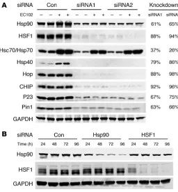
[image:3.585.44.318.82.260.2]The processing pathway of client proteins of the chaperone network is shown in Figure 2: Hsp40 and Hsp70 form the initial recognition complex to prevent the aggregation of aberrant proteins. CHIP is likely involved early in the cycle because of its interaction with Hsp70 (19). Hsp70 client proteins may then be transferred to the Hsp90 complex, a process depen-dent upon Hsp70/Hsp90-organizing protein (Hop; refs. 20–23). Subsequently, the Hsp90 complex can be refolded and dephosphorylated by binding with cochaperones (such as P23 and Pin1, respectively; refs. 24, 25) or degraded via the ubiquitin-proteasome sys-tem. In the latter pathway, Hsp90 is degraded. Hsp90 also suppresses HSF1 activity by binding the factor. Following Hsp90 degradation, free HSF1 translocates to the nucleus and initiates transcription of other Hsps. Chemical inhibition of Hsp90 promotes both of the above functions. Using siRNAs to target several of the above mediators of the chaperone network,
Figure 1
EC102 crosses the blood-brain barrier and reduces tau levels in cells after 24 hours. (A) CD-1 micewere injected i.p. with the indicated doses of EC102 and harvested 1, 3, 6, and 24 hours after injection. Brain levels of EC102 were assessed by HPLC analysis. Greater than 50% concentration was maintained for 3 hours with 200 mg/kg without detectable toxicity. (B) CD-1 mice were injected i.p. with 200 mg/kg EC102 or equivalent vehicle control (Con) to demonstrate the latency in Hsp70 induction following Hsp90 inhibition. After 6 hours, a slight increase in Hsp70 levels was observed in EC102-treated brain tissue, followed by a robust induction at 24 hours compared with vehicle-treated brain tissue. (C) HeLa cells overexpressing V5-tau were treated with a 1-μM concentration of EC102 for the indicated time points. p-tau, Hsp70, and GAPDH levels were assessed by Western blot. p-tau levels were modestly decreased 6 hours after treatment, with maximal reduction seen at 24 hours after treatment. Hsp70 lev-els were increased in a time-dependent manner. (D) In-cell Western analysis showed that 60%–65% of total tau was converted to p-tau in HeLa cells 24 hours after transfection with V5-tau.
Figure 2
[image:3.585.99.480.530.684.2]we attempted to elucidate whether a similar mechanism could be applied to the degradation of p-tau.
Two siRNAs (QIAGEN) per target gene were characterized for knockdown potency in the presence and absence of EC102. Figure 3A shows that both agents demonstrated similar knockdown effi-ciency regardless of EC102 exposure. These siRNAs were pooled in all subsequent studies, consistent with the work of others (26, 27). In order to determine the most appropriate time to administer EC102 to HeLa cells expressing V5-tau, we assessed the inhibitory time course of the above siRNAs for Hsp90 and HSF1 expression. Protein
suppression was apparent as early as 24 hours, but was optimal at 72 and 96 hours for both inhibitory RNAs (Figure 3B). On this basis, the 72-hour time point was used in all subsequent studies.
The constitutive chaperone complex mediates tau degradation by Hsp90 inhibition. In order to investigate the mechanism by which Hsp90 inhibition reduced p-tau expression, EC102 was used to assess HSF1 and Hsp90 expression in HeLa cells overexpressing tau. In cells transfected with nonsilencing siRNA, EC102 reduced p-tau levels by 35%–40%. In contrast, siRNA suppression of constitu-tive cytosolic Hsp90 expression completely abrogated
EC102-Figure 4
[image:4.585.44.300.81.357.2]Reductions in p-tau by Hsp90 inhibition are primarily mediated by a constitutive, not an inducible, chaperone response. (A) HeLa cells were transfected in duplicate with nonsilencing control, Hsp90, or HSF1 siRNA pools and incubated for 72 hours. The cells were then transfected with V5-tau and harvested after 24 hours’ EC102 exposure. EC102 caused robust reductions in p-tau and V5 immunoreactivity in cells transfected with a nonsilencing control or HSF1 siRNA (approximately 35%), while Hsp90 knockdown prevented this reduction. Densitometric values for Hsp90 and HSF1 siRNA pools are represented in separate graphs as a percentage of the optical density for nontransfected, vehicle-treated control cells after GAPDH normalization (dashed line). (B) HSF1 knockdown prevented Hsp40 and Hsp70 induction by EC102, while Hsp90 knockdown had no effect on either of these Hsps. Hsp90 levels were unaffected by HSF1 knockdown. EC102 treatment caused a shift in the distribution of HSF1 species, suggestive of phosphodependent activation.
Figure 3
[image:4.585.117.473.510.652.2]Figure 5
mediated tau reductions (Figure 4A). Despite incomplete Hsp90 knockdown, its suppression still attenuated EC102-mediated p-tau degradation (Figure 4A). Suppression of HSF1 expression by RNA interference prevented upregulation of both Hsp40 and Hsp70 (Figure 4B), yet had no effect on EC102-mediated p-tau reductions. In control studies, the same expression cassette had no effect on α-synuclein expression (Supplemental Figure 1). Taken together, these data suggest that p-tau was processed via the con-stitutive Hsp90 refolding system rather than being dependent on de novo transcription of Hsp chaperones stimulated by HSF1.
Cochaperones involved in the refolding and dephosphorylation of aber-rant proteins initially prevent degradation. In order to assess the role of other components of the chaperone network in the degradation of p-tau, siRNAs were used to knock down constitutive expression of CHIP, heat shock cognate 79 (Hsc79)/Hsp70, Hsp40, and Hop. Figure 5A shows that specific RNA interference for each of these proteins blocked EC102-mediated p-tau degradation, further suggesting a role for the cognate chaperone system in preventing p-tau degradation (quantification of these changes are presented in Figure 5B as a percentage of nontransfected, vehicle-treated cells). In control studies, drug efficacy was substantiated by ele-vated Hsp70 levels (Supplemental Figure 2) and reduced p-tau levels in cells receiving nonsilencing siRNA. In addition, elevation in Hsp70 expression was not altered by suppression of these chap-erones (excluding siRNA directly targeting Hsc70/Hsp70, Figure 3A, and HSF1, Supplemental Figure 2). Finally, suppression of the inducible chaperone Hsp27 had no effect on Hsp90 inhibi-tor–mediated degradation of p-tau (data not shown) despite its association with tau in AD brain (10).
Suppression of the cochaperones P23 and Pin1 functionally mimicked EC102 activity by causing significant reductions in p-tau levels. Figure 5C shows that P23 siRNA reduced p-tau lev-els to an extent similar to that of EC102, and Pin1 knockdown reduced p-tau levels to an even greater extent than did EC102. Fig-ure 5D shows the effect of each siRNA on total V5 immunoreactiv-ity. In these experiments, suppression of Hsp90, CHIP, Hop, and Hsp40 caused total tau levels to increase by approximately 40%. Neither HSF1 nor Hsp 70 affected tau levels. P23 and Pin1 reduced total tau levels by 32% and 76%, respectively. The implications of these results, shown in Figure 5E, suggest that damaged client proteins (i.e., p-tau) are preferentially repaired through restorative chaperone activity, rather than by protein degradation pathways.
CHIP is a critical regulator of the chaperone response and Hsp90 inhibi-tor–mediated p-tau degradation. We previously demonstrated that CHIP is a central component for the degradation of p-tau in vivo (16). CHIP is also a functional binding partner of Hsp70 and Hsp90 (11, 28). We therefore wanted to better understand both the role that CHIP was playing in Hsp90 inhibitor–mediated p-tau degradation and the interactions between this ubiquitin ligase and other chaperone components.
In order to assess the role of CHIP in Hsp90 inhibitor–mediated p-tau degradation, HeLa cells were treated with Hsp90 siRNA and transfected with V5-tau in the presence or absence of myc-tagged CHIP (myc-CHIP). Tau accumulated when Hsp90 expression was blocked. In the presence of CHIP overexpression, tau accumulation was prevented. In the presence of Hsp90 inhibition, V5-tau from cells overexpressing CHIP was coimmunoprecipitated, revealing that the number of CHIP/p-tau complexes was increased. These data indicate that CHIP can complex with tau, despite reductions in Hsp90 levels (Figure 6A).
In order to assess the interactions between CHIP and other chap-erone proteins, we assessed the levels of several other members of the chaperone network in brain homogenates from CHIP–/– mice.
Using this model, we have previously shown that CHIP–/– mice have
increased p-tau (14). In the present work, Figure 6, B and C, dem-onstrate that these mice showed significantly reduced levels of Hop, Hsp90, and Hsp40. Levels of the cochaperone P23 were unchanged.
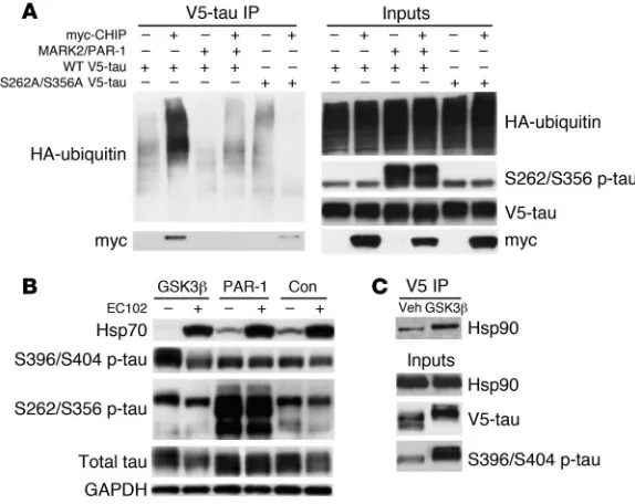
[image:6.585.87.246.83.411.2]CHIP fails to recognize tau phosphorylated at normal serine residues; evi-dence for selective degradation. The above data suggest that p-tau can be targeted for degradation by inhibition of Hsp90. On the other hand, we previously reported that tau phosphorylated by micro-tubule-affinity regulating kinase 2/PAR-1 serine/threonine kinase
Figure 6
The unique cochaperone CHIP is essential for Hsp90 inhibitor–medi-ated tau degradation and regulates the levels of other chaperones as well as Hsp90. (A) HeLa cells transfected with Hsp90 siRNA were subsequently transfected with V5-tau with or without myc-CHIP. Tau accumulated when Hsp90 expression was reduced; however, this accumulation was abrogated by CHIP and the amount of coimmuno-precipitated tau/CHIP complexes increased in the absence of Hsp90. (B) Chaperone protein levels were assessed in CHIP–/– brain tissue by
Western blot analysis. The absence of CHIP and elevation in total tau levels were confirmed. Both Hsp40 and Hsp90 levels were decreased. In addition, the non–HSF1-mediated cochaperone, Hop, was also sig-nificantly decreased in CHIP–/– mice. P23 levels remained unchanged
compared to GAPDH levels. (C) Quantification was assessed by stan-dard densitometry. Error bars represent SD of the 4 CHIP–/– mice.
(MARK2/PAR-1 in Drosophila) at S262/S356 was protected from Hsp90 inhibitor–mediated degradation (16). The demonstration that CHIP regulated tau degradation by Hsp90 inhibition sug-gests that phosphorylation at the S262/S356 site could prevent CHIP ubiquitination. In order to address this question, transfec-tion studies using constructs expressing either wild-type tau or a double serine-to-alanine mutant tau at 262/356 were performed to assess ubiquitination and degradation by CHIP or Hsp90 inhibi-tion. CHIP binding and ubiquitination were completely abrogated by PAR-1–mediated phosphorylation of tau at S262/S356. More-over, S262A/S356A tau mutants had reduced CHIP binding and no polyubiquitination similar to that of PAR-1 overexpression, suggesting that alterations in these residues, regardless of polar-ity, prevent CHIP ubiquitination and binding (Figure 7A).
In order to determine the impact of phosphorylation at the S262/S356 sites on Hsp90 inhibitor–mediated p-tau degradation,
tau was overexpressed with either a glycogen synthase kinase 3β (GSK3β) construct encoding a primary pro-line-directed serine/threonine tau kinase or a PAR-1 construct in HeLa cells overexpressing V5-tau. These cells were then treated with EC102 or vehicle. Despite GSK3β-induced increases in tau phosphorylation at S396/S404, EC102 still reduced tau levels (Figure 7B). Conversely, PAR-1–mediated phosphorylation of tau at S262/S356 prevented degradation despite EC102 treatment and resulted in increased total tau. Drug efficacy was confirmed by Hsp70 induction. These findings further suggest the necessity of functional CHIP in the mechanism of Hsp90 inhibitor–medi-ated p-tau degradation. Finally, coimmunoprecipita-tion of V5-tau showed that overexpression of GSK3β
enhanced Hsp90 binding to tau, further demonstrat-ing that when compared with normal tau, p-tau is a preferred client of Hsp90 (Figure 7C).
Peripheral administration of an Hsp90 inhibitor reduces aberrant p-tau species. In order to characterize the effi-cacy of EC102 in vivo, we used i.p. injections of EC102 into 2 groups of nontransgenic mice at 200 mg/kg per day for 7 days. Animals were sacrificed and their brain tissue was harvested 1 and 24 hours after the final injection. Hsp70 levels were significantly elevated in all EC102-treated mice at 1 hour after injection and persisted for 24 hours (Figure 8, A and B). In contrast, suppression of Hsp90 levels was only demonstrated at the 1-hour time point, with the levels recovering by 24 hours. These findings demonstrated that EC102 both activated HSF1 and promoted degradation of Hsp90 in the brain.
In order to assess the effect of EC102 inhibition of Hsp90 on p-tau accumulation, 8 age-matched (12–13 months) transgenic mice humanized for the tau gene (Htau mice; ref. 29) were treated with EC102 at 200 mg/kg per day or vehicle for 7 days. Twenty-four hours following the last injection, all animals were sacrificed, and Western blot analysis was used to assess brain tau levels. Figure 8C shows Western blots of soluble tau species from the forebrain compartments of EC102-treated mice. EC102 treatment resulted in a reduction of aberrant p-tau species, including a sub-stantial decrease in tau phosphorylated at S396/S404 and S202/T205 (Figure 8, C and D). S262/S356 p-tau was also decreased, but only to the same extent as total tau levels. Abnormal tau species with molecular weights of approximately 64 kDa (30, 31) were decreased by nearly 50%, but the normal tau species — in the 45–55 kDa range — were largely unaffected. Increased Hsp70 levels demonstrated drug efficacy in treated mice. In addition, nei-ther Cdk5 nor Akt levels were affected by EC102 treatment. Both are recognized Hsp90 client proteins and have been implicated in tau phosphorylation (32, 33).
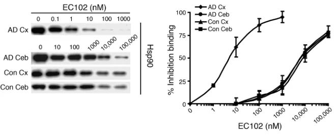
[image:7.585.43.330.83.311.2]A high-affinity Hsp90 complex exists in affected regions of the AD brain. In order to determine whether Hsp90 from AD brain tissue has a higher binding affinity in EC102 than does control brain tissue, we performed competitive binding assays using a biotin-labeled GA (biotin-GA). Hsp90 complexes precipitated with biotin-GA from AD and control brains were incubated with increasing con-centrations of EC102 (Figure 9). EC102 displaced biotin-GA from
Figure 7
Hsp90 complexes in affected (temporal cortex) and unaffected (cerebellum) regions of AD brain at mean IC50 values of 6 and
6,000 nM, respectively (Figure 9). Control cases from the same areas, temporal cortex and cerebellum, had mean IC50 values of
6,000 and 7,333 nM, respectively (Figure 9). Hsp90 levels were sim-ilar among all tissues, which suggests that the observed differences were not caused by different concentrations of Hsp90 (Figure 9). Taken together, these results suggest that the Hsp90 complex in affected areas of AD brain, where pathologic protein accumulation is found postmortem, has a significantly higher binding affinity (approximately 1,000-fold) for EC102 than does Hsp90 derived from unaffected brain tissue of the same patients or from brain tissue of control cases. This observation is similar to that found in mutation-linked solid tumors (15, 17), which largely served as the basis for the introduction of Hsp90 inhibitors at the clinical level as an anticancer therapeutic.
Discussion
Recent evidence suggests that neuronal protein degradation machinery may be impaired in neurodegenerative diseases such as AD, triggering the aberrant accumulation of tau proteins. This has been attributed, in part, to a decline in proteasomal and lysosomal function (11, 34). An additional potential mechanism, however,
is age-associated loss of activity and expression of chaperone and associated proteins (35). The results of the present study support this hypothesis and suggest that enhancing the activity of endog-enous chaperones through Hsp90 inhibition facilitates reductions in p-tau accumulation and selectively targets aberrant p-tau spe-cies that are associated with neurotoxicity. These data suggest that the surveillance mechanism of the chaperone network is highly sensitive and tightly regulated, rather than simply an unregu-lated “garbage disposal” for aberrant or unfolded protein species (36, 37). In addition, the results of these studies suggest that the administration of Hsp90 inhibitors may provide a useful clinical approach to the treatment of AD and other tauopathies.
[image:8.585.102.490.82.333.2]The dynamic and conserved chaperone complex (Figure 2) that facilitates the removal of abnormal proteins such as p-tau typically works in concert with various interchangeable components (i.e., E3 ubiquitin ligases, prolyl isomerases, and other cochaperones). This process culminates either in the partial or complete refolding of the substrate or in its proteasomal degradation. Our results indicate that p-tau is a client of Hsp90 and its chaperone network. Constitu-tive levels of Hsp90 were required for p-tau degradation (Figure 4A). In addition, abnormal tau (hyperphosphorylated) facilitated bind-ing to Hsp90 (Figure 7C). We have previously shown that phos-phorylation of tau is constitutive, and neuronal processing of the
Figure 8
aberrant protein is a continuous process (12). Taken together, these results suggest that subtle impairment in the chaperone system that may occur with aging, coupled with even minor overproduc-tion of abnormal client proteins, may result in aberrant accumula-tion and aggregaaccumula-tion client proteins such as p-tau.
Our results demonstrate that the degradation of p-tau follows a processing sequence through the chaperone cycle depicted in Figure 2. CHIP plays a central role in this succession of events: it has been shown to degrade Hsp70 client proteins as well as Hsp70 itself, thus acting as both a ubiquitin ligase and a stress sensor (19). CHIP also positively regulates HSF1 activity (38). In the pres-ent study, CHIP was shown to be a cpres-entral regulator of constitutive and inducible chaperones. Deletion of CHIP in vivo reduced con-stitutive expression of the Hop chaperone (Figure 6B), an essen-tial component for transferring proteins from the Hsp70 machine to the Hsp90 complex (23). Postdevelopmental alterations of a single chaperone could interrupt renaturation of a client protein and subsequently lead to neurotoxic protein accumulation. Our results suggest that CHIP plays a central role in mediating proper protein triage decisions, given its vast regulatory role through pro-tein interaction and ubiquitination mechanisms.
Cellular processing of aberrant proteins includes repair, deg-radation, or both. The selected pathway may be lineage specific. For example, in nondividing neurons, in which apoptosis is not a viable option, the chosen protein processing pathway must be stable and sustainable for decades. Combinations of repair and degradation may also be used. Our present results and others’ indi-cate that Hsp90 serves a somewhat passive scaffolding function, allowing other cochaperones (including P23 and Pin1) to interact with damaged client proteins. P23 maintains mature Hsp90/cli-ent complexes in a partially active state and can ultimately lead to complete refolding of the client protein (25, 39). Pin1 is a tau-specific prolyl isomerase involved in tau dephosphorylation (24). When these cochaperones are individually suppressed, they promote p-tau degradation (Figure 5). These results provide the
first evidence to our knowledge that pro-teins involved in the refolding of abnor-mal Hsp90 substrates may compete with chaperones involved in degradation. In addition, Hsp90 inhibitors and both P23 and Pin1 siRNAs promote a similar fate for client proteins in that they subvert attempts to refold proteins, thereby favor-ing degradation. This work suggests that the chaperone/client protein complex ini-tially attempts to restore protein function, but should that fail, the system resorts to a protein degradation pathway.
It was previously demonstrated that CHIP antagonizes the interaction of P23 with Hsp90 (40). The present results sug-gest that by removing P23, CHIP is able to freely and noncompetitively bind Hsp90, thus facilitating degradation. In addition, Pin1–/– mice demonstrate pathological tau
accumulation (24), suggesting that without Pin1 and its associated dephosphorylation and refolding, the degradation machinery may become overburdened, leading to accumulation of tau. These findings pro-vide epro-vidence for new substrates potentially involved in tau biology and imply a bifurcation of the chaperone network.
These previous findings, as well as our present results, demon-strate how the cellular chaperone machinery may mediate p-tau degradation; however, it remains unclear how the chaperone com-plex is able to distinguish between aberrant and normal p-tau spe-cies. Tau is normally a natively unfolded protein, yet early in the pathogenesis of AD, the microtubule-binding domain (MTBD) and the N terminus of tau interact to form a hairpin motif recog-nized by the Alz-50 and MC-1 antibodies (41). This serves as a tau misfolding event and is thought to be initiated by phosphorylation (42). We previously reported that CHIP binds and ubiquitinates tau within the MTBD (11). Phosphorylation of tau by MARK2/ PAR-1 occurs at the KXGS motifs (S262 and S356). Both sites are also within the MTBD (16). In addition, p-tau from AD brains was recently shown to be polyubiquitinated at lysine residues that lie in close proximity to the MARK2/PAR-1 consensus sites (43).
[image:9.585.43.372.83.216.2]The present study demonstrates (Figure 7) that CHIP is unable to bind or ubiquitinate tau that is phosphorylated or mutated at these KXGS residues, regardless of polarity (phosphorylated serine being hydrophilic, while alanine is hydrophobic). There-fore, KXGS may be a recognition consensus for both MARK2/ PAR-1 and CHIP, which would result in potential competitive binding. On the other hand, recent data suggest that nitration of N-terminal tyrosines promotes aggregation and misfolding of tau protein (44). Tau folding in AD may normally occur by a sterically favorable interaction of the nitrated tyrosine with the –OH group of serine residues within the KXGS motif. Disruption of these serines could therefore prevent misfolding and conse-quently also block chaperone/CHIP binding and activity. Regard-less, tau phosphorylated by MARK2 has an extended half-life (45, 46), perhaps due to inhibition of degradation mechanisms. This species may thus serve as a catalyst for subsequent pathogenic events. This could suggest an antagonistic relationship between CHIP and MARK2 with regard to tau turnover.
Figure 9
Hsp90 expressed in affected tissue of AD brains has significantly increased binding affinity for EC102. Brain homogenates from affected (temporal cortex, Cx) or unaffected (cerebellar cortex, Ceb) areas of 3 AD patients’ brains and homogenates from the same areas in brains of 3 control cases were evaluated for binding affinity to Hsp90 inhibitors in a competitive binding assay using a biotin-GA probe and increasing concentrations of EC102. Hsp90 derived from the temporal cortices of each AD patient showed 1,000-fold greater binding affinity for EC102, with an IC50
of 6 ± 3.6 nM, compared with Hsp90 from the cerebella, which had an IC50 of 6,000 ± 1,000 nM
(P < 0.01). Controls had an IC50 of 6,000 ± 2,000 nM and 7,333 ± 2,081 nM in the temporal and
Our results demonstrated that Hsp90 inhibition did not affect degradation of S262/S356 p-tau. These results suggest that (a) CHIP is intimately linked to tau degradation following Hsp90 inhibition and that (b) the involvement of CHIP in this process makes the pharmacologic inhibition of Hsp90 (with drugs such as EC102) extremely specific for promoting degradation of only aberrant p-tau species, leaving normal p-tau species unaffected. This is supported further by a similar pattern of specificity of the chaperones facilitating Hsp90 inhibitor–mediated degradation for mutant huntingtin protein in Huntington’s disease (47) and the in vivoefficacyof Hsp90 inhibitors in a polyglutamine expansion disorder with mutant androgen receptors (48).
In the present study, we extended these observations and showed that following systemic administration, a novel Hsp90 inhibitor (EC102) can cross the blood-brain barrier and rapidly reduce levels of soluble p-tau species (16, 30). The Htau mouse model we used produces all 6 splice variants of the human tau protein (29), repre-sents a rodent model for the study of the progressive pathogenesis of tauopathies such as AD, and may serve as a reasonable in vivo model for testing pharmacologic inhibitors of soluble p-tau accumulation.
Our results demonstrated rapid and selective degradation of aberrant p-tau in the brains of Htau mice following administra-tion of EC102 (Figure 8). Neither normal tau species nor other Hsp90 client proteins (Cdk5 or Akt) were affected by the drug. In addition, these results suggest that an intermittent dosing scheme may be possible, but exact treatment regimens remain to be deter-mined. The potential utility of EC102 is further supported by the results of binding affinity studies in human AD brain tissue, in which EC102 demonstrated 1,000-fold greater affinity for Hsp90 from affected areas (temporal cortex) than from unaffected areas (cerebellar cortex) and controls from the same areas. Thus, the higher affinity present in AD-affected brains is a result of disease (abnormal protein accumulation) rather than an intrinsic dif-ference between brain regions. Our results demonstrate a poten-tial for localization of therapeutic agents to areas of disease and suggest that clinically, lower concentrations of Hsp90 inhibitors could be used to treat AD, potentially limiting unwanted effects on Hsp90/client complexes in normal tissues. Finally, the presence of Hsp90 complexes with high affinity for EC102 in other neu-rodegenerative disorders may justify the use of Hsp90 inhibitor therapy in other diseases.
In summary, this work demonstrates that CHIP and the Hsp90 chaperone system are essential for the proper degradation of p-tau,
a protein species that undergoes con-tinuous turnover in the neuron (12). Minor perturbations in the chaper-one system with aging may retard turnover of tau, leading to aggrega-tion and subsequent neurodegenera-tion. The demonstration that Hsp90 inhibitors enhance Hsp90/CHIP-mediated p-tau degradation helps to clarify the mechanisms of tau metabolism and provides a possible therapeutic strategy for the clinical management of tauopathies.
Methods
Plasmids. PAR-1 was provided by B.-W. Lu, School of Medicine, Stanford Uni-versity, California, USA. Constitutively active GSK3β was provided by B. Wolozin, Boston University Medical Campus, Boston, Massachusetts, USA. Myc-CHIP, V5-tau, and HA-ubiquitin were generated by our lab. V5-tau S262A/S356A mutants were generated by our lab using the QuickChange Mutagenesis kit (Stratagene) following the manufacturer’s protocol.
Antibodies, siRNAs, and chemicals. We obtained 12E8 (anti–S262/S356 p-tau) from P. Seubert, Elan Pharmaceuticals, San Francisco, California, USA. PHF1 (anti–S396/S404 p-tau) and CP13 (S202/T205 p-tau) were provided by P. Davies, Albert Einstein College of Medicine, Yeshiva Uni-versity, New York, New York, USA. Tau 5 (anti–total tau) was provided by L. Binder, Northwestern University Medical School, Chicago, Illinois, USA. JJ3 (anti-P23) and F5 (anti-Hop) were provided by D.O. Toft, Mayo Clinic, Rochester, Minnesota, USA. Anti-V5, anti-myc, and anti-HA were obtained from Invitrogen. Anti-Hsp70, anti-Hsp40, anti–BAG-1, anti-Hsp90α, and anti-HSF1 were obtained from Stressgen Biotechnologies. Anti-GAPDH was obtained from BIODESIGN International. Anti-Hsc90 was obtained from BD Biosciences. Anti-Cdk5 was obtained from Santa Cruz Biotech-nology Inc. Anti–α-synuclein was obtained from Cell Signaling Technol-ogy. Anti-CHIP was generated by our group. Secondary antibodies were obtained from Southern Biotechnology Associates and Jackson Immuno-chemicals. All antibodies were used at a 1:1,000 dilution with the exception of PHF1 and CP13, which were used at a dilution of 1:100. All siRNAs were obtained from QIAGEN, and their sequences are listed in Table 1. siRNA efficiency for protein knockdown was validated by Western blot (Figure 3A). EC102 was generated by Conforma Therapeutics.
[image:10.585.55.384.108.223.2]Cell culture and transfections. HeLa and HEK293 cells were grown in Opti-Mem plus 10% FBS and passaged every 3–5 days based on 90% confluence. siRNA experiments were carried out in 6-well plates using human gene-spe-cific validated and genome-wide siRNAs. Final siRNA concentration per well was 20 nM in Opti-Mem, with 2 μl of siLentFect transfection reagent (Bio-Rad) used per well. This mixture was incubated in a final volume of 500 μl for 20 minutes and then added to 40%–50% confluent HeLa cells in 6-well dishes plated the previous day for a final in-well volume of 2.5 ml. Forty-eight hours after transfection, complete medium was removed and replaced with Opti-Mem (without serum) for subsequent plasmid trans-fection and drug treatment. For plasmid transtrans-fections, 1 μg V5-tau or V5-tagged α-synuclein was combined with Lipofectamine 2000 reagent for 15 minutes in 500 μl Opti-Mem, and this mixture was added to the siRNA-transfected cells for 4 hours. The transfection mixture was then replaced with fresh complete media, and EC102 was added to the cells for 18–24 hours at a final concentration of 1 μM. For coimmunoprecipita-tion studies, HEK293 and HeLa cells were treated as previously described (11). Briefly, 90% confluent cells in 10-cm dishes were transfected using
Table 1
siRNA sequences used in the present study
Gene name Common name Target sequence 1 Target sequence 2
HSP90AA1/HSPCA Hsp90 CTCGGAGGATCTCCCTCTAAA TCCCGACGATATTACTAATGA
HSF1 HSF1 CAGTGACCACTTGGATGCTAT CAGGTTGTTCATAGTCAGAAT
HSPA1A Hsp70 TCCTGTGTTTGCAATGTTGAA TCGAGTTTCCGGCGTCCGGAA
DNAJB1 Hsp40 CAGGACGATACCCGTCGTATT ACCCGTCGTATTCAAAGATGT
STIP1 Hop CCCGAAAGATGCCAAATTATA ATGACTTACATTACCAATCAA
STUB1 CHIP GCGCTCTTCGAATCGCGAAGA TGCCGCCACTATCTGTGTAAT
TEBP P23 CAGCTTAGGGAAAGAGAATAA CACTGTATTGATCCAAATGAT
PIN1 Pin1 si02662128A si02662667A
Nonsilencing control – AATTCTCCGAACGTGTCACGT –
ASequences for Pin1 were validated by Qiagen; the catalog number is provided in lieu of the sequence,
Lipofectamine 2000 with combinations of HA-ubiquitin, wild-type V5-tau, serine-to-alanine mutant tau, myc-CHIP, PAR-1, and GSK3β for 4 hours and then maintained in complete media for 48 hours.
In-cell Western assay. In-cell Western analysis of p-tau levels was performed as previously described (16). Briefly, HeLa cells overexpressing V5-tau were directly fixed with formaldehyde in 96-well plates following 72 hours of growth. Plates were blocked and incubated with either (a) total tau (diluted 1:500; Dako) or GAPDH antibodies (diluted 1:1,000; Biodesign or Biosource) or (b) PHF1 (diluted 1:50; provided by P. Davies) and GAPDH antibodies overnight. Plates were washed and incubated with appropriate species-specific secondary antibodies labeled for dual near-infrared fluores-cence at 700 and 800 nm (diluted 1:500; Rockland Immunochemicals and Invitrogen). After washing, plates were scanned using the Odyssey scanner, and relative fluorescent units were quantified. Background fluorescence was subtracted, and values for tau were normalized to GAPDH levels.
Coimmunoprecipitation with V5 tau and Hsc90/Hsp90 monoclonal antibodies. Coimmunoprecipitation to assess tau ubiquitination levels in HeLa and HEK293 cells was performed as previously described (11, 49). Briefly, cell supernatants following preclearing with protein G were incubated with anti-V5 antibody or anti-Hsc90/Hsp90 antibody and 50 μl protein G over-night at 4°C. Supernatants were washed 3 times in coimmunoprecipita-tion buffer, resuspended in sample buffer, and subjected to Western blot analyses following SDS-PAGE electrophoresis.
Rodent husbandry, treatment strategy, and tissue preparation. Htau mice (STOCK Mapttm1(EGFP)Klt Tg(MAPT)8cPdav/J) were obtained from The
Jack-son Laboratory and generated as previously described (29). Briefly, these mice are transgenic for the entire human tau locus using a PAC strategy, while being null for murine microtubule associated protein tau by enhanced GFP (EGFP) insertion into exon 1. This line was preserved on a mixed 129S4/SvJae × Swiss Webster × (C57BL/6 × DBA)F1 background, and genotype was assessed by PCR from tail clip digestions (29). All procedures involving transgenic mice, including treatment with Hsp90 inhibitors, were approved by the Mayo Clinic College of Medicine. An age-matched cohort (15–16 months) consisting of 8 Htau mice was injected i.p. with either a 200 mg/kg dose of EC102 or vehicle once daily for a single week and humanely euthanized following the seventh injection. For pharma-cokinetic studies, EC102 was injected i.p. at doses of 200 and 400 mg/kg into 8 groups of 3 Swiss outbred CD-1 mice (Charles River Laboratories). The animals were anesthetized with isoflurane and perfused with 10 ml sterile saline in the left ventricle. Brain tissues were collected at the
indi-cated time points (Figure 1A) and flash frozen in liquid nitrogen. The brain tissues were homogenized in methanol/acetonitrile (3:2 ratio), and the homogenates were centrifuged and analyzed, after standard extraction procedures, using a Hewlett-Packard HP 1100 Series HPLC equipped with autosampler, DAD detector, and HP ChemStation software (version 2.0). A similar dosing strategy was employed for studies involving 3-month-old nontransgenic C57BL/6 mice, except that a cohort of 10 mice (5 receiv-ing EC102 versus 5 receivreceiv-ing vehicle) were euthanized 1 hour followreceiv-ing the final injection to capture reductions in Hsp90 levels based on our pharmacokinetic data. The brains were frozen on dry ice for subsequent biochemical analyses. CHIP–/– mice were generated as previously described (38). CHIP–/– and CHIP+/+ mice were humanely euthanized at P30, and their brains were quickly removed for dissection and frozen on dry ice for subsequent biochemical analyses. Brain tissue was homogenized in buffer containing 50 mM Tris, 274 mM NaCl, 5 mM KCl, 100 mM PMSF, and a protease and phosphatase inhibitor cocktail (pH 8.0). Homogenates were centrifuged at 15,400 g for 15 minutes, and supernatants were collected.
Western blotting. Cells from siRNA transfection studies were harvested for subsequent Western blot analysis in lysis buffer containing 50 mM Tris-HCl pH 7.4, 1 M NaCl, 0.1% Triton-X, 5 mM EDTA plus 1% SDS, PMSF, and both a protease and phosphatase inhibitor cocktail followed by sonica-tion. Tissue samples were homogenized and processed as described above. Protein concentrations were measured by a standard BCA assay (Pierce Biotechnology). Cell lysate samples and tissue homogenates were then heated in Laemmli’s buffer, and equal amounts of protein were loaded into 10- or 15-well 10% Tris-Glycine gels (Novex). Following transfer, blots were blocked with Blotto (5% non-fat dry milk in TBS plus 0.1% Triton-X 100) for 2 hours, then antibodies were applied at appropriate dilutions in Blotto overnight at 4°C. Membranes were washed 3 times for 10 minutes in Tris-buffered saline + 0.1 tritonX-100 and incubated with secondary antibody for 1 hour. Membranes were then washed 3 times for 10 minutes, and protein expression was visualized by ECL treatment and exposure to film. Bands were quantified using Scion Image by analyzing pixel density. Semiquantitative analysis was performed by densitometry, correcting pro-tein levels for GAPDH. In order to more quantitatively assess the effect of EC102 and siRNAs on p-tau levels, the Odyssey scanner was used again for Western blot, as previously described for total tau (12). For optimal quan-titative interpretation, each membrane can only be scanned by this method on the first pass. Therefore, PHF1 and GAPDH were used on the same blot in Figures 4 and 6. Quantitation was performed using Scan Image software (version Beta 3b), and PHF1 immunoreactivity was normalized to GAPDH immunoreactivity to control for loading discrepancies.
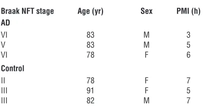
Hsp90 binding assay. Hsp90 binding assays were performed as previously described (15, 50). Brain homogenates from 3 AD cases and control sub-jects (obtained from Mayo Clinic Jacksonville Brain Bank; Table 2) from affected (temporal cortex) and unaffected (cerebellar cortex) areas were pre-pared by dounce homogenization in lysis buffer (20 mM HEPES, pH 7.3, 1 mM EDTA, 5 mM MgCl2, and 100 mM KCl), incubating with or without EC102 at 0.1, 1, 10, 100, and 1,000 nM for 30 minutes at 4°C, and then incubating with biotin-GA linked to BioMag streptavidin magnetic beads (QIAGEN) for 1 hour at 4°C. Tubes were placed on a magnetic rack, and the unbound supernatant was removed. The magnetic beads were washed 3 times in lysis buffer and heated for 5 minutes at 95.8°C in SDS-PAGE sample buffer. Samples were analyzed on SDS protein gels, and Western blots were performed. Bands in the Western blots were quantified using the Bio-Rad Fluor-S MultiImager, and the percentage inhibition of binding of Hsp90 to biotin-GA was calculated. The IC50 reported is the concentration of EC102 needed to cause half-maximal inhibition of binding.
[image:11.585.53.272.123.234.2]Statistics. Significance was determined using a 2-tailed Student’s t test. *P < 0.05, **P < 0.01, and ***P < 0.001 were assessed.
Table 2
Characteristics of human brains used for the Hsp90 inhibitor binding study
Braak NFT stage Age (yr) Sex PMI (h) AD
VI 83 M 3
V 83 M 5
VI 78 F 6
Control
II 78 F 7
III 91 F 5
III 82 M 7
Acknowledgments
We would like to thank D.O. Toft for his provision of antibod-ies against Hop and P23 as well as for his invaluable insights in interpretation of our results with respect to mechanisms of chaperone networks. We would also like to thank B.-W. Lu, B. Wolozin, L. Binder, P. Davies, and P. Seubert for providing antibodies, plasmids, and Htau mice. This work was supported by NIH grants P50-NS40256 and P01-AG17216 and by the Insti-tute for Study of Aging.
Received for publication July 16, 2006, and accepted in revised form December 19, 2006.
Address correspondence to: Leonard Petrucelli, Mayo Clinic, 4500 San Pablo Road, Jacksonville, Florida 32224, USA. Phone: (904) 953-2855; Fax: (904) 953-7370; E-mail: [email protected]. Chad Dickey’s present address is: University of South Florida Col-lege of Medicine, Tampa, Florida, USA.
1. Hutton, M., et al. 1998. Association of missense and 5′-splice-site mutations in tau with the inher-ited dementia FTDP-17. Nature.393:702–705. 2. Feany, M.B., and Dickson, D.W. 1996.
Neurode-generative disorders with extensive tau pathol-ogy: a comparative study and review. Ann. Neurol.
40:139–148.
3. Gomez-Isla, T., et al. 1997. Neuronal loss corre-lates with but exceeds neurofibrillary tangles in Alzheimer’s disease. Ann. Neurol.41:17–24. 4. Braak, H., and Braak, E. 1991. Neuropathological
staging of Alzheimer-related changes. Acta. Neuro-pathol. (Berl.).82:239–259.
5. Katsuno, M., et al. 2005. Pharmacological induc-tion of heat-shock proteins alleviates polygluta-mine-mediated motor neuron disease. Proc. Natl. Acad. Sci. U. S. A.102:16801–16806.
6. Waza, M., et al. 2006. Modulation of Hsp90 func-tion in neurodegenerative disorders: a molecular-targeted therapy against disease-causing protein.
J. Mol. Med. 84:635–646.
7. Sahara, N., et al. 2005. In vivo evidence of CHIP up-regulation attenuating tau aggregation. J. Neu-rochem.94:1254–1263.
8. Hamos, J.E., et al. 1991. Expression of heat shock proteins in Alzheimer’s disease. Neurology.
41:345–350.
9. Perez, N., et al. 1991. Increased synthesis and accu-mulation of heat shock 70 proteins in Alzheimer’s disease. Brain Res. Mol. Brain Res.11:249–254. 10. Shimura, H., Miura-Shimura, Y., and Kosik, K.S.
2004. Binding of tau to heat shock protein 27 leads to decreased concentration of hyperphosphory-lated tau and enhanced cell survival. J. Biol. Chem.
279:17957–17962.
11. Petrucelli, L., et al. 2004. CHIP and Hsp70 regulate tau ubiquitination, degradation and aggregation.
Hum. Mol. Genet.13:703–714.
12. Dickey, C.A., et al. 2006. Deletion of the ubiquitin ligase CHIP leads to the accumulation, but not the aggregation, of both endogenous phospho- and caspase-3–cleaved tau species. J. Neurosci.
26:6985–6996.
13. Panaretou, B., et al. 1998. ATP binding and hydroly-sis are essential to the function of the Hsp90 molec-ular chaperone in vivo. EMBO J.17:4829–4836. 14. Ciechanover, A., and Brundin, P. 2003. The
ubiq-uitin proteasome system in neurodegenerative dis-eases: sometimes the chicken, sometimes the egg.
Neuron.40:427–446.
15. Kamal, A., et al. 2003. A high-affinity conforma-tion of Hsp90 confers tumour selectivity on Hsp90 inhibitors. Nature.425:407–410.
16. Dickey, C.A., et al. 2006. HSP induction mediates selective clearance of tau phosphorylated at pro-line-directed Ser/Thr sites but not KXGS (MARK) sites. FASEB J.20:753–755.
17. Biamonte, M.A., et al. 2006. Orally active purine-based inhibitors of the heat shock protein 90.
J. Med. Chem.49:817–828.
18. Dickey, C.A., et al. 2005. Development of a high throughput drug screening assay for the detection of changes in tau levels — proof of concept with HSP90 inhibitors. Curr. Alzheimer Res.2:231–238. 19. Qian, S.B., McDonough, H., Boellmann, F., Cyr,
D.M., and Patterson, C. 2006. CHIP-mediated stress recovery by sequential ubiquitination of substrates and Hsp70. Nature.440:551–555. 20. Chang, H.C., Nathan, D.F., and Lindquist, S. 1997.
In vivo analysis of the Hsp90 cochaperone Sti1 (p60). Mol. Cell. Biol.17:318–325.
21. Frydman, J., and Hohfeld, J. 1997. Chaperones get in touch: the Hip-Hop connection. Trends Biochem. Sci.22:87–92.
22. Chen, S., and Smith, D.F. 1998. Hop as an adaptor in the heat shock protein 70 (Hsp70) and hsp90 chap-erone machinery. J. Biol. Chem.273:35194–35200. 23. Johnson, B.D., Schumacher, R.J., Ross, E.D., and
Toft, D.O. 1998. Hop modulates Hsp70/Hsp90 interactions in protein folding. J. Biol. Chem.
273:3679–3686.
24. Liou, Y.C., et al. 2003. Role of the prolyl isomerase Pin1 in protecting against age-dependent neurode-generation. Nature.424:556–561.
25. Grenert, J.P., et al. 1997. The amino-terminal domain of heat shock protein 90 (hsp90) that binds geldanamycin is an ATP/ADP switch domain that regulates hsp90 conformation. J. Biol. Chem.
272:23843–23850.
26. Li, T., et al. 2004. Identification of the gene for vita-min K epoxide reductase. Nature.427:541–544. 27. Soifer, H.S., Zaragoza, A., Peyvan, M., Behlke, M.A.,
and Rossi, J.J. 2005. A potential role for RNA interfer-ence in controlling the activity of the human LINE-1 retrotransposon. Nucleic Acids Res.33:846–856. 28. Grelle, G., et al. 2006. Identification of VCP/p97,
carboxyl terminus of Hsp70-interacting protein (CHIP), and amphiphysin II interaction partners using membrane-based human proteome arrays.
Mol. Cell. Proteomics.5:234–244.
29. Andorfer, C., et al. 2003. Hyperphosphorylation and aggregation of tau in mice expressing normal human tau isoforms. J. Neurochem.86:582–590. 30. Santacruz, K., et al. 2005. Tau suppression in a
neu-rodegenerative mouse model improves memory function. Science.309:476–481.
31. Tronjanowski, J.Q., and Lee, V.M. 2005. Pathologi-cal tau: a loss of normal function or a gain in toxic-ity? Nat. Neurosci. 8:1136–1137.
32. Kobayashi, S., et al. 1993. A cdc2-related kinase PSSALRE/cdk5 is homologous with the 30 kDa subunit of tau protein kinase II, a proline-directed protein kinase associated with microtubule. FEBS Lett.335:171–175.
33. Hong, M., and Lee, V.M. 1997. Insulin and insu-lin-like growth factor-1 regulate tau phosphory-lation in cultured human neurons. J. Biol. Chem.
272:19547–19553.
34. Barrachina, M., Maes, T., Buesa, C., and Ferrer, I. 2006. Lysosome-associated membrane protein 1
(LAMP-1) in Alzheimer’s disease. Neuropathol. Appl. Neurobiol.32:505–516.
35. Cohen, E., Bieschke, J., Perciavalle, R.M., Kelly, J.W., and Dillin, A. 2006. Opposing activities protect against age-onset proteotoxicity. Science.
313:1604–1610.
36. Goldberg, A.L., Stein, R., and Adams, J. 1995. New insights into proteasome function: from archaebac-teria to drug development. Chem. Biol.2:503–508. 37. Dalton, W.S. 2004. The proteasome. Semin. Oncol.
31(Suppl. 16):3–9; discussion 33.
38. Dai, Q., et al. 2003. CHIP activates HSF1 and confers protection against apoptosis and cellular stress. EMBO J.22:5446–5458.
39. Grenert, J.P., Johnson, B.D., and Toft, D.O. 1999. The importance of ATP binding and hydrolysis by hsp90 in formation and function of protein hetero-complexes. J. Biol. Chem.274:17525–17533. 40. Connell, P., et al. 2001. The co-chaperone CHIP
regulates protein triage decisions mediated by heat-shock proteins. Nat. Cell Biol.3:93–96. 41. Weaver, C.L., Espinoza, M., Kress, Y., and Davies, P.
2000. Conformational change as one of the earliest alterations of tau in Alzheimer’s disease. Neurobiol. Aging.21:719–727.
42. Ghoshal, N., et al. 2001. Tau-66: evidence for a novel tau conformation in Alzheimer’s disease.
J. Neurochem.77:1372–1385.
43. Cripps, D., et al. 2006. Alzheimer’s disease–spe-cific conformation of hyperphosphorylated phf-tau is polyubiquitinated through lys-48, lys-11, and lys-6 ubiquitin conjugation. J. Biol. Chem.
281:10825–10838.
44. Reynolds, M.R., Berry, R.W., and Binder, L.I. 2005. Site-specific nitration differentially influences tau assembly in vitro. Biochemistry.44:13997–14009. 45. Biernat, J., et al. 2002. Protein kinase MARK/PAR-1
is required for neurite outgrowth and establishment of neuronal polarity. Mol. Biol. Cell.13:4013–4028. 46. Mandelkow, E.M. 2006. The modes and pathways
of Tau’s toxicity to neurons in neurodegeneration. Paper presented at the Keystone Symposia Confer-ence on Alzheimer’s Disease: Genes, Cellular Path-ways and Therapies. February 21–26. Breckenridge, Colorado, USA.
47. Sittler, A., et al. 2001. Geldanamycin activates a heat shock response and inhibits huntingtin aggre-gation in a cell culture model of Huntington’s dis-ease. Hum. Mol. Genet.10:1307–1315.
48. Waza, M., et al. 2005. 17-AAG, an Hsp90 inhibitor, ameliorates polyglutamine-mediated motor neu-ron degeneration. Nat. Med.11:1088–1095. 49. Petrucelli, L., and Dawson, T.M. 2004. Mechanism
of neurodegenerative disease: role of the ubiquitin proteasome system. Ann. Med.36:315–320. 50. Castro, J.E., et al. 2005. ZAP-70 is a novel