Biology Dissertations Department of Biology
12-14-2017
Ion Channel SUMOylation: a Novel Role for
SUMO in Homeostatic Regulation of Multiple
Ionic Conductances
Anna Parker
Follow this and additional works at:https://scholarworks.gsu.edu/biology_diss
This Dissertation is brought to you for free and open access by the Department of Biology at ScholarWorks @ Georgia State University. It has been accepted for inclusion in Biology Dissertations by an authorized administrator of ScholarWorks @ Georgia State University. For more information, please [email protected].
Recommended Citation
Parker, Anna, "Ion Channel SUMOylation: a Novel Role for SUMO in Homeostatic Regulation of Multiple Ionic Conductances." Dissertation, Georgia State University, 2017.
by
ANNA R. PARKER
Under the Direction of Deborah J. Baro, PhD
ABSTRACT
Neurons can adjust their ionic currents to maintain a stable output. The homeostatic
mechanisms that produce compensatory changes in ionic currents operate over multiple time
scales. The rapid mechanisms that act over minutes are mostly unknown. We have been
characterizing a fast homeostatic mechanism that stabilizes activity phase in the rhythmically
active lateral pyloric neuron (LP) of the crustacean stomatogastric ganglion. LP activity phase is
invariant. It is determined, in part, by the balance between the hyperpolarization-activated
current (Ih) and the transient potassium current (IA). When LP IA is experimentally decreased,
activity phase is initially disrupted, but then it recovers over minutes. This is because the
dependent fashion. We have found that neuronal activity adjusts the level of ion channel
post-translational modification by Small Ubiquitin-like Modifier (SUMO), a peptide which when
conjugated to target proteins alters their protein-protein interactions. Using a heterologous
expression system, we showed that enhancing SUMOylation of HCN or Kv4 ion channels that
mediate Ih and IA, respectively, produced opposite effects on the amplitudes of Ih and IA. We also
demonstrated that a given change in activity produced the opposite effect on SUMOylation
levels associated with each current. Thus, activity-dependent regulation of ion channel
SUMOylation specified a positive correlation between the two currents. We have also
demonstrated that activity-dependent regulation of ion channel SUMOylation is conditional; it
only occurs in the presence of the appropriate modulatory tone. We showed this is because
modulators, like dopamine, specify the targets of the SUMOylation machinery. In sum, we have
discovered a novel mechanism that acts over minutes to correlate ionic conductances and thereby
stabilize neuronal output.
INDEX WORDS: SUMOylation, Dopamine, Activity-Dependent, Ion Channel, HCN, Kv4,
by
ANNA R. PARKER
A Dissertation Submitted in Partial Fulfillment of the Requirements for the Degree of
Doctor of Philosophy
in the College of Arts and Sciences
Georgia State University
Copyright by Anna Rachel Parker
by
ANNA R. PARKER
Committee Chair: Deborah J. Baro
Committee: Chun Jiang
Aaron Roseberry
Astrid Prinz
Electronic Version Approved:
Office of Graduate Studies
College of Arts and Sciences
Georgia State University
DEDICATION
I would like to dedicate my dissertation to my family. My parents, Calvin and Mendy
Huff, have always encouraged me to strive for my dreams (no matter how many years it takes). It
is because they nurtured and supported my love of science that I chose to pursue my Ph.D. and a
career doing what I love. They have loved me and supported through the many ups and downs
that have come with graduate school, and there aren’t enough words to express how grateful I am
for them. My husband, Christian Parker, whom I met and married while I was in graduate school,
has been a daily source of encouragement and inspiration for me. He has been incredibly patient
with my late hours, long weekends of work, and occasional bouts of neurotic stress. I can’t wait
to close this chapter and start the next one with him and the family we plan to start. My two older
brothers, Bradley and Kelly Huff, and my sister-in-law, Jana Huff, have always been a
significant source of support for me, without their love, encouragement, and shared moments of
much-needed laughter during times of stress, I would not have been able to accomplish what I
have. My aunt and uncle, Harry and Rachel Kelly, have been wonderfully supportive and have
never missed an opportunity to tell me how proud they are of me. Thank you so much.
ACKNOWLEDGEMENTS
First, I would like to thank my Ph.D. advisor, Dr. Deborah Baro. She has not only helped
to mold me as a successful researcher but has encouraged to become a more confident woman.
Dr. Baro nurtured my scientific curiosity and thanks to her I have learned more about research
and hard work than I ever could have asked for and I couldn’t be more grateful!
Second, I would like to thank the members of my committee, Dr. Chun Jiang, Dr. Aaron
Roseberry, and Dr. Astrid Prinz. They have provided me with much-needed guidance and
support as I progressed towards obtaining my Ph.D. I would also like to thank Dr. Vincent
Rehder for his advice during my qualifying exams and his continued support following the
completion of my exam. Thank you to Dr. David Blaustein and Nancy Russell along with the
professors that I worked with as a teaching assistant, Dr. Paul Ulrich, and Dr. Rebekah Chapman,
for providing me with a wonderful experience as a teaching assistant. Also, a sincere thank you
to all of the professors that have taught and supported me in my graduate career; Dr. Paul Katz,
Dr. Sarah Pallas, Dr. Donald Edwards, and Dr. Anne Murphy. Thank you also to the Brains and
Behavior Fellowship Program for the support it has been an honor to be a fellow.
Next, I would like to thank all of the students I have worked with over the years in the
Baro lab, for their friendship, the help they provided me with my project and the opportunity
many provided me to develop my skills as a teacher and mentor. They include but are in no way
limited to Meghyn Welch, Lori Forster, Saline Atlas, Leslie-Anne Jansen, Janhavi Dubhashi,
Sarah Tasneem, Kristin Jackson, Sasha Guillory, Melissa Issa-Boube, and Ayisha Mcintyre.
Also, I would like to express my appreciation for Dr. Wulf-Dieter Christian Krenz, Dr. Edmond
Rodgers, and Tim Dever, who worked in the lab when I first started graduate school and played a
to give special thanks to Meghyn Welch, you have not only helped me immensely in
accomplishing what I have with my research, but you have become a real friend.
Finally, I would like to thank the many members of other labs. A special thank you to
Liana Artinian from the Rehder Lab and Dr. Ningren Cui of the Jiang Lab for their help in
establishing our patch clamping rig. Thank you to the members of the Roseberry lab, Anna
Dunigan, and Katherine Stuhrman, for the help and advice you provided. Thank you to the Jiang
lab, Dr. Weiwei Zhong, Dr. Max Oginsky, Dr. Shuang Zhang and Christopher Johnson, for
TABLE OF CONTENTS
ACKNOWLEDGEMENTS ... V
LIST OF TABLES ... XII
LIST OF FIGURES ... XIII
LIST OF ABBREVIATIONS ... XV
1 INTRODUCTION... 1
1.1 Stomatogastric Nervous System ... 1
1.2 Dopamine ... 4
1.3 Dopamine in the STG ... 5
1.4 SUMOylation mediates activity-dependent regulation of ion channels ... 9
1.5 Hypothesis ... 11
2 DETAILED MATERIALS AND METHODS ... 15
2.1 Hek cells ... 15
2.1.1 Tissue Culture ... 15
2.1.2 Cryopreservation ... 15
2.2 Calcium Phosphate Transfection... 16
2.3 GFP Immunoprecipitation ... 16
2.3.1 Hek Cell Lysates ... 16
2.3.2 Co-Immunoprecipitation ... 17
2.5 Whole Cell Patch Clamping of Hek Cells ... 19
2.6 Tat-SUMO Peptide ... 20
2.6.1 Tat-SUMO Construct ... 20
2.6.2 Tat-SUMO Peptide Synthesis ... 21
3 CHAPTER 1: SUMOYLATION OF THE HYPERPOLARIZATION-ACTIVATED CYCLIC NUCLEOTIDE-GATED CHANNEL 2 INCREASES SURFACE EXPRESSION AND THE MAXIMAL CONDUCTANCE OF THE HYPERPOLARIZATION-ACTIVATED CURRENT ... 22
3.1 Abstract ... 23
3.2 Introduction ... 24
3.3 Methods ... 27
3.3.1 Drugs ... 27
3.3.2 Mouse Brain Membrane Preparations... 27
3.3.3 Plasmids and Antibodies ... 28
3.3.4 Site Directed Mutagenesis ... 28
3.3.5 Cell culture, Stable and Transient Transfections ... 29
3.3.6 Immunoprecipitations ... 30
3.3.7 Western Blotting ... 31
3.3.8 Patch Clamping Electrophysiology ... 32
3.3.10 Image analysis and Quantification ... 34
3.3.11 Statistical analysis ... 35
3.4 Results ... 35
3.4.1 Mouse HCN2 is SUMOylated In vivo ... 35
3.4.2 GFP-HCN2 Channels are SUMOylated in a Heterologous Expression System 36 3.4.3 Transient Transfection of SUMO2 and Ubc9 Increases HCN2 Channel SUMOylation in a Hek Cell Line Stably Expressing Mouse HCN2 ... 37
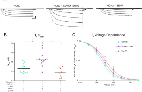
3.4.4 HCN2 Channel SUMOylation Increases Ih Gmax ... 39
3.4.5 SUMOylation Increases HCN2 Channel Surface Expression ... 40
3.4.6 Only One of Six Putative SUMOylation Sites is Necessary for the Increase in GFP-HCN2 Surface Expression and Ih Gmax Elicited by Overexpression of SUMO and Ubc9 41 3.5 Discussion... 43
3.5.1 Ion Channels are SUMOylated In vivo ... 44
3.5.2 HCN channels contain multiple SUMOylation Consensus Sequences ... 45
3.5.3 Potential SUMO-dependent HCN2 Channel Interactions ... 48
3.5.4 HCN Channel SUMOylation and Neurological Disorders ... 49
4.1 Abstract ... 67
4.2 Introduction ... 68
4.3 Methods ... 71
4.3.1 Animals ... 71
4.3.2 Chemicals ... 72
4.3.3 Antibodies ... 72
4.3.4 STNS Dissection and LP Identification ... 73
4.3.5 Somatic Two-Electrode Voltage Clamp (TEVC) ... 73
4.3.6 SUMO cloning... 74
4.3.7 Tat-SUMO Peptide Synthesis ... 75
4.3.8 Immunoprecipitation and Western Blots ... 76
4.3.9 Statistical Analysis ... 77
4.4 Results ... 77
4.4.1 DA reconfigures the activity-dependence of LP Ih and IA ... 77
4.4.2 Logic supporting link between SUMOylation and activity-dependent regulation of LP IA and Ih ... 79
4.4.3 HCN and Kv4 channels are SUMOylated in vivo ... 80
4.4.4 SUMOylation is necessary for activity-dependent changes in LP Ih and IA
4.4.5 Enhanced SUMO availability converts bi-directional into one-way
DA-enabled LP Ih activity-dependence ... 81
4.4.6 DA blocks the effect of enhanced SUMO availability on LP IA ... 84
4.5 Discussion... 85
5 GENERAL DISCUSSION ... 102
5.1 SUMO regulation of neuronal homeostasis ... 102
5.2 The function of tonic DA ... 103
5.2.1 Tonic spiking of DA neurons... 104
5.2.2 Dopaminergic tone stabilizes neuronal activity states ... 105
5.2.3 Dysfunctions associated with dopaminergic tone ... 107
5.3 Limitations of Exogenous Expression and Heterologous Expression Systems 109 5.4 Conclusions ... 110
REFERENCES ... 111
LIST OF TABLES
Table 3.1 Primary Antibodies ... 64
Table 3.2 Site directed mutagenesis primers ... 65
Table 4.1 Lobster SUMO Primers ... 101
LIST OF FIGURES
Figure 1.1 SUMOylation ... 12
Figure 1.2 The Pyloric Network ... 13
Figure 3.1 Mouse HCN2 is SUMOylated in vivo... 51
Figure 3.2 Establishing a culture system to investigate HCN2 channel SUMOylation ... 52
Figure 3.3 GFP-HCN2 channels are SUMOylated in Hek-HCN2 cells ... 53
Figure 3.4 Transient transfection with SUMO and Ubc9 leads to an increase in GFP-HCN2 channel SUMOylation ... 55
Figure 3.5 Increased HCN2 channel SUMOylation augments Ih Gmax ... 56
Figure 3.6 Increased SUMOylation augments HCN2 channel surface expression ... 58
Figure 3.7 Identification of HCN2 channel SUMOylation sites... 59
Figure 3.8 Overexpression of SUMO and Ubc9 enhances SUMOylation at K669... 60
Figure 3.9 Enhanced SUMOylation at K669 augments Ih Gmax ... 61
Figure 3.10 Enhanced SUMOylation at K669 increases GFP-HCN2 channel surface expression ... 63
Figure 4.1 Tonic 5nM DA reconfigures the activity-dependence of LP IA and Ih ... 90
Figure 4.2 Panulirus interruptus HCN and Kv4 channels are SUMOylated in vivo ... 92
Figure 4.3 SUMOylation is necessary for the maintenance and activity-dependence of LP Ih and IA ... 93
Figure 4.4 Enhancing SUMO availability converts DA-enabled, LP Ih activity-dependence from bi-directional to uni-directional ... 95
Figure 4.6 Summary of SUMO-mediated, activity-dependent regulation of LP Ih and IA ... 97
Figure 4.7 Supplemental 1 - Validation for Tat-SUMO experiments ... 98
Figure 4.8 Supplemental 2 - Validation of antibodies used for immunoprecipitation and western
blotting ... 100
LIST OF ABBREVIATIONS
ADHD Attention Deficit Hyperactivity Disorder
cAMP Cyclic adenosine monophosphate
CNBD Cyclic Nucleotide Binding Domain
CRMP Collapsin Response Mediator Proteins
DA Dopamine
HCN Hyperpolarization-Activated Cyclic Nucleotide-Gated Channel
Hek Cells Human Embryonic Kidney Cells
IA Transient Potassium Current
Ih Hyperpolarization-Activated Current
LP Lateral Pyloric Neuron
LTD Long-Term Depression
LTP Long-Term Potentiation
NMDA N-methyl-D-aspartate
PIR Post-Inhibitory Rebound
PKA Protein Kinase A
SENP Sentrin Specific Protease
SIM SUMO-Interaction Motif
STDP Spike Timing-Dependent Plasticity
STG Stomatogastric Ganglion
STNS Stomatogastric Nervous System
1 INTRODUCTION
Rhythmically active networks drive such functions as walking, swallowing, breathing,
and swimming. These types of networks require that the precise timing of component neuron
firing remain stable while still being flexible enough to respond to external stimuli. Thus, a
mechanism is needed to stabilize neuronal excitability and maintain activity within a specific
range. In the rhythmically active pyloric network of the spiny lobster, the timing of network
neuron firing (phase) is maintained within a narrow range across individual animals (Bucher,
Prinz et al. 2005, Goaillard, Taylor et al. 2009). Maintenance of pyloric neuron phase has been
attributed to multiple cellular and synaptic parameters including the correlation of specific ionic
conductances (Soofi, Archila et al. 2012, Zhao and Golowasch 2012). Researchers have
described a long-term mechanism that acts through transcription and translation to maintain these
correlations. However, we have published evidence that alterations in phase can be restored over
minutes in the presence of tonic Dopamine (DA) (Krenz, Rodgers et al. 2015), indicating that a
fast activity-dependent mechanism also exists to maintain phase. This dissertation will
investigate a novel role for Small Ubiquitin-like Modifier (SUMO) in the rapid coregulation of
the transient potassium current (IA) and the hyperpolarization-activated current (Ih). We will also
begin to characterize the involvement of tonic DA in the SUMO-mediated activity-dependent
regulation of ionic conductances.
1.1 Stomatogastric Nervous System
The crustacean Stomatogastric Ganglion (STG) is a component of the Stomatogastric
Nervous System (STNS), a well-characterized central pattern generator that drives the rhythmic
contraction of the gut and foregut of the spiny lobster. The STG has long been an ideal model
circuit. Modulatory input is sent to the STG via descending inputs through the stn (Fig 1.2A;
CoG), as well as being present in the hemolymph that is continuously bathing the ganglion
(Oginsky, Rodgers et al. 2010, Hedrich, Diehl et al. 2011). Our work will focus on the pyloric
circuit of the STG, a 14-neuron recurrent inhibitory circuit that drives the rhythmic contraction
of the foregut (Fig 1.2C). The pyloric circuit produces a triphasic rhythm, that can be observed in
vivo and in situ, through the patterned bursting of six different cell types. Each neuron fires in a
particular phase relative to the other neurons. The firing pattern is set by the electrically coupled
pacemaker kernel, composed of the anterior burster (AB) and the two pyloric dilator (PD)
neurons, which simultaneously inhibit follower neurons (LP, PY, IC, and VD) (Fig 1.2C)
(Marder and Bucher 2007). Through post-inhibitory rebound (PIR) the follower neurons
repolarize and fire a burst of action potentials. The varying rates of PIR together with the
synaptic architecture determine the order and timing of each follower neuron’s burst. The lateral
pyloric (LP) and inferior cardiac (IC) neurons recover first following pacemaker inhibition. Next
the pyloric (PY) and ventricular dilator (VD) neurons fire to complete the triphasic cycle
(Fig1.2B).
The LP neuron will be the primary focus of our research. LP functions in the circuit to
slow increasing cycle frequencies. It sets an upper limit on cycle frequency by inhibiting PD.
The timing of LP inhibition is critical because it maintains the narrow range of frequencies the
network experiences. Without LP feedback on the pacemaker, frequencies could increase outside
of that range and disrupt network activity (Weaver and Hooper 2003). The timing of LP
inhibition is determined by its PIR (Johnson, Brown et al. 2011). The ratio of two opposing ionic
conductances, IA and Ih, regulates the rate of PIR (Harris-Warrick, Coniglio et al. 1995).
et al. 2014). Upon depolarization IA is activated, producing an inward current that slows
depolarization (Tierney and Harris-Warrick 1992, Birnbaum, Varga et al. 2004). In part, it is the
balance between these two currents, one depolarizing the cell, the other slowing that
depolarization, in part regulate the time at which the neuron fires its burst of action potentials.
The distribution, function, and regulation of IA and Ih have been extensively studied in the
STG. In the pyloric network, IA plays an important role in determining neuron firing phase, cycle
frequency, and spike frequency (Tierney and Harris-Warrick 1992, Harris-Warrick, Coniglio et
al. 1995). The shal channel that mediates IA undergoes extensive alternative splicing with the
biophysical properties of the different spliceforms showing significant variability (Baro, Levini
et al. 1997, Baro, Ayali et al. 2000, Baro, Quinones et al. 2001). Shal channels are primarily
localized to the somatic membrane, primary neurites, and the neuropil of STG neurons (Baro,
Ayali et al. 2000). Ih plays a critical role in neuronal excitability, rhythmic activity, and synaptic
function (Robinson and Siegelbaum 2003, Goeritz, Ouyang et al. 2011, Kase and Imoto 2012).
The Hyperpolarization-activated Cyclic Nucleotide-gated (HCN) channel that mediates Ih in the
STG is also subject to alternative splicing, and the identified spliceforms show large variability
in activation kinetic and voltage dependences (Ouyang, Goeritz et al. 2007). HCN channels
primarily localize to the fine neural in the STG, in close proximity to synapses (Goeritz, Ouyang
et al. 2011). Researchers have overexpressed shal in identified pyloric neurons and observed a
corresponding increase in expression of Ih that prevented any significant changes in network
activity (MacLean, Zhang et al. 2003, MacLean, Zhang et al. 2005). Interestingly, when the
reciprocal experiment was performed, overexpressing Ih did not lead to a corresponding increase
in IA expression (Zhang, Oliva et al. 2003). However, additional studies have also demonstrated
network activity (Khorkova and Golowasch 2007, Hudson and Prinz 2010, Temporal, Desai et
al. 2012, Zhao and Golowasch 2012, Golowasch 2014).
Phase refers to the timing of network neuron firing relative to the other network
components. We study LP-on phase, which refers to the timing of LP firing relative to the
pacemaker, and is calculated by dividing the LP-on delay by the cycle period (Fig 1.2B; b÷a)
(Rodgers, Fu et al. 2011). Despite there being several-fold variability in conductance levels and
synaptic strengths across individuals, phase and the IA:Ih ratio are both highly conserved,
underscoring their importance (Bucher, Prinz et al. 2005, Goaillard, Taylor et al. 2009).
Maintaining IA:Ih requires a regulatory mechanism or homeostat (MacLean, Zhang et al. 2003,
MacLean, Zhang et al. 2005), and recent evidence indicates that tonic DA modulation may be
part of that mechanism (Rodgers, Fu et al. 2011, Krenz, Hooper et al. 2013, Krenz, Parker et al.
2015).
1.2 Dopamine
DA is a neuromodulator that plays an essential role in many neurological processes such
as motor control, reward, and motivation. Dysfunction of the dopaminergic system can result in a
variety of conditions including Parkinson’s Disease, Schizophrenia, Restless Leg Syndrome, and
Attention Deficit Disorder (ADHD)(Schultz 2007). DA is released from dopaminergic neurons
through volume transmission in many model systems, including the STNS (Oginsky, Rodgers et
al. 2010). As opposed to a classical synapse where receptors are located within the synaptic cleft,
directly adjacent to where the neuromodulator is being released, DA releases far away from its
receptor and diffuses to reach them or reach distant reuptake mechanisms (Schultz 2007).
Tonically active DA neurons produce a constant presence of DA at nanomolar (nM) levels,
et al. 2013). Upon stimulation, dopaminergic neurons will release DA in bursts. This produces a
transient increase in DA concentration that can reach micromolar (μM) to millimolar (mM)
levels near the dopaminergic neuron at the time of release, and is referred to as phasic DA
(Justice 1993, Goto, Otani et al. 2007, Rice, Patel et al. 2011). DA receptors can be present in
either a high or low-affinity states. Tonic DA will activate high-affinity receptors, while phasic
DA will activate low-affinity DA receptors, producing different responses in the target neuron.
Tonic DA plays an enabling role and is known to enable motor, cognitive, and motivational
processes (Bilder, Volavka et al. 2004, Krenz, Hooper et al. 2013, Krenz, Rodgers et al. 2015).
On the other hand, phasic DA modifies these processes and many others (Schultz 2007,
Bromberg-Martin, Matsumoto et al. 2010, Lisman, Grace et al. 2011). DA activates either type-1
or type-2 receptors (D1R and D2R respectively). Both receptors couple with G proteins and
activation of these receptors have opposing effects. D1R’s, acting through Gs increase adenylyl
cyclase activity while D2R’s acting through Gi/o decrease it (Clark and Baro 2006, Clark, Khan
et al. 2008). LP exclusively expresses D1Rs, therefore, the work presented here will focus on the
canonical D1R pathway.
1.3 Dopamine in the STG
In the STG, phasic and tonic DA can affect many aspects of the network including
rhythmic activity, the firing properties of specific neurons, synaptic strengths, and ionic
conductances. Bath applying phasic DA to the STG alters the firing properties of each pyloric
neuron differently (Harris-Warrick, Johnson et al. 1998). Since these early findings, additional
studies have characterized the specific effects of DA on network output, synaptic strengths, and
specific ionic conductances. In a study that examined the effects of phasic DA on the cycle
concentrations of DA slightly decreased the cycle frequency of the pyloric rhythm (Ayali and
Harris-Warrick 1999). This overall decrease in cycle frequency closely resembled the DA
modulated decrease in the isolated AB-2xPD pacemaker kernel, resulting from the combination
of an increase in AB frequency paired with a decrease in the frequencies of both PD neurons.
Researchers have also characterized the pre- and postsynaptic effects of DA modulation at many
of the pyloric synapses. As was mentioned before the synapse between LP and PD is particularly
important because LP directly inhibits the pacemaker and can function to slow the network
rhythm. DA modulation enhances LP pre-synaptic transmitter release through an increase in
calcium levels at the presynaptic terminals and decreases the PD post-synaptic responsiveness to
the transmitter (Johnson, Peck et al. 1995, Cleland and Selverston 1997, Johnson and
Harris-Warrick 1997, Johnson, Kloppenburg et al. 2003, Kloppenburg, Zipfel et al. 2007, Johnson,
Brown et al. 2011). The net outcome of these opposing effects is an overall enhancement of the
inhibitory LP/PD synapse. Common among many pyloric neurons is DA modulation of IA and Ih,
although the directionality of this modulation can differ between neuron types. For instance,
phasic concentrations of DA reduce IA in the AB, PY, and IC neurons (Peck, Nakanishi et al.
2001), while it increases IA in the PD neuron (Kloppenburg, Levini et al. 1999). Phasic DA
significantly shifted the voltage of activation in the depolarized direction without changing Gmax
in LP, AB, PY, and VD neurons, with no effect on PD and IC Ih (Harris-Warrick, Coniglio et al.
1995, Peck, Gaier et al. 2006).
Our work focuses on the mechanisms that regulate LP neuron activity, and bath
application of phasic µM DA decreases LP IA, disrupting the LP IA:Ih ratio and producing an
advance in LP-on phase (Rodgers, Fu et al. 2011). The phase advance is accompanied by an
exposure to phasic µM levels of DA, LP phase recovers while cycle frequency and burst duration
remained altered (Rodgers, Fu et al. 2011, Krenz, Hooper et al. 2013). Additional studies have
begun to describe both long and short-term mechanisms that regulate IA and Ih and the role of
tonic DA in this regulation.
To date, studies examining the correlation between Ih and IA have focused long-term
mechanisms that coregulate the two conductances through transcription and translation (Schulz,
Goaillard et al. 2007, Temporal, Desai et al. 2012). We have demonstrated that tonic DA can
regulate both LP IA and Ih over the long-term through transcription and translation. Tonic DA
persistently increases LP IA Gmax through a translation-dependent mechanism (Rodgers, Krenz et
al. 2011, Rodgers, Krenz et al. 2013). We also showed that when LP-on phase is advanced in a
manner that is thought to be mediated by an increase in IA, phase can recover in the presence of
tonic DA over the course of an hour (Rodgers, Fu et al. 2011). When changes in LP Ih were
prevented phase recovery was no longer observed in the presence of tonic DA (Rodgers, Fu et al.
2011, Krenz, Hooper et al. 2013). These findings indicate that tonic DA may stabilize network
activity through translation-dependent long-term regulation of LP IA and Ih, likely to maintain the
previously described IA:Ih.
Although a majority of the literature examining the coregulation of ionic conductances
describes a long-term mechanism acting over hours, logic would dictate that a short-term
mechanism may also exist to restore conductance correlations and maintain stable network
activity rapidly. Our recent findings have begun to describe an activity-dependent mechanism
that can regulate ionic currents over minutes to restore LP-on phase. Rapid recovery of LP-on
phase was demonstrated by pharmacologically decreasing IA, advancing LP-on phase after only
not the absence of tonic DA. Additional experiments that prevented changes in LP Ih with Cs
showed that phase was no longer recovered in the presence of tonic (Krenz, Hooper et al. 2013,
Krenz, Rodgers et al. 2015). We found that in fact tonic DA LP Ih Gmax to be bidirectionally
regulated by changes in duty cycle.
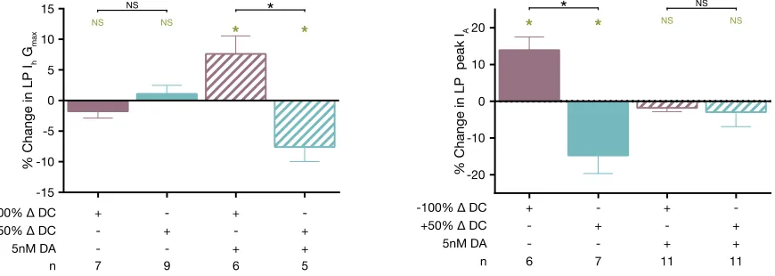
In the presence but not the absence of tonic DA (5nM), bidirectional changes in LP duty
cycle (LP burst duration ÷ cycle period) produce corresponding bidirectional changes in LP Ih
Gmax (Krenz, Rodgers et al. 2015). In these experiments, the STG was superfused with saline
containing or lacking (control) 5nM DA and slow wave and spike activity were blocked with
TTX. LP activity, measured before TTX application, was used to construct a voltage step
protocol to mimic the slow wave oscillations of LP. This voltage step protocol was then used to
increase or decrease LP duty cycle and Ih was measured before and 10min after applying the
voltage protocol. When the change in LP Ih Gmax (Ih Gmax after voltage protocol ÷ LP Ih Gmax
before voltage protocol) was plotted against the change in LP duty cycle, we observed that LP Ih
Gmax changed bidirectionally with changes in duty cycle (Krenz, Rodgers et al. 2015). Note that
activity-dependent changes in LP Ih were only observed in the presence of tonic DA and that in
the absence of DA there is no change in LP Ih Gmax regardless of activity.
These experiments were followed by experiments that would begin to uncover the
mechanism mediating LP Ih Gmax activity-dependent regulation (Krenz, Rodgers et al. 2015). We
found that tonic DA acting through D1Rs activates PKA which has two simultaneous effects; 1-
PKA produces an activity-independent increase in LP Ih Gmax, 2 – PKA permits calcineurin to
decrease LP Ih Gmax in an activity-dependent manner (Krenz, Rodgers et al. 2015). The specific
examine SUMO mediated activity-dependent coregulation of LP Ih and IA and the role of tonic
DA in selecting the ionic conductance target of SUMOylation.
1.4 SUMOylation mediates activity-dependent regulation of ion channels
SUMO is a small peptide that is post-translationally added to target proteins, altering the
properties of the modified protein in diverse ways. SUMOylation was first described over 20
years ago for its role in regulating the cell cycle and was thought to be restricted to the nucleus
(Matunis, Coutavas et al. 1996, Mahajan, Delphin et al. 1997). Since then SUMO substrates have
been identified throughout the cell and have been shown to regulate a wide variety of processes
including DNA repair, transcription, protein trafficking, cell migration (Hay 2005). Recent
studies have implicated SUMOylation in the regulation of neuronal proteins with functional
consequences that include the regulation of synaptic plasticity, and altering the biophysical
properties and trafficking of ion channels (Flotho and Melchior 2013).
There are four SUMO homologs in mammals (SUMO1-4). SUMO2 and 3 share ~97%
homology and are often referred to as SUMO2/3 because they are not readily distinguishable by
commercially available antibodies. Despite SUMO1 sharing only ~50% sequence identity with
SUMO2/3 there is significant overlap in target proteins between SUMO1-3. The distribution and
function of SUMO4 remains widely uncharacterized (Henley, Craig et al. 2014). SUMO is
reversibly conjugated to the lysine residue of a target protein through an enzymatic cascade that
closely resembles that of ubiquitin. Precursor SUMO requires proteolytic processing by a
member of the sentrin protease (SENP) family to exposed the C-terminal diglycine motif before
SUMO can be conjugated to a target protein. Mature SUMO is then activated through the
formation of a thioester bond with the E1 SUMO-activating enzyme (SAE1/SAE2) (Fig 1.1).
the formation of an isopeptide bond between the C-terminal diglycine of SUMO and the lysine
of a target protein (Fig1.1). In some cases, SUMO conjugation by Ubc9 is facilitated by the
presence of an E3 SUMO-ligase (Flotho and Melchior 2013). In most, but not all cases,
SUMOylation occurs at a SUMO consensus sequence, ΨKXE (Ψ is a large hydrophobic residue
and X can be any residue), that can be recognized by Ubc9. Conjugated SUMO can be cleaved
from a target protein by SENP (Fig 1.1). Members of the SENP family of proteases differ in their
affinity for different SUMO homologs as well as their ability to perform both hydrolase
(maturing of precursor SUMO) and isopeptidase (deSUMOylation) functions (Mukhopadhyay
and Dasso 2007, Nayak and Muller 2014).
Once conjugated to a target protein, SUMO acts like a docking station, promoting
interaction between the SUMOylated protein and other proteins that contain a SUMO Interacting
Motif (SIM) domain (Hay 2013). By promoting protein-protein interactions, SUMO can
coordinately regulate functionally related proteins. In neurons, SUMO is emerging as an
important regulator of activity and modulator-dependent processes (Lu, Liu et al. 2009, Loriol,
Khayachi et al. 2013, Loriol, Casse et al. 2014). Numerous reports have described SUMOylation
as being necessary for the regulation of different ion channels, including Na+ channels
(Dustrude, Wilson et al. 2013, Dustrude, Moutal et al. 2016, Plant, Marks et al. 2016), K+
channels (Benson, Li et al. 2007, Dai, Kolic et al. 2009, Plant, Dowdell et al. 2011, Qi, Wang et
al. 2014), and multiple glutamate receptors subunits (O'Brien, Kamboj et al. 1998, Konopacki,
Jaafari et al. 2011, Chamberlain, Gonzalez-Gonzalez et al. 2012, Jaafari, Konopacki et al. 2013,
Choi, Park et al. 2016). SUMO regulation can alter the biophysical properties or trafficking of
ion channels. SUMO-mediated regulation of these channels plays an important role in neuronal
neuronal homeostasis (Craig, Jaafari et al. 2012), LTP (Jaafari, Konopacki et al. 2013, Nair, Patil
et al. 2017), and LTD (Chamberlain, Gonzalez-Gonzalez et al. 2012, Luo, Ashikaga et al. 2013).
SUMO has also been implicated in several neuronal disease states including seizures (Qi, Wang
et al. 2014), Parkinson’s Disease (Eckermann 2013, Guerra de Souza, Prediger et al. 2016),
Alzheimer’s Disease (Lee, Dale et al. 2014, Hoppe, Salbego et al. 2015), Huntington’s Disease
(Ochaba, Monteys et al. 2016), and Down Syndrome (Schorova and Martin 2016). Ion channels
can be regulated by SUMO through either direct modification of the channel or indirect
modification of an interacting protein. For example, direct modification of the Nav1.2 channel
results in a shift of voltage-dependence of activation to a more negative voltage (Plant, Marks et
al. 2016). On the other hand, indirect SUMOylation of CRMP2 promotes CRMP2 interaction
with NaV1.7 leading to an increase in channel externalization (Dustrude, Wilson et al. 2013,
Dustrude, Moutal et al. 2016).
1.5 Hypothesis
The work presented here will seek to address the question, “what are the mechanisms that
mediate fast activity-dependent regulation of correlated ionic conductances?” We hypothesize
that SUMOylation mediates fast activity-dependent regulation of correlated ionic conductances.
We test this hypothesis by first characterizing SUMO regulation of the HCN channel, that
mediates Ih, using a heterologous expression system. Next, we will then look to see if SUMO
mediates activity-dependent regulation of LP Ih Gmax in a similar manner to what was observed in
the heterologous expression system. Finally, we will characterize activity-dependent regulation
Figure 1.1 SUMOylation
Figure 1.2 The Pyloric Network
(A) The STNS is dissected and pinned into a dish. A Vaseline well is used to enclose the STG
(blue box). Saline is continuously superfused through the well (blue arrows). The commissural
ganglia (CoGs) contain DA neurons that project through the stn to the STG. Two of the 14 STG
neurons are depicted here, PD (red) and LP (blue). PD and LP axons project through the dvn and
lvn to reach their target muscles. (B) Extracellular recordings were taken from the lvn and pdn
using pin electrodes. Intracellular recordings were also taken from LP and PD using
micropipettes. LP on-phase is defined as LP on-delay (b) divided by cycle period (a). (C) The
2 DETAILED MATERIALS AND METHODS
2.1 Hek cells
2.1.1 Tissue Culture
Cells were maintained in EMEM media (Corning; cat#10-009-CV) supplemented with
10% Fetal Bovine Serum (Sigma Aldrich; cat#F6178) and 1% Penecillin Streptomycin (Sigma
Aldrich; cat#P4333) at 37℃ and 5%CO2. For plates that needed to be split and passaged, media
was aspirated and 1-2ml (for 100mm plates) of Trypsin-EDTA 0.25% (Gibco; cat#25200-056)
was added to the plate and incubated for 2-3min, monitoring for cell detachment from the plate.
Using a Pasteur pipette, any cells still attached to the plate were gently washed into one corner of
the dish and the trypsin/cells suspension was transferred to of 15ml tube containing 5ml of
complete media. Cells were gently pelleted at 2,000rpm for 2-3min. The liquid was aspirated
from the cell pellet, the cells were gently resuspended in 1ml of complete media, an appropriate
number of cells were dropped on a plate containing fresh media and gently swirled to distribute
the cells evenly. Plates were returned to the 37℃ and 5%CO2 incubator. If a specific number of
cells needed to be plated a hemocytometer was used to determine a cell count. Briefly,
suspended cells were diluted in 500µl of complete media (1:25), 100µl of Trypan blue was
added, gently mixed and incubated for 5min at room temperature. Cells were then pipetted into
the hemocytometer and counted according to the instructions included with the hemocytometer.
2.1.2 Cryopreservation
For long-term storage of Hek cells, a confluent 100mm plate was trypsinized, cells were
cryotube (Thermo Fisher; cat#368632) containing 75ul (7.5%) DMSO, inverted to mix and
quickly placed in a styrofoam box inside of a -80℃ freezer. Once frozen, usually 24hr later, the
cryotube was transferred to a dewar filled with liquid nitrogen.
2.2 Calcium Phosphate Transfection
Approximately 24hr before transfection, enough cells were seeded onto a 60mm culture
dish to obtain ~60% confluence the next day (~1.9x10⁶ cells). The next day, the media was
changed 1hr before transfecting. For a 60mm plate, 25µg total of plasmid DNA (divided equally
for co-transfections) was combined with TE Buffer (10mM Tris-HCl, 1mM EDTA) to obtain a
total volume of 440µl. Next, 60µl of 2M CaCl2 was added drop wise to the DNA mixture, while
gently flicking the tube. Next, 500µl of 2xHBS (275mM NaCl, 10mM KCl, 12mM dextrose,
1.4mM Na2HPO4, 40mM HEPES, pH 7.05-7.10, sterile filtered) that had been slowly thawed at
room temperature was added dropwise while mixing. DNA mixture was immediately added to
the cells by carefully dropping it evenly over the plate and gently rocking the plate to distribute
the DNA. Plates were returned to the incubator for 4hr, and then the media was changed. The
transfection efficiency was checked 48hr later, and only plates with >80% efficiencies were used
for western blotting experiments.
2.3 GFP Immunoprecipitation
2.3.1 Hek Cell Lysates
To obtain a lysate, media was aspirated from a 100mm plate, and the plate was gently
washed twice with cold PBS (137mM NaCl, 2.7mM KCl, 10mM Na2HPO4, 1.8mM KH2PO4, pH
7.4). Cells were lysed by adding 1ml of RIPA Buffer (50mM Tris-HCl pH7.4, 150mM NaCl,
0.1% SDS, 0.5% Deoxycholate, 1% NP-40, 20mM EDTA, 20mM N-Ethylmaleimide, 1:100
occasionally. A cell scraper was used to scrape the contents of the plate into one corner where it
could be collected and transferred to a microcentrifuge tube. Cell debris was pelleted at
14,000rpm at 4℃ for 10min, and the supernatant (cell lysate) was transferred to a clean
microcentrifuge tube. Protein concentration was determined by BCA Assay. Briefly, 25µl of
lysate (diluted 1:1, 1:5, and 1:10) and Bovine Serum Albumin standards (0, 125, 250, 500, 750,
1000, 1500, and 2000µg/ml) were added to the wells of a 96 well plate, noting of the order of the
samples, 200µl of Reagent A:B (50:1) was added to each well, the plate was covered with plastic
wrap and incubated at 37℃ for 30min. The absorbance was measured at 562nm on a plate reader
and used with the standard concentrations to construct an xy-plot that was fit with a linear
trendline. The equation for the trendline was then used to calculate the concentration of the lysate
samples.
2.3.2 Co-Immunoprecipitation
Co-immunoprecipitation experiments of Hek cell lysates were performed using the Pierce
Classic Magnetic IP & Co-IP Kit (Thermo Scientific cat# 88804). For GFP Co-IPs, 1mg of
protein was combined with 2µl of an anti-GFP antibody (Abcam, cat# ab290) in a
microcentrifuge tube with Pierce IP Lysis/Wash Buffer to a total volume of 500µl.
Antibody/antigen complex was incubated together overnight at 4℃ with agitation. Magnetic
beads were prepared by combining 25µl of the beads with 175µl of Pierce IP Lysis/Wash Buffer,
collecting the beads on a magnetic stand, removing the supernatant, and repeating the wash with
an additional 500µl of Pierce IP Lysis/Wash Buffer. The antigen/antibody complex was then
added to the washed beads and incubated for 1hr at room temperature with agitation. The beads
were then collected and washed twice with Pierce IP Lysis/Wash Buffer. To elute the
room temperature, the beads were then collected, and the eluate was retained and neutralized
with 10µl of Neutralization Buffer.
2.4 Biotinylation
Hek cells on 60mm plates were washed twice with 1ml of cold PBS-CM (PBS - 137mM
NaCl, 2.7mM KCl, 10mM Na2HPO4, 1.8mM KH2PO4, pH 7.4 supplemented with 0.2mM CaCl2
and 1.5mM MgCl2). Biotin was added to the plate (2ml of 1mg/ml in PBS-CM; Thermo Fisher;
cat#21331) and incubated for 30min at 4℃. Cells were washed twice with ice cold PBS-CM, and
then 2ml of Quench Buffer (PBS-CM, 1.5mM MgCl2, 0.2mM CaCl2, 100mM Glycine) was
added to the plate and incubated for 15min at 4℃. Next, the cells were lysed in 500µl RIPA
Buffer (1% NP40, 50mM Tris pH 7.4, 150mM NaCl, 0.1% SDS, 0.5% DOC, 2mM EDTA,
protease inhibitor cocktail at 1:100) for 30min on ice, occasionally rocking the plate. Contents of
the plate were scraped into one corner using a cell scraper (Thermo Fisher; cat#08100241), the
lysate was transferred to 1.5ml microcentrifuge tube, and the cell debris was pelleted by
centrifuging for 10min at 14,000rpm at 4℃. NeutrAvidin beads (Thermo Fisher; cat#29201)
were prepared by adding 100µl of NeutrAvidin slurry to a spin column (Thermo Fisher;
cat#69725) and washing three times with 500µl of PBS, centrifuging for 1min at 500xg between
each wash. The bottom of the spin column was capped, and the cleared lysate was then incubated
with the beads for 2hr at 4℃ with agitation. Columns were uncapped and centrifuged for 2min at
1000xg; flow-through was retained as “intracellular fraction”. Beads were washed 3 times with
500µl of Wash Buffer 1 (PBS supplemented with 1% NP40, and 0.1% SDS), 3 times with 500µl
of Wash Buffer 2 (PBS supplemented with 0.1% NP40 and 0.5M NaCl), and then the tube was
capped and 50µl of SDS Buffer (50mM Tris-HCl pH 6.8, 100mM DTT, 2% SDS, 0.1%
extracellular protein. Eluted protein (extracellular fraction) was centrifuged from the column into
a clean tube at 1000xg for 2min.
Intracellular and Extracellular fractions were run together on a western blot, and the blot
was cut just above the 50kDa ladder marker. The portion of the blot containing proteins >50kDa
was dually probed with anti-GFP (Santa Cruz Biotechnology; cat#sc-8334; rabbit polyclonal;
1:2000) and anti-Na/K Pump (Abcam; cat#ab7671; mouse monoclonal; 1:3000) antibodies; with
expected bands corresponding to GFP-HCN2 being ~150-250kDa and Na/K Pump being
~100kDa. The portion of the blot containing proteins <50kDa was probed with an anti-Actin
antibody (Santa-Cruz Biotechnology; cat#sc-1616-R; rabbit polyclonal; 1:2000) to confirm no
intracellular contamination was present in extracellular samples.
2.5 Whole Cell Patch Clamping of Hek Cells
Poly-L-Lysine coated cover slips were prepared as follows: 20mm round glass coverslips
(Fisher Scientific; cat#12-546-2) were sterilized with ethanol and placed in the wells of 8 well
dishes (Thermo Fisher; cat#267062) to air-dry; enough 50µg/ml Poly-L-Lysine (Sigma Aldrich;
cat#P1524; prepared at 1mg/ml and sterile filtered) was added to each well to cover the
coverslips; coverslips were incubated with Poly-L-Lysine for 1hr at 37℃; Poly-L-Lysine was
aspirated; coverslips were washed once with dH2O; allowed to air-dry and stored at 4℃. For
whole-cell patch clamping, ~7x10⁴ cells were seeded onto each coverslip 24-48hr before
recording. Coverslips containing cells were perfused with extracellular saline (138mM NaCl,
5.6mM KCl, 2.6mM CaCl2, 1.2mM MgCl2, 10mM HEPES, 10mM Glucose, pH 7.4). Fire
polished patch pipettes (Sutter Instruments; cat#B120-69-15; 5-8MΩ) were filled with
intracellular saline (10mM NaCl, 145mM KCl, 1mM MgCl2, 5mM HEPES, pH 7.2) and
form a ≥1GΩ seal between the cell membrane and the opening of the micropipette. Suction was
used to rupture the cell membrane and recordings were taken from cells that maintained a seal
≥700 MΩ following rupture. PClamp software implemented voltage protocols that were used to
elicit the current being studied.
2.6 Tat-SUMO Peptide
2.6.1 Tat-SUMO Construct
Activated Panulirus interruptus SUMO (ending in diglycine) cDNA was obtained by
PCR using specific primers that included EcoRI restriction sites on the 5’ and 3’ ends. Briefly,
lobster cDNA was amplified using SUMO Forward (5’-TTTTGAATTCGATGTCCGAGGAAG
CCAAGAA-3’) and SUMO Reverse Primers (5’-GGGGGAATTCTCACCCACCAGTTTGCTG
CTGGAACAC-3’) with the following cycling parameters: 1cycle – 95℃ for 1min; 35℃ cycles
– 95℃ for 30sec, 61℃ for 1min, 68℃ for 1min; 1 cycle – 68℃ for 5min. The ~275bp PCR
product was gel isolated, and both the SUMO cDNA and pcDNA3 Tat HA vector (a gift from
Matija Peterlin (Cujec, Okamoto et al. 1997); Addgene plasmid #14654) were cut with EcoRI
(1ug of DNA to 1U of enzyme). The exposed ends of the cut Tat-HA vector were
dephosphorylated using Calf Intestine Phosphatase (CIP), then the SUMO cDNA and Tat-HA
vector were ligated at a 1:1, insert to vector ratio. The ligation product was transformed into
competent cells, and the presence of colonies containing Tat-SUMO was identified by PCR
using Tat vector-specific primers (For: 5’-GGGGGAATTCTCACCCACCAGTTTGCTGCTGG
AACAC-3’). DNA from colonies containing Tat-SUMO were sequenced to confirm proper
2.6.2 Tat-SUMO Peptide Synthesis
To synthesize the Tat-SUMO peptide, the plasmid was transformed into
BL21-CodonPlus (DE3)-RIPL E. coli (Agilent; cat#230280) and plated on NYZ agar plates. A single
isolated colony was grown overnight in 200ml of NYZ broth containing ampicillin (100µg/ml) at
37◦C with agitation. The 200ml overnight culture was then added to 1L of broth containing
500µM IPTG to induce expression of the peptide, and further incubated for 5hr. Cells were
divided among 250ml autoclaved bottles and pelleted at 8,000rmp for 10min at 4◦C, and the
pellet was washed with cold PBS (137mM NaCl, 2.7mM KCl, 10mM Na2HPO4, 1.8mM
KH2PO4, pH7.4). Pelleted cells were resuspended in 20ml of Buffer Z (8M Urea, 100mM NaCl,
20mM HEPES, pH8), transferred to autoclaved 50ml centrifuge tubes and sonicated on ice using
a 10sec “on” 30sec “off” protocol at 15% amplitude for a total of 10min. Sonicate was cleared by
centrifuging at 12,000rpm for 10mins at 4◦C. The supernatant was transferred to a clean tube and
the cleared sonicate was equilibrated with 10mM imidazole. Next, 10ml of Ni-NTA agarose
resin (Qiagen; cat#1018244) was equilibrated with Buffer Z containing 10mM imidazole and
then incubated with the sonicate at 4◦C for 1hr. The resin mixture was added to a
chromatography column (Bio-Rad; cat#7372512) and washed with 100ml of Buffer Z with
10mM imidazole. The peptide was eluted with incrementally increasing concentrations of
imidazole (100, 250, 500mM, and 1M; 10ml each) and the buffer was exchanged for PBS with
10% glycerol using PD-10 desalting columns (GE Healthcare; cat#17085101). Peptide
3 CHAPTER 1: SUMOYLATION OF THE HYPERPOLARIZATION-ACTIVATED
CYCLIC NUCLEOTIDE-GATED CHANNEL 2 INCREASES SURFACE
EXPRESSION AND THE MAXIMAL CONDUCTANCE OF THE
HYPERPOLARIZATION-ACTIVATED CURRENT
Publication: Parker, A. R., M. A. Welch, L. A. Forster, S. M. Tasneem, J. A. Dubhashi and D. J.
Baro (2016). "SUMOylation of the Hyperpolarization-Activated Cyclic Nucleotide-Gated
Channel 2 Increases Surface Expression and the Maximal Conductance of the
Hyperpolarization-Activated Current." Front Mol Neurosci 9: 168.
Contribution Disclosure: Authors A. Parker and D. Baro were responsible for the conception and
design of the research presented here. Authors A. Parker, M. Welch, L. Forster, S. Tasneem, J.
Dubhashi, and D. Baro provided substantial contribution to the acquisition and analysis of the
data presented here. All authors also provided input during the drafting and revision of the
3.1 Abstract
SUMO is a ~10kDa peptide that can be post-translationally added to a lysine (K) on a
target protein to facilitate protein-protein interactions. Recent studies have found that
SUMOylation can be regulated in an activity-dependent manner and that ion channel
SUMOylation can alter the biophysical properties and surface expression of the channel. HCN
channel surface expression can be regulated in an activity-dependent manner through unknown
processes. We hypothesized that SUMOylation might influence the surface expression of HCN2
channels. In this manuscript, we show that HCN2 channels are SUMOylated in the mouse brain.
Baseline levels of SUMOylation were also observed for a GFP-tagged HCN2 channel stably
expressed in Hek cells. Elevating GFP-HCN2 channel SUMOylation above baseline in Hek cells
led to an increase in surface expression that augmented Ih, mediated by these channels. Increased
SUMOylation did not alter Ih voltage-dependence or kinetics of activation. There are five
predicted intracellular SUMOylation sites on HCN2. Site-directed mutagenesis indicated that
more than one K on the GFP-HCN2 channel was SUMOylated. Enhancing SUMOylation at one
of the five predicted sites, K669, led to the increase in surface expression and Ih Gmax. The role of
SUMOylation at additional sites is currently unknown. The SUMOylation site at K669 is also
conserved in HCN1 channels. Aberrant SUMOylation has been linked to neurological diseases
that also display alterations in HCN1 and HCN2 channel expression, such as seizures and
Parkinson’s Disease. This work is the first report that HCN channels can be SUMOylated and
3.2 Introduction
Post-translational modifications can rapidly regulate proteins. SUMOylation is one such
modification that is essential for most organisms (Flotho and Melchior 2013). SUMO (a.k.a.
Sentrin) is a ~100 amino acid peptide that is covalently added to a lysine (K) residue on a target
protein. The addition involves several steps. First, the immature SUMO protein is cleaved by a
SENP, exposing a C-terminal diglycine (Mukhopadhyay and Dasso 2007). The SUMO E1
activating enzyme then transfers the mature SUMO peptide to the E2 conjugating enzyme, Ubc9,
which will then add SUMO to the target protein (Desterro, Thomson et al. 1997). SUMO can
also be deconjugated from a target protein by SENP (Hickey, Wilson et al. 2012). In mammals,
SUMO is encoded by a family of four genes, termed SUMO1-4 (Flotho and Melchior 2013).
SUMO2 and SUMO3 proteins are ~97% identical and are not readily distinguishable in most
experiments. SUMO 1 and SUMO2/3 proteins share ~46% identity, and their sets of targets
greatly overlap. SUMO4 cannot be cleaved into the mature form by Ubc9, and its function is
unclear.
Modification by SUMO is subject to numerous forms of regulation. The
availability of the SUMOylation site on a target protein can be regulated by the presence of other
post-translational modifications. For example, phosphorylation near the SUMOylation site can
either inhibit or enhance SUMOylation (Bossis and Melchior 2006, Konopacki, Jaafari et al.
2011). SUMOylation can also be regulated by the availability of SUMO and/or SUMOylation
enzymes (Loriol, Khayachi et al. 2013).
SUMOylation mediates protein-protein interactions (Makhnevych, Sydorskyy et
al. 2009, Flotho and Melchior 2013). As such, SUMO can coordinately regulate many diverse
membrane (Hickey, Wilson et al. 2012). The effects of SUMO are observed throughout the
neuron (Henley, Craig et al. 2014). SUMOylation can influence neuronal transcription by
controlling the stability of transcription factors; for example, BMAL1 ubiquitination and
degradation is enhanced when it is SUMOylated (Lee, Lee et al. 2008). SUMO can also regulate
synaptic release. SUMOylation of the synaptic vesicle protein, RIM1α, promotes its direct
interaction with Cav2.1 channels, causing them to form clusters, which leads to rapid exocytosis
of synaptic vesicles (Girach, Craig et al. 2013). In addition, SUMOylation plays a role in the
trafficking of integral membrane proteins such as kainate receptors. SUMOylation of GluK2 is
required for agonist-induced internalization of the kainate receptor, likely by facilitating its
interaction with scaffolding and/or trafficking proteins (Konopacki, Jaafari et al. 2011,
Chamberlain, Gonzalez-Gonzalez et al. 2012). However, the distinct interaction that leads to
GluK2 internalization is still unknown. Voltage-gated ion channels are fundamental constituents
of the neuronal membrane. Their trafficking is complex and highly regulated. The functions of
SUMOylation in voltage-gated ion channel trafficking are largely unknown. We are interested in
the role of SUMOylation in the regulated trafficking of Hyperpolarization-activated Cyclic
Nucleotide gated (HCN) ion channels.
HCN channels play a pivotal role in shaping neuronal excitability and synaptic
integration by influencing several neuronal activity features including membrane potential, firing
threshold, resonance frequency, temporal summation and synaptic strength (Hutcheon and
Yarom 2000, Wahl-Schott and Biel 2009, Shah 2014). The mammalian HCN1-4 gene family
encodes distinct channel isoforms (He, Chen et al. 2014). All isoforms are permeable to K+ and
Na+, activate upon hyperpolarization, and mediate a slowly depolarizing current termed the
HCN channels can also be gated by the binding of cyclic nucleotides to the C-terminal CNBD
found in all isoforms (Robinson and Siegelbaum 2003, He, Chen et al. 2014).
HCN isoforms differ in their biophysical properties and modulation by cAMP. Under
basal conditions, the CNBD inhibits hyperpolarization-gating to a different extent in each
isoform due to its isoform-specific interactions with the core transmembrane domain and the
C-linker that connects the CNBD to the transmembrane domain (Wang, Chen et al. 2001). This
variable inhibition results in isoform-specific steady-state activation curves. For example, the
steady-state activation curve of HCN2 channels is 20mV more hyperpolarized compared with
HCN1 channels. Binding of cAMP to the CNBD relieves its inhibition on
hyperpolarization-gating. Because of the isoform-specific interactions between the CNBD and the C-linker, the
effect of cAMP binding will vary with the isoform (Wang, Chen et al. 2001). For example,
binding of cAMP to the CNBD of HCN2 and HCN1 channels shifts their respective activation
curves to more positive potentials, but HCN2 channels display a 17mV shift while HCN1
channels display a 4mV shift. Maximal effects of cAMP binding are observed for HCN2 and
HCN4 isoforms. The activation kinetics also varies between isoforms with HCN1 and HCN4
having the fastest and slowest activation kinetics, respectively.
In addition to their distinct biophysical properties and cAMP modulation, each of the four
isoforms also displays a unique expression pattern in the nervous system (He, Chen et al. 2014).
HCN1 is highly enriched in the neocortex, hippocampus, cerebellar cortex and brainstem. HCN2
is widely expressed in most brain regions, while HCN3 has low expression levels in the nervous
system. HCN4 expression mirrors HCN1 and is also selectively expressed in several thalamic
nuclei and neuronal populations in the basal ganglia and habenular cortex. The formation of
HCN channel surface expression throughout the nervous system can be adjusted over
several time courses (Zha, Brewster et al. 2008, Shah 2014, Furst and D'Avanzo 2015, Smith, Al
Otaibi et al. 2015, Brennan, Baram et al. 2016). Regulated SUMOylation could play a role in the
trafficking of HCN channels. Since the trafficking of HCN2 channels has been fairly well
studied (see Discussion), here we examine SUMOylation of HCN2 channels.
3.3 Methods
3.3.1 Drugs
All drugs were obtained from Sigma-Aldrich with the exception of Tween 20 (Fisher
Scientific).
3.3.2 Mouse Brain Membrane Preparations
Nondenaturing: A single mouse forebrain was homogenized on ice in homogenization
buffer (5mM NaH2PO4 buffer, 0.32M sucrose) supplemented with protease inhibitor cocktail
(1:100, Sigma cat. #P8340) and 20mM N-Ethylmaleimide (NEM) to prevent SUMO
deconjugation (Suzuki, Ichiyama et al. 1999). Cell debris was pelleted at 5,000rpm for 10min at
4°C, and the supernatant was retained and further centrifuged at 40,000rpm for 90min at 4°C to
pellet membrane-bound proteins. The supernatant was removed and the pellet containing
intracellular and extracellular membrane bound proteins was resuspended in 1ml of resuspension
buffer (0.5% SDS, 5nM NaH2PO4, protease inhibitor cocktail at 1:100) followed by shaking at
4°C for 1hr. Denaturing: Tissue homogenization and centrifugation were the same as for the
nondenaturing preparation. The membrane pellet was resuspended in 100µl of denaturing buffer
(2% SDS, 50mM Tris-HCl pH7.5, 5mM DTT) followed by shaking at 4°C for 1hr. The
preparation was then diluted to 1ml total volume with dH2O and boiled for 10min. In all cases
Protein Assay Kit). Mouse brain membrane fractions were obtained from whole mouse forebrain
tissue generously provided by Dr. Chun Jiang. All animal procedures were conducted in
compliance with the regulation of the Institutional Animal Care and Use Committee of Georgia
State University.
3.3.3 Plasmids and Antibodies
A previously described mouse GFP-HCN2 fusion plasmid (Santoro, Wainger et al. 2004)
was generously provided by Dr. Bina Santoro’s Lab. Plasmids for transient transfection include
the following: mCherry2-C1 was a gift from Michael Davidson (Addgene plasmid #54563),
Ubc9 (Yasugi and Howley 1996) was a gift from Peter Howley (Addgene plasmid #14438),
SENP1 (Cheng, Kang et al. 2007) was a gift from Edward Yeh (Addgene plasmid #17357), and
SUMO2 (Kamitani, Nguyen et al. 1998) was a gift from Edward Yeh (Addgene plasmid
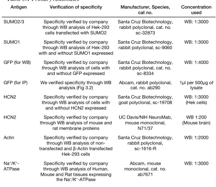
#17360). Antibodies used are shown in Table 1, and the specificity of each antibody was verified
as indicated in the table.
3.3.4 Site Directed Mutagenesis
PCR was used to create two site-directed mutations in the GFP-HCN2 fusion plasmid
described above: K534R and K669R. The two sets of primers are listed in Table 2. PrimeStar
GXL polymerase (Takara) was used along with the buffer, nucleotides, and instructions supplied
by the manufacturer. Typically, 10ng of plasmid DNA served as the template in a 50μl reaction.
The cycling conditions were: 1x 98°C,1min; 30x 98°C,30sec, 68°C, 7min; 1x 68°C, 5min. Upon
completion, 20 units of DpnI (Clontech) were added to the reaction, which was incubated at
37°C for 1-2hr to digest the template DNA. Afterward, 1-2μl of the reaction was added to a 50μl
aliquot of subcloning grade competent XL1blue cells (Agilent) and incubated on ice for 30min.
were incubated at 37°C for 20min to allow for expression of kanamycin resistance. Cells were
then plated on NZY plates containing 30μg/ml kanamycin and incubated at 37°C overnight.
Plasmid DNA from resultant colonies was isolated (Qiagen), the insert was sequenced in its
entirety, and analyzed with Lasergene software (DNAstar) to ensure that only the intended
mutation was generated. All sequencing was done by the Georgia State University Cell Protein
and DNA Core Facilities. To create multiple mutations, a previously mutated GFP-HCN2 fusion
plasmid was used as the template for a PCR with a different primer.
3.3.5 Cell culture, Stable and Transient Transfections
Hek-293 cells and all cell culture reagents were obtained from American Type Culture
Collection (ATCC; Manassas, VA). Hek-293 cells were cultured at 37°C, and 5% CO2 in EMEM
media supplemented with 10% fetal bovine serum and 1% Penicillin-Streptomycin.
Transfections to produce stable lines: A cell line stably expressing a GFP-HCN2 fusion protein
(Hek-HCN2) was generated by transfecting a 60mm plate of Hek-293 cells with the GFP-HCN2
plasmid using Lipofectamine 2000 (Invitrogen) according to instructions provided by the
manufacturer. After two days, transfected cells were trypsinized, resuspended and replated to
60mm plates (5-20l of a 1ml cell resuspension per 60mm plate) and the selection agent, G418
(Geneticin, Gibco, 500µg/ml), was added to the media. After 3-5 weeks, individual colonies
were selected using cloning rings. Stable expression was checked by fluorescence and whole cell
patch clamping for Ih. In all cases, a colony was selected only if GFP and Ih was observed in
every cell; however, the intensity of GFP expression and Ih maximal conductance could vary
between colonies from the same transfection by a factor of 10, presumably due to differences in
the number of copies of plasmid integrated into the host cell genome. Stable cell lines expressing
our hands, Lipofectamine transfection efficiencies ranged from 5-25%. This was not adequate for
transient transfection experiments, which required 80-100% transfection efficiency. Therefore, a
high efficiency calcium phosphate transfection was employed for immunoprecipitation,
biotinylation and patch clamping experiments using transient transfection to increase or decrease
SUMOylation. Briefly, cells were plated at ~60% confluence on 100mm or 60mm cell culture
dishes 24hr before transfection. The media was changed at least 1hr before transfection. For a
single 100mm or 60mm dish, 25µg or 10µg of total plasmid DNA, respectively, was diluted in
TE buffer (10mM Tris-HCl pH8, 1mM EDTA), 2M CaCl2 was added dropwise to the DNA
followed by the addition of 2x HBS (275mM NaCl, 10mM KCl, 12mM Dextrose, 1.4mM
Na2HPO4, 40mM HEPES, pH 7.1). Calcium Phosphate/DNA precipitate was then immediately
dropped onto cells. Cells were returned to 37°C and 5% CO2 for 4hr followed by either a media
change or passaging to 20mm Poly-L-Lysine coated coverslips and allowed to grow another
24-48hr. Cells were transfected with plasmid DNA expressing either mCherry or a mixture of
mCherry+SUMO+Ubc9 or mCherry+SENP1. For co-transfection of multiple plasmids, the total
amount of plasmid DNA remained the same (25µg or 10µg) but comprised equal amounts of
each of the different plasmids (1:1:1 or 1:1). Transfection efficiency was determined to be the
percentage of cells expressing mCherry as judged by fluorescence microscopy. The transfection
efficiency of every plate was checked for each experiment, and a plate was only used if the
transfection efficiency exceeded 80%.
3.3.6 Immunoprecipitations
GFP-HCN2 IP from Hek cells: 100mm plates containing Hek cells were washed twice
with ice-cold PBS (137mM NaCl, 2.7 mM KCl, 10 mM Na2HPO4, 1.8 mM KH2PO4, pH 7.4)
SDS, 0.5% DOC, 20mM EDTA, protease inhibitor cocktail at 1:100 and 20mM NEM) to the
plate. Cells were lysed on the plate for 30min on ice, rocking occasionally. A disposable cell
scraper was used to scrape any remaining cell debris from the bottom of the plate, and the lysate
was collected and transferred to a sterile tube. Cell debris was pelleted at 12,000rpm for 10min at
4°C, and the supernatant was retained. Protein concentration was determined with a BCA assay.
Immunoprecipitations (IP) were performed using the Classic Magnetic Co-IP Kit (Pierce),
following the manufacturer’s instructions. Typically 1mg of protein was added to the beads and
the protein was eluted from the beads in a volume of 100µl. HCN2 IP from mouse forebrain:
1.5mg of the mouse membrane preparation with 5ug of anti-HCN2 antibody (Table 1) or IgG
(Santa Cruz Biotechnology; sc-2028) were used in combination with the Classic Magnetic Co-IP
Kit (Pierce), following the manufacturer’s instructions. Protein was eluted from the beads in a
volume of 100µl and 20µl was used for one lane of an SDS-PAGE gel.
3.3.7 Western Blotting
Protein samples were run on an SDS-PAGE gel and transferred to a PDVF membrane
using a semidry electroblotting system. A membrane was blocked for 1hr in a solution of 5%
non-fat powdered milk in TBS (10mM Tris-HCl pH7.5, 150mM NaCl), washed for 10min in
TTBS (1x TBS, 0.1% Tween 20), and then incubated overnight at 4°C with the primary
antibody, diluted as described in Table 1, in 1% non-fat powdered milk in TTBS. The membrane
was then washed 3x 5min each in TTBS and then incubated with the appropriate alkaline
phosphatase conjugated secondary antibody, diluted in 1% non-fat powdered milk in TTBS for
2hr at room temperature. Following incubation with the secondary antibody, the membrane was
washed 3x 10min each and Immunstar AP substrate (BioRad) was added to the surface of the