NAN O R EVIE W
Open Access
A review on potential neurotoxicity of
titanium dioxide nanoparticles
Bin Song
1,2, Jia Liu
2, Xiaoli Feng
2, Limin Wei
2and Longquan Shao
2*Abstract
As the rapid development of nanotechnology in the past three decades, titanium dioxide nanoparticles (TiO2NPs), for their peculiar physicochemical properties, are widely applied in consumer products, food additives, cosmetics, drug carriers, and so on. However, little is known about their potential exposure and neurotoxic effects. Once NPs are unintentionally exposed to human beings, they could be absorbed, and then accumulated in the brain regions by passing through the blood–brain barrier (BBB) or through the nose-to-brain pathway, potentially leading to dysfunctions of central nerve system (CNS). Besides, NPs may affect the brain development of embryo by crossing the placental barrier. A few in vivo and in vitro researches have demonstrated that the morphology and function of neuronal or glial cells could be impaired by TiO2NPs which might induce cell necrosis. Cellular components, such as mitochondrial, lysosome, and cytoskeleton, could also be influenced as well. The recognition ability, spatial memory, and learning ability of TiO2NPs-treated rodents were significantly impaired, which meant that accumulation of TiO2 NPs in the brain could lead to neurodegeneration. However, conclusions obtained from those studies were not consistent with each other as researchers may choose different experimental parameters, including administration ways, dosage, size, and crystal structure of TiO2NPs. Therefore, in order to fully understand the potential risks of TiO2 NPs to brain health, figure out research areas where further studies are required, and improve its bio-safety for applications in the near future, how TiO2NPs interact with the brain is investigated in this review by summarizing the current researches on neurotoxicity induced by TiO2NPs.
Keywords:Titanium dioxide; TiO2; Anatase; Rutile; Nanoparticles; Brain; CNS; Neurotoxicity
Review Introduction
Nanomaterial, with one dimension in the range of 1 to 100 nm at least, possesses unique physicochemical [1], optical [2], and electrical properties [3]. Because of its peculiar features, nanomaterial is widely applied in cos-metics [4], food and personal care products [5], medical devices [6], and so on. As nanotechnology is advancing rapidly, more concerns on health risks about exposure to nanoparticles have been arising. The TiO2 particles are believed to possess low toxicity and thus are widely used in biomedical applications for their excellent bio-compatibility [7–9]. However, when the size of TiO2 is diminished to nanoscale, the bioactivity and physio-chemical properties of nano-sized TiO2are significantly different from the properties of their bulk analogue. As a
consequence, the toxic effects of TiO2 NPs on human beings could not be simply determined by traditional methods. What’s more, the understanding about the risk assessments of NPs is insufficient and often lags behind their rapid advancement and widespread applications [10–13]. TiO2NPs-containing products are widely used as well, which could unintentionally lead to human ex-posure and environmental pollution. The TiO2 NPs might be potentially absorbed mainly through inhalation, indigestion, and skin penetrations into the circulation of human beings. And then they may be redistributed into other tissues (such as the liver, heart, lung, etc.), which could induce impairments on organs after unintentional exposure. As the concerns about unintentional exposure of NPs on human beings arise, an increasing number of researches have been performed to study the potential toxic effects of TiO2NPs in recent years. Several in vivo researches adopted rats or mice for the experimental models, and they were exposed to TiO2 NPs for bio-safety * Correspondence:[email protected]
2Nanfang Hospital, Southern Medical University, Guangzhou 510515, China
Full list of author information is available at the end of the article
assessment. The main administration routes in in vivo studies included inhalation [14], intratracheal [15] or nasal instillation [16], oral gavage and dermal expos-ure [17], intragastric feeding [18], intraperitoneal [19], and intravenous injection [15]. Numerous reports have revealed that when the TiO2 NPs were administrated and transported into second targets, they could induce renal fibrosis, change cell cycle of lung epithelial cell, disturb the metabolism of hepatocytes, and impair the spleen [20–23].
The central nervous system (CNS), including the brain and spinal cord, is an extremely important system for human beings. Its functions are mainly composed of the following: (1) to transfer, store, and process information; (2) to generate a variety of psychological activities; and (3) to command and control all the behaviors of human beings. Several in vivo studies have investigated the bio-distribution of TiO2 NPs. After rats or mice were ex-posed to TiO2 NPs, the NPs were capable of reaching most parts of the brain zones, and the Ti contents in the brain were increased. The main pathways for TiO2NPs to be transported into the brain included (1) the blood– brain barrier (BBB) pathway [24], (2) the olfactory nerve translocation pathway [25], and (3) the placental barrier pathway [26]. However, the processes of translocation of TiO2 NPs into the brain would be regulated by several parameters, such as administration routes, size, and sur-face modification. Once the TiO2NPs were transported into the brain regions, major cells in the CNS, including the neurons and the glial cells, would be affected by NPs. Reactive oxygen species (ROS), apoptosis, and in-flammation would be induced by TiO2NPs, which may lead to cell death and disturb the CNS functions or even induce neurodegenerative disease. Some in vitro studies also indicated that when neurons or glial cells were incu-bated with TiO2NPs, the viability, cell cycle, cell morph-ology, antioxidant capability, and cellular components would be affected [27–30].
However, current knowledge about neurotoxicity in-duced by TiO2NPs is insufficient and more detailed and standardized researches are needed. Therefore, in order to fully understand the potential risks of TiO2 NPs to brain health, figure out research areas where further studies are required, and improve its bio-safety for appli-cations in the near future, how TiO2NPs can be translo-cated into the brain and how they influenced the CNS function are investigated in this review via summarizing relevant in vivo and in vitro researches.
The main routes of TiO2NPs into the brain
Due to peculiar physicochemical properties of NPs [31], TiO2NPs are widely used in many fields, such as photo-voltaic appliances [32], sensors [33], renewable energy devices [34], functional building blocks [35], textiles
[10], sunscreens [11], food [13], and medical applications [36]. These widespread applications, however, put hu-mans at a high risk of getting exposed to TiO2 NPs, probably through inhalation, ingestion, skin penetration, medical applications, and so on. Therefore, it is urgent to evaluate the bio-safety of TiO2 NPs at length. The CNS is an extremely important system for human be-ings. Numerous studies have already demonstrated that once NPs were absorbed, they were able to be trans-ported to the second targets, including the brain, liver, lung, spleen, and so on. In in vivo studies, after the ex-perimental rodents were exposed to TiO2 NPs, these NPs can be transported into the brain regions mainly through the following routes.
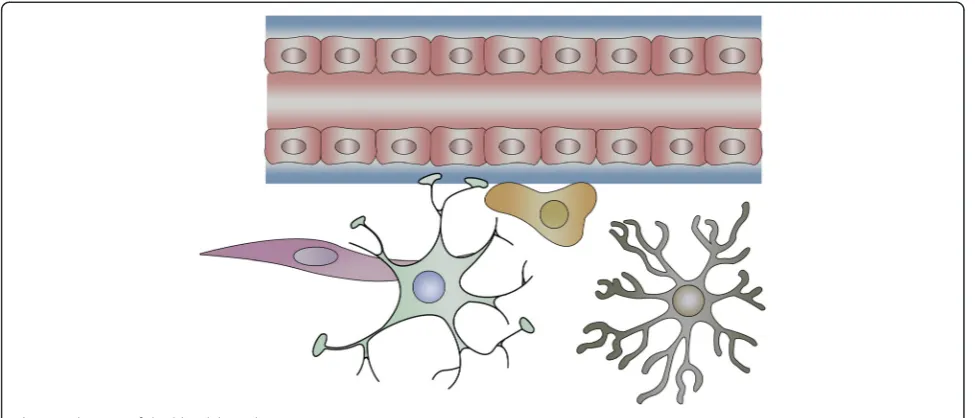
Translocation of TiO2NPs from the blood to the brain The BBB is an effectively protective structure, which is mainly composed of endothelial cells, astrocytes, and peri-cytes [37]. The endothelial cells are connected with each other through complicated tight junctions, while the con-nections are supported by the astrocytes and pericytes. On account of this sophisticated structure, only specific sub-stances with small size or low-molecular weight (MW) could be allowed to pass through the BBB by means of three main transport patterns (passive diffusion, active transport, and endocytosis). In another word, BBB is cap-able of protecting the healthy and functional CNS from being affected by harmful chemicals, toxins, and drugs in the circulatory system. Whereas, NPs possess unique chemical–physical characteristics and tiny size which make them be similar to biomolecule. Therefore, they are able to pass through the BBB and enter into the CNS [38–40] (Fig. 1). On the other hand, the permeability of BBB can be altered by NPs, which could assist in influx of exogenous substances into the brain. As a result, NPs in-duced inflammation, edema, and cell injury or even cell death in brain regions.
NPs with diameters of 10 and 20 nm were both trans-ported into the brain, inducing cerebral injury in a dosage-dependent way. However, TiO2NPs with diameter of 200 nm did not cause any significant changes in the brain. From these results, we could infer that the ability of passing through the BBB for TiO2 NPs was associated with the nanoparticle diameter.
TiO2 NPs could not only pass through the BBB but also disrupt the integrity of the BBB. The toxic effects of early, acute, and long-term exposures of TiO2 NPs on the BBB were investigated on the basis of an in vitro BBB model. The model, mimicking the specific charac-teristics of an in vivo one [43], was composed of rat pri-mary endothelial cells (BECs) and astrocytes. After the BBB model was treated with TiO2NPs for acute or long-term exposure, the expression levels of P-glycoprotein (P-gp), claudin 5, caveolin-1, and caveolin-2, which regu-lated the integrity of BBB, were reduced. These results indicated that direct harmful impacts of TiO2 NPs on BBB integrity were presented during the acute and long-term exposure. While in this study, authors still discov-ered that after exposure for 4 h the mRNA expression levels of CXC chemokines, CC chemokines, ADAM17, Ccl2, Tgfβ1, ICAM, and VCAM were increased, paral-leled by the decreased mRNA expression levels of ABC transporters. The upregulated expression levels of those target genes were reported to be related with the de-creased permeability of BBB [44–50], which could facili-tate the transportation of other exogenous substances into brain. These findings meant that besides direct im-pairments, the indirect harmful impacts (inflammatory effects) of TiO2NPs on BBB integrity also occurred. On the other hand, if the NPs were eliminated from the
brain slowly, they might induce long-term adverse ef-fects after the exposure.
Axonal translocation of TiO2NPs from the nose to the brain
Axonal transport is defined as the process that proteins and other substances synthesized in neurosome which are transported to the nerve endings through the cyto-skeleton [51]. However, some low-molecular weight or small-size substances such as NPs could be taken up by the nerve ending, and then transported to the neuro-some. This process is called retrograde axonal transport [52]. The olfactory and trigeminal nerve endings are abundant in the nasal areas. Once NPs were instilled through the nose, they can enter the circulation system and pass through the BBB, or they can bypass the BBB to be transported into the brain regions along the axons. The second direct pathway is reported to be the major route for NPs of being transported to the brain zones after intranasal instillation (Fig. 2).
Wang et al. [16] confirmed that after the female mice were exposed to TiO2 NPs of different sizes with two crystal types (80 nm for rutile and 155 nm for anatase) through intranasal instillation, the Ti concentration was significantly increased in the brain as compared with the control. Wang et al. [25] discovered that when female mice were treated with TiO2NPs (80 nm rutile, 155 nm anatase) through intranasal instillation every other day, the titanium content was detected in the brains after 30 days. The Ti contents determined by synchrotron ra-diation X-ray fluorescence (SRXRF) analysis and ICP– MS were significantly upregulated in the hippocampus, followed by the olfactory bulb, cerebellum, and cerebral
[image:3.595.56.544.89.298.2]cortex. These findings suggested that the TiO2 NPs could be transported to the mice brain through the olfactory bulb after intranasal instillation. The same re-search group conducted another study [53], which also detected Ti contents in the hippocampus, olfactory bulb, cerebellum, and cerebral cortex by ICP-MS in a time-dependent way. Both the two studies indicated that there was a high Ti content in the hippocampus, so this brain area would be easily affected by TiO2NPs. It is generally accepted that the hippocampus is mainly in charge of memory and learning [54, 55]. Therefore, impairments on it might probably induce neurodegenerative diseases, such as Alzheimer’s diseases [56–58].
In another in vivo study [59], the female mice were treated with TiO2NPs by intranasal instillation. The ex-perimental mice were divided into four groups according to different sizes, coatings, and shapes of the TiO2 parti-cles. Groups A and B shared the same insoluble prop-erty, but with various size (micro- and nano-sized TiO2 particles) and no surface coatings. Groups C and D were hydrophilic NPs and silica-coated with different shapes. After treatment every other day for 30 days, the titanium contents in the brain were determined by ICP-MS. These data demonstrated that groups C and D showed higher Ti concentration in the cerebral cortex and stri-atum than groups A and B, while group A was detected with no Ti content in the sub-brain zones. These results indicated that the size, surface modification, and shape of TiO2NPs played an important role on their transport to the brain from the nose. In this study [60], CD-1 (ICR) female mice were exposed to TiO2 NPs with dif-ferent dosages (2.5, 5, and 10 mg/kg body weight). After nasal administration for 90 consecutive days, the TiO2 NPs were detected in the brain, and this accumulation could induce CNS dysfunctions.
Translocation into the brain of offspring through the placental barrier
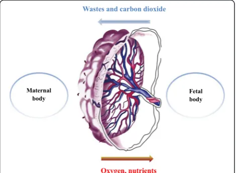
Placental barrier, composed of both maternal and fetal tissues, is another internal barrier that can protect the development of embryo [61]. It could protect the fetus from being affected by harmful substances in maternal blood circulation, while the fetus could get nutrients and oxygen from the mother via the placenta (Fig. 3). How-ever, a great number of studies [62, 63] have already re-vealed that after pregnant mice/rats were exposed to exogenous substances, such as nanoparticles, those sub-stances could be detected in the brain of fetus, and then they can disturb the homeostasis of CNS or even induce neuronal death. Those harmful impacts on fetus brain have been demonstrated to be related with psychiatric disorders such as autism, schizophrenia, depression, and so on in their later life [64, 65]. As a consequence, those
Fig. 2A simple diagram of nose–brain pathway after intranasal administration
[image:4.595.59.540.88.268.2] [image:4.595.305.540.531.703.2]findings suggest that placental barrier plays an important part on fetal growth and development.
In this study [66], pregnant Wistar rats were adminis-trated with TiO2NPs intragastrically daily from gestational day 2 to 21. Then, the 1-day-old neonates were sacrificed, and the TiO2 NPs concentration in the hippocampus, determined by ICP-MS, was significantly upregulated as compared with the control group. Yamashita et al. [67] also detected silica (70 nm) and TiO2(35 nm) NPs in the placenta and fetal brain after the pregnant mice were injected intravenously with these NPs, which led to the pregnancy complication.
In another study [68], the authors discovered that when pregnant ICR mice were treated with TiO2 NPs, the levels of dopamine and its metabolites were in-creased in some regions of the fetus brain on postnatal day 21 as compared with the control group. Another study [69] adopted microarray to assess gene expression changes in the brains of male fetus and pups after the pregnant ICR mice were exposed to TiO2 NPs. Data showed that the expression levels of genes related with dopamine neuron system were altered. Microarray was also applied in this study [70], and pregnant ICR mice were administrated with anatase TiO2NPs. By analyzing the gene expression alternations in the brains of male fetus and pups, data obtained revealed that the expres-sion levels of genes related with oxidative stress, neuro-transmitters, and psychiatric diseases were dysregulated. The neurobehavioral performance of the offspring might be moderately altered due to maternal exposure to TiO2 NPs [71]. Similarly, Cui et al. [72] discovered that when Sprague–Dawley rats were injected subcutaneously with TiO2NPs, the antioxidant ability of pups’brain was im-paired. Although these researches did not measure the contents of TiO2 NPs in the brain directly, those data collected indirectly demonstrated that the TiO2 NPs in maternal circulation system would affect the develop-ment of embryo. Then, they could impair the brain de-velopment and finally lead to CNS dysfunctions in their later life.
Bio-distribution and elimination of TiO2NPs from
the brain
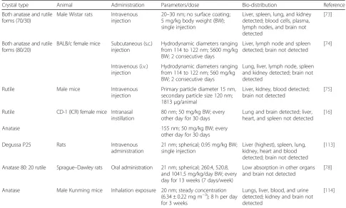
When TiO2 NPs were absorbed into circulation, they were capable of being redistributed to second organs (Fig. 4). At present, several researches have been per-formed to study the bio-distribution of TiO2 NPs after administrations (Table 1). In this study [73], when rats were treated with TiO2 NPs (5 mg/kg body weight) by intravenous injection, TiO2NPs can be detected in the liver, spleen, lung, and kidney except blood cells, plasma, brain, and lymph nodes. The BALB/c female mice were exposed to TiO2 NPs at a dose of 560 mg/kg by intra-venous injection (i.v.) or 5600 mg/kg by subcutaneous
injection (s.c.). The TiO2 NPs were detected by energy dispersive X-ray spectroscopy (EDS) in the lung, liver, lymph node, spleen, and kidney from i.v.-administrated mice but only in the liver, lymph node, and spleen of s.c.-administrated mice, while the content of NPs was not detected in the brain [74]. Another study [75] also did not detect TiO2 NPs in the brain of male mice ex-cept blood and liver after i.v. injection. However, after hairless mice were treated with TiO2 NPs (21 nm) by dermal exposure for 60 days, significant pathological alter-ations were presented in the skin and liver and the NPs were also detected in the brain without pathological changes [76]. Wang et al. [16] studied the bio-distribution of TiO2 NPs (50 mg/kg) after female mice were treated with NPs by intranasal instillation every other day for 30 days. The biochemical parameters of the liver, spleen, heart, and serum were not affected by NPs as compared with the control group; while the concentration of NPs was apparently enhanced in the lung and brain regions. Another study investigated the bio-distribution of TiO2 NPs after rats were repeatedly orally administrated for 13 weeks. Even in the highest dosage group (1041.5 mg/kg BW), the Ti content in the brain was minimal with no statistical significance. Geraets et al. [77] compared the different distributions of TiO2NPs in rats after oral and intravenous administration. The data obtained demon-strated that the Ti concentrations were not detectable in tissues, including the brain after oral administration. How-ever, the Ti contents were detected in the liver, spleen, kidney, lung, heart, brain, thymus, and reproductive or-gans after intravenous injection. It could be inferred from those studies that (1) intranasal instillation might be the most effective routes for TiO2 NPs transported to the brain and (2) Ti content could be undetectable in the brain regions after intravenous injection. Undoubtedly, those conclusions drawn from abovementioned in vivo re-searches might not be convincing, because the transloca-tions of TiO2NPs into the brain would be influenced by several parameters, such as administration routes, size, dosage, and so on, which would be discussed in later chapters.
period, Ti concentrations in the feces and urine demon-strated no increase as compared with control group. Also, Ti contents in the brain demonstrated no detectable alterations.
It can be inferred from the results that (1) the excre-tion of TiO2NPs from the brain is limited and (2) even negligible dosage of TiO2NPs could be accumulated in the brain with minimal elimination, so chronic or long-term exposure might be potentially risky for the brain health. TiO2NPs are thought to be low toxic. However, the long-term, repeated exposure, and durative existence of TiO2 NPs in the brain regions made it necessary to
evaluate their bio-safety. Therefore, more in vivo re-searches are needed to further investigate the parameters of TiO2 NPs which might influence their elimination rates from the brain. Then, the health risks of TiO2NPs to the brain are expected to be lowered.
Neurotoxicity of titanium dioxide nanoparticle
[image:6.595.58.540.89.235.2]Once the TiO2NPs are translocated into the CNS through the abovementioned pathways, they may accumulate in the brain regions. For their slow elimination rates, those NPs could remain in the brain zones for a long period, and the Ti contents would gradually increase with
Table 1Bio-distribution of TiO2NPs after rat/mice were administrated by different routes
Crystal type Animal Administration Parameters/dose Bio-distribution Reference
Both anatase and rutile forms (70/30)
Male Wistar rats Intravenous injection
20–30 nm; no surface coating; 5 mg/kg body weight (BW); single injection
Liver, spleen, lung, and kidney detected; blood cells, plasma, lymph nodes, and brain not detected
[73]
Both anatase and rutile forms (80/20)
BALB/c female mice Subcutaneous (s.c.) injection
Hydrodynamic diameters ranging from 114 to 122 nm; 5600 mg/kg BW; 2 consecutive days
Liver, lymph node and spleen detected; brain not detected
[74]
Intravenous (i.v.) injection
Hydrodynamic diameters ranging from 114 to 122 nm; 560 mg/kg BW; 2 consecutive days
Lung, liver, lymph node, spleen and kidney detected; brain not detected
Rutile Male mice Intravenous
injection
Primary particle diameter 15 nm, secondary particle size 120 nm; 1813μg/animal
Liver, kidney, blood detected; brain not detected
[75]
Rutile CD-1 (ICR) female mice Intranasal instillation
80 nm; 50 mg/kg BW; every other day for 30 days
Lung and brain detected; liver, heart, and spleen not detected
[16]
Anatase 155 nm; 50 mg/kg BW; every
other day for 30 days
Degussa P25 Rats Intravenous
administration
21 nm; spherical; 0.95 mg/kg BW; single injection
Liver (highest), spleen, lung, kidney, heart and blood detected; brain not detected
[113]
Anatase 80: 20 rutile Sprague–Dawley rats Oral administration 21 nm; spherical; 260.4, 520.8, and 1041.5 mg/kg/day BW; every day for 13 weeks (7 days/week)
Low absorption in other organs and brain not detected
[78]
Anatase Male Kunming mice Inhalation exposure 20 nm; steady concentration (6.34 ± 0.22 mg m−3); 8 h per day
for 3 weeks
Lungs, liver, blood, and urine detected; kidney and brain not detected
[114]
[image:6.595.54.540.440.732.2]repeated exposure. This would induce pathologic changes, such as inflammation, immunological response, edema, cell injury, cell necrosis, and so on, which would ultim-ately lead to CNS dysfunctions, including neurodegene-rative diseases and psychiatric disorders. Generally, the neurons and glial cells are the main cell types in the CNS. Therefore, the toxic effects of TiO2NPs on them would lead to impairments on the brain as a consequence.
Toxic effects on CNS in in vivo studies
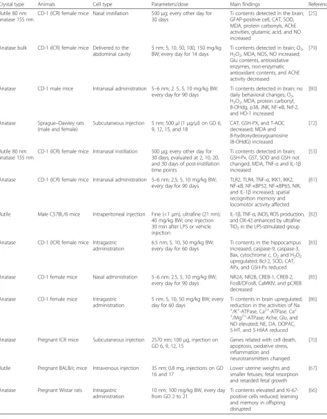
Several in vivo studies have demonstrated that the TiO2 NPs could be transported into the brain regions, and then accumulated in the CNS, eventually leading to CNS dysfunctions (Table 2).
Wang et al. [25] revealed that after the female mice were treated with TiO2NPs (80 nm, rutile and 155 nm, anatase; 500μg) through intranasal instillation every other day for 30 days, the Ti contents were detected in the brain re-gions, including the olfactory bulb, hippocampus, cerebel-lum, and cerebral cortex. This accumulation induced increased glial fibrillary acidic protein (GFAP)-positive cell, CAT activity, MDA content, protein carbonyls con-tent, AChE activities, glutamic acid, and NO concon-tent, accompanied by cell lost in both experimental groups. While, SOD level was increased only in the 155 nm group. In another study [79], the ICR female mice were injected with TiO2NPs (anatase; 5 nm; 5, 10, 50, 100, 150 mg/kg BW) into the abdominal cavity every day for 14 days. The coefficients of the brain-to-body weight, antioxidative en-zymes (SOD, CAT, APx, GSH-Px), non-enzymatic antioxi-dant contents (ASA/DASA,GSH/GSSG), Glu contents, and AChE activity were decreased, but the Ti contents in the brain, the levels of O2, H2O2, MDA, NOS, and NO were increased in a dose-dependent way. Ze et al. [80] dis-covered that when CD-1 male mice were intranasally treated with TiO2 NPs (5–6 nm; 2.5, 5, 10 mg/kg BW) every day for 90 days, the mRNA expressions of genes regulating oxidative stress, including p38, JNK, NF-κB, Nrf-2, and HO-1, were increased besides upregulated Ti concentrations and levels of O2, H2O2, MDA, protein car-bonyl, and 8-OHdg,.
However, in another study [53], after the CD-1 (ICR) female mice were exposed to TiO2 NPs (rutile 80 nm and anatase 155 nm) every other day for 30 days by in-tranasal instillation, the activities of GSH-Px, GST, SOD, and GSH level in the brain were not changed at 30 days. Yet, the level of MDA was enhanced all the same. The Ti contents were highest in the hippocampus, followed by olfactory bulb, cerebellum, and cerebral cortex. How-ever, the levels of TNF-α and IL-1β in brain were up-regulated in the 155 nm group, which indicated that intranasal instillation with anatase TiO2NPs would induce inflammation in the brain of mice. Ze et al. [81] also dem-onstrated that TiO2 NPs could induce inflammation in
mice brain. In their study, the mRNA and protein levels of Toll-like receptor (TLR)2, TLR4, TNF-α, IKK1, IKK2,
NF-κB, NF-κBP52, NF-κBP65, NIK, and IL-1β in the brain were enhanced after the female mice were treated with TiO2NPs (5–6 nm; 2.5, 5, 10 mg/kg BW) every day for 90 days. However, the mRNA expression and protein level of IκB were downregulated significantly. Moreover, the spatial recognition memory and locomotor activity were impaired mostly due to the inflammation response to TiO2 NPs. In this study [82], ultrafine TiO2 (21 nm, 40 mg/kg BW, one injection 30 min after vehicle adminis-tration) could not induce detectable impairments on the brain when the male C57BL/6 mice were exposed by in-traperitoneal injection. However, when mice were pre-treated with lipopolysaccharide (LPS), the mRNA expression levels of IL-1β, TNF-α, and iNOS in the cor-tex and hippocampus were markedly upregulated at 2 h after LPS injection, accompanied by significantly in-creased protein level of IL-1βat 6 h after the usage of LPS pretreatment in the LPS-stimulated group. The ROS production and the expression of OX-42 were sig-nificantly increased in the cortex and hippocampus by ultrafine TiO2 at 24 h after LPS injection in the LPS-treated group. It was inferred from these findings that ultrafine TiO2 could augment the damage in the pre-inflamed but not the healthy brain.
Hu et al. [83] revealed that after CD-1 (ICR) female mice were exposed to TiO2NPs (6.5 nm; 5, 10, 50 mg/kg BW) by intragastric administration every day for 60 days, the expressions of apoptosis-related genes were affected. The mRNA and protein expression levels of caspase-9, caspase-3, Bax, and cytochrome c were upregulated with downregulated level of Bcl-2. However, the caspase-8 level was not affected, which indicated that this apoptosis of the hippocampus induced by TiO2NPs might result from intrinsic pathway. Moreover, levels of O2 and H2O2 were increased and the activities of SOD, CAT, APx, and GSH-Px were downregulated. The ratios of AsA to DAsA and GSH to GSSGG were decreased as well. These results indicated that the antioxidant capabilities of the brain were impaired by TiO2NPs. The time spending exploring the novel arm was significantly reduced in the experimen-tal groups as compared with the control one, which in-ferred that the spatial recognition memory of mice was impaired for the apoptosis of neurons in hippocampus. In this study [84], when Wistar rats were injected intraven-ously with nano-TiO2(21 nm; 5, 25, and 50 mg/kg BW) once a week for 4 weeks, the Ti contents were detected in the brain regions. Moreover, oxidative stress was induced, which led to inflammation and changed levels of neuro-transmitters. Ultimately, mitochondria-mediated apoptosis were found.
Table 2Toxic effects of TiO2 NPs on CNS in in vivo studies
Crystal type Animals Cell type Parameters/dose Main findings Reference
Rutile 80 nm anatase 155 nm
CD-1 (ICR) female mice Nasal instillation 500μg; every other day for 30 days
Ti contents detected in the brain; GFAP-positive cell, CAT, SOD, MDA, protein carbonyls, AChE activities, glutamic acid, and NO increased
[25]
Anatase bulk CD-1 (ICR) female mice Delivered to the abdominal cavity
5 nm; 5, 10, 50, 100, 150 mg/kg BW; every day for 14 days
Ti contents detected in brain; O2,
H2O2, MDA, NOS, NO increased;
Glu contents, antioxidative enzymes, non-enzymatic antioxidant contents, and AChE activity decreased
[79]
Anatase CD-1 male mice Intranasal administration 5–6 nm; 2. 5, 5, 10 mg/kg BW. every day for 90 days
Ti contents detected in brain; no daily behavioral changes; O2,
H2O2, MDA, protein carbonyl,
8-OHdg, p38, JNK, NF-κB, Nrf-2, and HO-1 increased
[80]
Anatase Sprague–Dawley rats (male and female)
Subcutaneous injection 5 nm; 500μl (1μg/μl) on GD 6, 9, 12, 15, and 18
CAT, GSH-PX, and T-AOC decreased; MDA and 8-hydroxydeoxyguanosine (8-OHdG) increased
[72]
Rutile 80 nm anatase 155 nm
CD-1 (ICR) female mice Intranasal instillation 500μg; every other day for 30 days; evaluated at 2, 10, 20, and 30 days of post-instillation time points
Ti contents detected in brain; GSH-Px, GST, SOD and GSH not changed; MDA, TNF-αand IL-1β increased
[53]
Anatase CD-1 (ICR) female mice Intranasal administration 5–6 nm; 2.5, 5, 10 mg/kg BW; every day for 90 days
TLR2, TLR4, TNF-α, IKK1, IKK2, NF-κB, NF-κBP52, NF-κBP65, NIK, and IL-1βincreased; spatial recognition memory and locomotor activity affected
[81]
Rutile Male C57BL/6 mice Intraperitoneal injection Fine (<1μm), ultrafine (21 nm); 40 mg/kg BW; one injection 30 min after LPS or vehicle injection
IL-1β, TNF-α, iNOS, ROS production, and OX-42 enhanced by ultrafine TiO2in the LPS-stimulated group
[82]
Anatase CD-1 (ICR) female mice Intragastric administration
6.5 nm; 5, 10, 50 mg/kg BW; every day for 60 days
Ti contents in the hippocampus increased; caspase-9, caspase-3, Bax, cytochrome c, O2and H2O2
upregulated; Bcl-2, SOD, CAT, APx, and GSH-Px reduced
[83]
Anatase CD-1 female mice Nasal administration 5–6 nm; 2.5, 5, 10 mg/kg BW; every day for 90 days
NR2A, NR2B, CREB-1, CREB-2, FosB/DFosB, CaMKIV, and pCREB decreased
[85]
Anatase CD-1 female mice Intragastric administration
5 nm; 5, 10, 50 mg/kg BW; every day for 60 days
Ti contents in brain upregulated; reduction in the activities of Na
+
/K+-ATPase, Ca2+-ATPase, Ca2
+/Mg2+-ATPase; Ache, Glu, and
NO elevated; NE, DA, DOPAC, 5-HT, and 5-HIAA reduced
[86]
Anatase Pregnant ICR mice Subcutaneous injection 2570 nm; 100μg, injection on GD 6, 9, 12, 15
Genes related with cell death, apoptosis, oxidative stress, inflammation and neurotransmitters changed
[70]
Rutile Pregnant BALB/c mice Intravenous injection 35 nm; 0.8 mg, injections on GD 16 and 17
Lower uterine weights and smaller fetuses; fetal resorption and retarded fetal growth
[67]
Anatase Pregnant Wistar rats Intragastric administration
10 nm; 100 mg/kg BW, every day from GD 2 to 21
Ti contents elevated and Ki-67-positive cells reduced; learning and memory in offspring disrupted
NPs might lead to dramatic damage to the brain. In Ze et al.’s study [85], the CD-1 female mice were treated with TiO2NPs (5–6 nm; 2.5, 5, 10 mg/kg BW) every day for 90 days. The histopathological changes, including rarefaction of glial cells, dispersive replication of pyram-idal cells, and reduced size of cell volume, were pre-sented in the hippocampus. The neuronal ultrastructure was found to be affected as well, such as mitochondrial swelling and nuclear membrane collapse. TiO2 NPs-treated mice learned the training task more slowly than the control group and showed apparently downregula-tion of LTP amplitudes of fEPSP, which were consistent with the reduction in mRNA and protein levels of NR2A, NR2B, CREB-1, CREB-2, FosB/DFosB, and CaM-KIV. These results suggested that the spatial recognition memory in mice should be impaired, due to long-term exposure of low-dose TiO2 NPs. Spatial recognition memory could be influenced by disturbance of the trace elements in mice’ s CNS due to chronic exposure to TiO2NPs as well. In this study [86], CD-1 female mice were exposed to TiO2NPs (5 nm; 5, 10, 50 mg/kg BW) every day for 60 days by intragastric administration. The Ti concentrations in the brain were upregulated in a dose-dependent way. The concentrations of Ca2+ and Na+ were markedly elevated, accompanied by de-creased levels of Mg2+, K+, Zn2+, and Fe3+, all of which were consistent with reduction in the activities of Na+/K+-ATPase, Ca2+-ATPase, and C3a2+/Mg2+-ATPase of the brain. The levels of Ach, Glu, and NO were elevated, while the contents of NE, DA, DOPAC, 5-HT, and 5-HIAA were reduced. The disturbance in such elements, neu-rotransmitters, and enzymes may potentially impair the spatial recognition memory of experimental mice. In another research [87], male Wistar rats were intraperi-toneally administrated with TiO2 NPs (20 mg/kg body weight) every 2 days for 20 days. This subacute expos-ure changed neurobehavioral performance of rats.
As mentioned above, TiO2NPs are able to be translo-cated through the placenta barrier, influencing the fetal development. The fetal brain is another target of TiO2 NPs. Shimizu et al. [70] adopted DNA microarrays to determine altered genes expressions in the brain of male mice fetuses (ED 2) and pups (PD 2, 7, 14, 21) after their mothers were injected subcutaneously with TiO2 NPs (2570 nm; 100μg, injection on gestational day (GD) 6, 9, 12, 15). They used GO category and MeSH term to analyze the microarray dataset and discovered that those changed genes were related with cell death, apoptosis, oxidative stress, inflammation, and neurotransmitters in the brain. This suggested that maternal exposure to TiO2NPs would affect the brain development of fetuses and pups of mice. Cui et al. [72] found out that when pregnant Sprague–Dawley rats were exposed to TiO2 NPs (5 nm; 500 μl (1μg/μl)) on gestational day (GD 6,
9, 12, 15, 18), the antioxidant capabilities of the brain in their pups would be affected. The levels of CAT, GSH-PX, and T-AOC in newborn pups were downregulated, while the level of MDA was enhanced in the experimen-tal groups. The 8-hydroxydeoxyguanosine (8-OHdG) content was also upregulated, which might indicate that the maternal exposure to TiO2NPs could induce oxida-tive stress to the nucleic acids in the fetal brain. More-over, behavioral tests were finished during postnatal day (PD) 40 to 44. The experimental groups spent less time exploring the novel object and consumed less sucrose water. The time of immobility in the force swimming test was increased. These results inferred that the im-pairments in the brain of the offspring due to prenatal exposure to TiO2 NPs could affect the antioxidant capabilities of the newborn pups’ brain, leading to depressive-like behaviors during adulthood. In this study [66], when pregnant Wistar rats were treated with TiO2 NPs (10 nm; 100 mg/kg BW) every day from GD 2 to 21, the Ti contents were significantly elevated and the number of Ki-67-positive cells were reduced significantly in the hippocampus of PD 1 newborn as compared with the control group. Moreover, the learning and memory in offspring (PD21, PD60) were markedly disrupted. Ya-mashita et al. [67] had also demonstrated that after preg-nant BALB/c mice were exposed to TiO2NPs (35 nm, 0.8 mg) by intravenous injection on GD 16 and 17, Ti contents were detected in the placenta and fetal brain. Their uterine weights and fetuses sizes were reduced as well, leading to retarded development of the fetal brain.
Toxic effects on CNS in in vitro studies
Moreover, as the concentrations of TiO2NPs increased, the percentage of dihydrodichlorofluorescein (DCF)-positive cells was elevated. Both the 10 and 50 μg/ml TiO2 NPs-treated groups demonstrated that there was an increased
[image:10.595.58.534.100.670.2]ratio of cell apoptosis after 24 h incubation. But pre-treated with N-MPG (ROS scavenger) could ameliorate the harmful effects on PC12 cells induced by TiO2NPs. Wu et al. [90] also adopted PC12 cells as an model of Table 3Toxic effects of TiO2 NPs on CNS in in vitro studies
Crystal type Cell type Parameters/dose Main findings Reference
Unknown Primary microglia from
Sprague–Dawley rats and PC12
20 nm; 0.25 or 0.5 mg/ml; 24 or 48 h
NO, iNOS, MCP-1, MIP-1α, and NF-κB binding activity increased and Th inhibited in microglia; marked cytotoxicity in PC12 after incubation with supernatant of NPs-treated microglia
[88]
Anatase PC12 Average 21 nm (range from
20 to 50 nm); 1, 10, 50, 100μg/ml; incubated for 6, 12, 24, 48 h
Viability of cells decreased except 1μg/ml group; DCF-positive cells and ratio of PC12 apoptosis elevated
[89]
Anatase and rutile PC12 20 nm; 25, 50, 100,
200μg/ml for 24 h
Apparent cytotoxicity; GSH, SOD, and mitochondrial membrane potential decreased; MDA and G2/M phase cells elevated; p-JNK, JNK, p-c-Jun, Jun, p-P53, p53, p21 GADD45, Bcl-2, and Bax disrupted
[90]
Anatase (96 %) C6 U373 40–200 nm; 2.5, 5, 10, 20,
40μg/ml; 24, 48 or 96 h
Apoptosis; cellular proliferation depressed; morphology and cytoskeleton changed; reduction in immune-location of F-actin fibers
[96]
Unknown C6 U373 50 nm; 20μg/cm2for 2,
4, 6, 24, 48, 72 h
Imbalance in GPx, SOD and catalase; fluorescence of cis-parinaric acid and Rh123 downregulated; H2DCFDA and MitoTracker Green FM staining elevated
[97]
Rutile coated by SiO2 Mouse NSCs line C17.2 80–100 nm; 50, 100, 150,
200, 250μg/ml exposed for 12, 24, 36, 48, 60, 72 h, or 7 days
Inhibition on cellular proliferation;β-tubulin positive cells detected; Cx43 elevated; PKCεreduced
[99]
Unknown HCECs (human cerebral
endothelial cells)
21 nm; 2 mg/ml; 0.12, 0.6, 3, 15, 75μg/cm2for 4, 24, 48, or 72 h
Significant cytotoxicity, ROS production, and marked DNA damage detected; cathepsin D and LC3-II upregulated
[98]
Anatase Primary hippocampal neurons 5 nm; 5, 15, 30, 40, 50μg/ml for 6, 12, 24, or 48 h
Cell viabilities and MMP reduced; LDH activities, apoptotic rate, and cytoplasmic Ca2+elevated;
ultrastructure of cells altered; apoptotic cytokine disturbed
[91]
Anatase (S) Anatase (80 %) + rutile (D)
Human SHSY5Y neuronal cells 25 nm; 20, 40, 60, 80, 100, 120, 140, 160μg/ml for 3, 6, 24 h
No cytotoxicity; cell cycle changed; apoptotic cells elevated; genotoxicity detected; no oxidative damage
[92]
Anatase Human neural stem cell line 80 nm; 0.01, 0.1, 1 mg/ml for 7 days
Morphology changed; mitochondrial activity not changed; Nestin, neurofilament heavy polypeptide, and high mobility group AT-hook 1 elevated
dopaminergic neurons to study the neurotoxicity caused by TiO2NPs on CNS. The PC12 cells were co-incubated with different concentrations of anatase or rutile TiO2NPs (20 nm; 25, 50, 100, 200μg/ml) for 24 h. The two crystal types induced apparent cytotoxicity. However, anatase TiO2NPs led to significant lactate dehydrogenase (LDH) leakage at all concentrations. It induced significant eleva-tion in DCF fluorescence intensity in 200 μg/ml anatase group, which was higher than rutile group with the same concentration. The levels of GSH and SOD were decreased by anatase (50, 100, 200 μg/ml) and rutile (200 μg/ml) TiO2NPs, while the level of MDA was elevated by anatase (100, 200μg/ml) and rutile (200μg/ml) TiO2NPs. Further-more, the reduction in mitochondrial membrane potential in anatase (200μg/ml) type was higher than that in rutile (200 μg/ml) one. Cell apoptosis and necrosis were also presented in TiO2NPs-treated groups at the concentration of 200 μg/ml. The elevated percentage of G2/M phase cells was caused by anatase (100, 200 μg/ml) and rutile (200μg/ml) TiO2NPs as well. The protein levels of p-JNK, JNK, p-c-Jun, Jun, p-P53, p53, p21 GADD45, Bcl-2, and Bax were disrupted, too. The activity of caspase-3 was increased by anatase (50, 100, 200 μg/ml) and rutile (200μg/ml) TiO2NPs. Those changes might contribute to apoptosis or necrosis and cell cycle arrest in PC12 cells. In this study [91], primary hippocampal neurons from 1-day-old Sprague–Dawley rat were incubated with TiO2 NPs (approximate 5 nm; 5, 15, 30, 40, 50μg/ml) for 6, 12, 24, or 48 h. The cytotoxicity, determined by MTT assay, dem-onstrated that the cell viabilities were reduced in a time-dependent and dose-time-dependent way. The LDH activity was significantly enhanced as well. Observed by TEM, it is revealed that changes of the ultrastructure of cells related with apoptosis were presented in cytoplasm and the apoptotic rate was elevated assessed by TUNEL method. Mitochondrial membrane potential (MMP) was markedly reduced, suggesting mitochondrial impairments. The Ca2+concentration in cytoplasm was also apparently increased. Apoptotic cytokine levels, including cyto-chrome c, caspase-3, Bax, caspase-12, GRP78, and CHOP, were markedly enhanced with significant re-duction in Bcl-2 level.
The function of human neuronal cells could be af-fected by TiO2 NPs. Valdiglesias et al. [92] treated hu-man SH-SY5Y neuronal cells with anatase TiO2 NPs (TiO2-S) and TiO2 NPs (anatase (80 %) + rutile) (TiO2 NPs-D) at different concentrations (20, 40, 60, 80, 100, 120, 140, 160 μg/ml) for 3, 6, and 24 h. Cytotoxicity, assessed by MTT and NRU assays, was not induced by both types of TiO2NPs, but NPs were apparently inter-nalized by cells, observed by flow cytometry. Cell cycle, determined by analyzing the relative DNA content, was changed in the TiO2-S group. The elevated percentage of apoptotic cells, measured by flow cytometry, and
genotoxicity, determined by Comet assay, were pre-sented in both experimental groups. Both TiO2-S and TiO2-D induced no oxidative stress in human SH-SY5Y neuronal cells. Mao et al. [93] also employed the SH-SY5Y to investigate the neurotoxicity of TiO2NPs. After the SH-SY5Y were exposed to TiO2NPs (0.1, 1, 10, and 100 μg/ml), the cell viability was not affected in all groups. However, the microtubules of cells were dis-rupted, which contributed to the neurotoxic effects of TiO2NPs. But a conflicting conclusion was obtained in a recent study [94]. After SH-SY5Y cell lines were treated with TiO2 NPs, the mitochondrial function was affected and cell membrane was damaged after both acute and chronic exposures. Hong et al. [95] reported that after the primary cultured hippocampal neurons were exposed to 5, 15, and 30 mg/ml nano-TiO2 for 24 h, the protein expressions of NMDAR were reduced. At the same time, the nitric oxide, nitrice synthase, and ADP/ATP ratios were upregulated. Those changes dis-rupted neurite outgrowth of hippocampal neurons.
results suggested that TiO2NPs were able to cause oxida-tive stress and the mitochondrion could be impaired in both C6 and U373 glial cell lines in vitro study.
Once NPs were absorbed into the circulation system after exposure, they have to cross the BBB to enter into the brain regions. Therefore, the integrity of the BBB could be affected by NPs as well. In this in vitro study [98], human cerebral endothelial cells (HCECs) were ex-posed to 21-nm TiO2 NPs at different concentrations for different times. After 24 h incubation, TiO2NPs were internalized by HCECs which were observed by TEM. Significant cytotoxicity reduction was determined by MTT assay after 72 h exposure. Carboxy-H2DCFDA demonstrated that TiO2NPs induced apparent elevation in ROS production after 4 h treatment. Marked DNA damage was also observed in cells by Comet assay after 24 and 48 h incubation. Moreover, the levels of activated cathepsin D and LC3-II were upregulated, which indi-cated that TiO2NPs induced autophagy.
The TiO2 NPs still have some positive effects on the brain in addition to neurotoxicity. In this research [99], mouse neural stem cells (NSCs) line C17.2 were incu-bated with TiO2NPs (coated with SiO2; 80–100 nm; 50, 100, 150, 200, 250μg/ml) for 12, 24, 36, 48, 60, 72 h, or 7 days to determine the effects of TiO2NPs on the dif-ferentiation trend of neural stem cells. It was discovered that after C17.2 cells were incubated with TiO2NPs for 7 days, the β-tubulin positive cells were obviously in-creased as compared with that in the control group. This finding indicated that the TiO2 NPs could induce the C17.2 differentiating into neurons. But once the HB1.F3 human neural stem cells (hNSCs) were incubated with TiO2 NPs (80 nm; 0.01, 0.1, 1 mg/ml) for 7 days, the cells were aggregated and the morphology of cells chan-ged with no change in mitochondrial activity. The levels of proteins, linked to hNSC differentiation including Nestin (stem cell marker) and neurofilament heavy poly-peptide (N-FH; neuron marker), were elevated with the increase in mRNA level of high mobility group AT-hook 1 (HMGA1) after 24 h exposure [100].
We can draw a conclusion from the abovementioned in vivo and in vitro researches that for the typical properties of TiO2NPs, exposure to them might pose a high risk on the brain health. Molecular mechanisms underlying the neurotoxicity of TiO2NPs might mainly include oxidative stress, apoptosis, inflammation, and disturbance of ATPases or neurotransmitters. Similarly, these sorts of mechanisms could be present in other types of nanomaterials besides TiO2NPs [101–104]. But what factors mainly influence the neurotoxicity of TiO2NPs are still unclear.
Major factors influence the neurotoxicity of TiO2NPs The neurotoxic effects of TiO2NPs could be modulated by its peculiar physicochemical characteristics, administration
routes, dosage, and so on. Therefore, although the above-mentioned studies were all focused on the harmful impacts of TiO2NPs on the brain, different conclusions had been obtained. As a consequence, for the purpose of assessing their neurotoxicity in a standard way, it is vital to discuss the major factors that might influence the neurotoxicity of TiO2NPs.
Crystal type
TiO2NPs, unlike other NPs, have two crystal types, i.e., the anatase and rutile [31]. Both of them possess subtle different physicochemical characteristics, which lead to different toxicities. It was reported that the toxicity of anatase form was higher than that of rutile [105–107]. However, some studies did not draw the same conclu-sions [108, 109]. Concerning the TiO2 NPs, how dif-ferent crystal forms of TiO2 NPs affect neurotoxicity is still unclear. In this study [25], CD-1 (ICR) female mice were treated with anatase (155 nm) or rutile (80 nm) TiO2 NPs. The levels of GFAP protein, MDA, AChE activity, and glutamic acid in anatase group were higher than that in rutile group. However, the numbers of cell lost in both groups were similar, with no statistical difference. In another report of the same research group [53], the elevated levels of GSH-Px, GST, GSH, and SOD in rutile group were significantly higher than that in control and anatase groups at the time point of 10 days after CD-1 (ICR) female mice were treated with TiO2NPs. But the levels of IL-1βand TNF-αin anatase group were apparently higher than that in the control and rutile groups after 30 days exposure. The rutile TiO2 NPs did not induced elevation in the levels of IL-1βand TNF-α.
PC12 cells were exposed to anatase or rutile TiO2NPs (20 nm), the reduction of cell viability, mitochondria membrane potential (MMP), and levels of GSH and SOD in the anatase group were remarkably higher than that in the rutile one at the concentration of 200μg/ml. The levels of LDH and MDA, the caspase 3 activity, and the percentage of necrosis in the anatase group were increased, which were higher than that in the rutile group at the concentration of 200 μg/ml [90]. However, an inconsistent conclusion was drawn from this study that the effects of both TiO2-S (100 % anatase NPs) and TiO2-D (80 % anatase + 20 % rutile) on the CNS were similar with no statistical difference [92].
Size of NPs
the toxicity of the micro-TiO2 with that of nanosized-TiO2. Micro-TiO2 was found to be not detected in the brain regions. Thus, it was demonstrated to have no toxic effects [59, 79, 82, 90, 100]. Therefore, for fully understanding the neurotoxicity of TiO2 NPs, more studies are needed to investigate the effects of TiO2NPs on the CNS with different sizes at the nanoscale level.
Administration route
As for in vivo studies, the administration routes play an important part on neurotoxicity of NPs [111, 112]. The Ti contents in the brain regions of mice/rats could not be detected when the TiO2 NPs were administrated via intravenous injection [73–75, 113]. But when the preg-nant mice were treated with TiO2 NPs, their uterine weights were lowered with smaller fetuses, which in-directly suggested that the TiO2NPs could induce fetal resorption and retard fetal growth via intravenous injec-tion [67]. Ti could not be detected in the brain when mice were exposed to TiO2 NPs by inhalation [114]. Based on current studies, the intranasal instillation or nasal administration was the most effective pathway for TiO2NPs to be translocated into the brain [16]. This might be due to the retrograde axonal translocation of NPs dir-ectly from the nose to the brain. Therefore, more researches are needed to investigate how and why the different admin-istration routes lead to different NP bio-distributions.
Shape and Surface modification
The morphology of NPs is crucial to their toxicity. The ef-fects of NPs on organisms might be regulated by morpho-logical structure [115–119]. However, how different shapes (mainly including nanobelts, nanorods, nanotubes, and nanospheres) of TiO2NPs modulate their transportation to the brain, how they get excretion from the CNS, and how they have toxic effects on neurons or glia cells are still largely unknown. Moreover, the surface coating can regu-late the physicochemical properties of TiO2 NPs, which might influence their toxicity [120, 121]. However, whether TiO2NPs coated with inorganic or organic mate-rials could alleviate or exacerbate the harmful impacts on the CNS is unclear as well. In this study [99], mouse NSCs line C17.2 were treated with SiO2-coated rutile TiO2NPs. This exposure induced elevation of theβ-tubulin positive cells, which indicated that the TiO2NPs can induce the C17.2 differentiating into neurons. In another study [100], the HB1.F3 human neural stem cells (hNSCs) were incu-bated with uncoated TiO2NPs for 7 days, the cells were aggregated and the morphology of cells was changed. Re-sults from the two researches might suggest that the coat-ing on TiO2 NPs could make NPs possess beneficial properties. Zhang et al. [59] investigated how different sizes, morphology, and surface modification of TiO2NPs in rutile form regulated their toxic effects on the brain
after CD-1 (ICR) female mice were exposed by intranasal instillation every other day for 30 days. The experimental groups included A (micro-sized, hydrophobic, rod-like, and no coating), B (nano-sized, hydrophobic, needle-like, and no coating), C (nano-sized, hydrophilic, needle-like, and coated with silica), and D (nano-sized, hydrophilic, rod-like, and coated with silica). Results collected from the study demonstrated that (1) micro-sized TiO2 could not be detected in the brain regions. The Ti contents in the cerebellum region demonstrated no significant differ-ence in the five groups (four experimental groups and one control group). Significantly increased Ti contents in the cerebral cortex were detected in C and D groups. In the striatum region, the Ti contents were markedly elevated in the B, C, and D groups; (2) the neuron loss in cerebral cortex and hippocampus CA1 region was significant in B, C, and D groups; (3) the levels of norepinephrine in the hippocampus, cerebral cortex, cerebellum, and striatum were significantly reduced in groups C and D; and (4) the levels of DA, DOPAC, HVA, 5-HT, and 5-HIAA in the four sub-brain regions were significantly affected in groups C and D. These findings suggested that the size, shape, and surface modification could modulate the toxic effects of TiO2 NPs in the brain. However, studies about effects of shape and surface modification on neurotoxicity of TiO2 NPs were limited, which needed further investigations.
Conclusions
As the rapid development of nanotechnology, numerous nanomaterial-based products are widely used at present, such as consumer products, food additives, cosmetics, drug carriers, and so on. Meanwhile, concerns on health risks about unexpected exposure to TiO2 NPs are arising. In in vivo studies, once animals were exposed to TiO2NPs, the NPs could be translocated into the brain mainly through the blood–brain barrier (BBB) and nose–brain pathway. Besides, TiO2 NPs may affect the brain development of embryo by crossing the placental barrier. The Ti contents accumulated in the brain re-gions are tiny at one exposure, but its elimination from the brain was limited. Therefore, long-term or chronic exposure to TiO2NPs could potentially lead to the grad-ually increased Ti contents in the brain, which may eventually induce impairments on the neurons and glial cells and lead to CNS dysfunction as a consequence.
assessing the neurotoxicity of TiO2NPs. In addition, all the research objects in those experiments were only consisted of animals (such as mice and rats) or cells (such as PC12, U373, C6). In consequence, neurotoxic data of TiO2NPs collected from those studies might be inappropriate to determine their neurotoxic effects on humans. Therefore, in order to fully understand the neurotoxicity of TiO2NPs, using human exposures or cells derived from humans to do experiment are needed. On the other hand, the toxic effects of different physicochemical characteristics of TiO2 NPs on the brain are unclear and should be investigated inten-sively, which includes the crystal forms, shape, size, surface modifications, and so on. In order to reduce translocation rate of TiO2 NPs into the brain and neurotoxicity, it is urgent to seek out the optimum pa-rameters of physicochemical properties of TiO2NPs to improve the bio-safety of TiO2 NPs-based products. Moreover, because TiO2NPs could induce neurons or glial cells death and disturb the homeostasis in the brain, the possible relationship between the TiO2NPs exposure and neurodegenerative diseases or psychiatric disorders needs further investigation.
Competing interests
The authors declare that they have no competing interests.
Authors’contributions
LS presented the idea. BS collected and reviewed the data and drafted the manuscript. JL, XF, and LW helped in modifying the draft in the first version and after revision. All authors approved the final manuscript.
Acknowledgements
This work was supported by the National Natural Science Foundation of China (31070857, 50973045, 51172283, 81400557) and the Project on the Integration of Industry, Education and Research of Guangdong Province, China (2012B091000147).
Author details
1Guizhou Provincial People’s Hospital, Guiyang 550002, China.2Nanfang
Hospital, Southern Medical University, Guangzhou 510515, China.
Received: 2 July 2015 Accepted: 27 July 2015
References
1. Aillon KL, Xie Y, El-Gendy N, Berkland CJ, Forrest ML. Effects of nanomaterial physicochemical properties on in vivo toxicity. Adv Drug Deliv Rev. 2009;61(6):457–66. doi:10.1016/j.addr.2009.03.010.
2. Murphy CJ, San TK, Gole AM, Orendorff CJ, Gao JX, Gou L, et al. Anisotropic metal nanoparticles: synthesis, assembly, and optical applications. J Phys Chem B. 2005;109(29):13857–70. doi:10.1021/jp0516846.
3. Wessels JM, Nothofer HG, Ford WE, von Wrochem F, Scholz F, Vossmeyer T, et al. Optical and electrical properties of three-dimensional interlinked gold nanoparticle assemblies. J Am Chem Soc. 2004;126(10):3349–56. doi:10.1021/ja0377605.
4. Auffan M, Pedeutour M, Rose J, Masion A, Ziarelli F, Borschneck D, et al. Structural degradation at the surface of a TiO2-based nanomaterial used in cosmetics. Environ Sci Technol. 2010;44(7):2689–94. doi:10.1021/es903757q. 5. Weir A, Westerhoff P, Fabricius L, Hristovski K, von Goetz N. Titanium dioxide
nanoparticles in food and personal care products. Environ Sci Technol. 2012;46(4):2242–50. doi:10.1021/es204168d.
6. Kerman K, Saito M, Yamamura S, Takamura Y, Tamiya E. Nanomaterial-based electrochemical biosensors for medical applications. Trac-Trends in Analytical Chemistry. 2008;27(7):585–92. doi:10.1016/j.trac.2008.05.004. 7. Logan N, Sherif A, Cross AJ, Collins SN, Traynor A, Bozec L, et al. TiO2-coated
CoCrMo: improving the osteogenic differentiation and adhesion of mesenchymal stem cells in vitro. J Biomed Mater Res Part A. 2015;103(3):1208–17. doi:10.1002/jbm.a.35264.
8. Wu Q, Li J, Zhang W, Qian H, She W, Pan H, et al. Antibacterial property, angiogenic and osteogenic activity of Cu-incorporated TiO2 coating. J Mat Chem B. 2014;2(39):6738–48. doi:10.1039/c4tb00923a.
9. Catauro M, Bollino F, Papale F, Marciano S, Pacifico S. TiO2/PCL hybrid materials synthesized via sol–gel technique for biomedical applications. Mater Sci Eng C. 2015;47:135–41. doi:10.1016/j.msec.2014.11.040. 10. Montazer M, Pakdel E. Functionality of nano titanium dioxide on textiles
with future aspects: focus on wool. J Photochem Photobiol C-Photochem Rev. 2011;12(4):293–303. doi:10.1016/j.jphotochemrev.2011.08.005. 11. Newman MD, Stotland M, Ellis JI. The safety of nanosized particles in
titanium dioxide- and zinc oxide-based sunscreens. J Am Acad Dermatol. 2009;61(4):685–92. doi:10.1016/j.jaad.2009.02.051.
12. Ma Y, Wang X, Jia Y, Chen X, Han H, Li C. Titanium dioxide-based nanomaterials for photocatalytic fuel generations. Chem Rev. 2014;114(19):9987–10043. doi:10.1021/cr500008u.
13. Martirosyan A, Schneider Y-J. Engineered nanomaterials in food: implications for food safety and consumer health. Int J Environ Res Public Health. 2014;11(6):5720–50. doi:10.3390/ijerph110605720.
14. Gustafsson A, Jonasson S, Sandstrom T, Lorentzen JC, Bucht A. Genetic variation influences immune responses in sensitive rats following exposure to TiO2 nanoparticles. Toxicology. 2014;326:74–85. doi:10.1016/j.tox.2014.10.004.
15. Shinohara N, Oshima Y, Kobayashi T, Imatanaka N, Nakai M, Ichinose T, et al. Dose-dependent clearance kinetics of intratracheally administered titanium dioxide nanoparticles in rat lung. Toxicology. 2014;325:1–11. doi:10.1016/ j.tox.2014.08.003.
16. Wang J, Li Y, Li W, Chen C, Li B, Zhao Y. Biological effect of intranasally instilled titanium dioxide nanoparticles on female mice. Nano. 2008;3(4):279–85. 17. Auttachoat W, McLoughlin CE, White Jr KL, Smith MJ. Route-dependent
systemic and local immune effects following exposure to solutions prepared from titanium dioxide nanoparticles. J Immunotoxicol. 2014;11(3):273–82. doi:10.3109/1547691x.2013.844750.
18. Hong F, Hong J, Wang L, Zhou Y, Liu D, Xu B, et al. Chronic exposure to nanoparticulate TiO2 causes renal fibrosis involving activation of the Wnt pathway in mouse kidney. J Agric Food Chem. 2015;63(5):1639–47. doi:10.1021/jf5034834.
19. Chen J, Dong X, Zhao J, Tang G. In vivo acute toxicity of titanium dioxide nanoparticles to mice after intraperitioneal injection. J Appl Toxicol. 2009;29(4):330–7. doi:10.1002/jat.1414.
20. Huang K-T, Wu C-T, Huang K-H, Lin W-C, Chen C-M, Guan S-S, et al. Titanium nanoparticle inhalation induces renal fibrosis in mice via an oxidative stress upregulated transforming growth factor-beta pathway. Chem Res Toxicol. 2015;28(3):354–64. doi:10.1021/tx500287f. 21. Medina-Reyes EI, Bucio-Lopez L, Freyre-Fonseca V, Sanchez-Perez Y,
Garcia-Cuellar CM, Morales-Barcenas R, et al. Cell cycle synchronization reveals greater G2/M-phase accumulation of lung epithelial cells exposed to titanium dioxide nanoparticles. Environ Sci Pollut Res. 2015;22(5):3976–82. doi:10.1007/s11356-014-3871-y.
22. Sheng L, Wang L, Sang X, Zhao X, Hong J, Cheng S, et al. Nano-sized titanium dioxide-induced splenic toxicity: a biological pathway explored using microarray technology. J Hazard Mater. 2014;278:180–8. doi:10.1016/j.jhazmat.2014.06.005.
23. Filippi C, Pryde A, Cowan P, Lee T, Hayes P, Donaldson K, et al. Toxicology of ZnO and TiO2 nanoparticles on hepatocytes: impact on metabolism and bioenergetics. Nanotoxicology. 2015;9(1):126–34. doi:10.3109/ 17435390.2014.895437.
24. Lockman PR, Koziara JM, Mumper RJ, Allen DD. Nanoparticle surface charges alter blood–brain barrier integrity and permeability. J Drug Target. 2004;12(9–10):635–41. doi:10.1080/10611860400015936.
25. Wang J, Chen C, Liu Y, Jiao F, Li W, Lao F, et al. Potential neurological lesion after nasal instillation of TiO2 nanoparticles in the anatase and rutile crystal phases. Toxicol Lett. 2008;183(1–3):72–80. doi:10.1016/j.toxlet.2008.10.001. 26. Tsyganova NA, Khairullin RM, Terentyuk GS, Khlebtsov BN, Bogatyrev VA,
rat placental barrier. Bull Exp Biol Med. 2014;157(3):383–5. doi:10.1007/ s10517-014-2572-3.
27. Gheshlaghi ZN, Riazi GH, Ahmadian S, Ghafari M, Mahinpour R. Toxicity and interaction of titanium dioxide nanoparticles with microtubule protein. Acta Biochim Biophys Sin. 2008;40(9):777–82. doi:10.1111/j.1745-7270.2008.00458.x. 28. Wu W-h, Sun X, Yu Y-p, Hu J, Zhao L, Liu Q, et al. TiO2 nanoparticles
promote beta-amyloid fibrillation in vitro. Biochem Biophys Res Commun. 2008;373(2):315–8. doi:10.1016/j.bbrc.2008.06.035.
29. Li X, Xu S, Zhang Z, Schluesener HJ. Apoptosis induced by titanium dioxide nanoparticles in cultured murine microglia N9 cells. Chin Sci Bull. 2009;54(20):3830–6. doi:10.1007/s11434-009-0548-x.
30. Long TC, Tajuba J, Sama P, Saleh N, Swartz C, Parker J, et al. Nanosize titanium dioxide stimulates reactive oxygen species in brain microglia and damages neurons in vitro. Environ Health Perspect. 2007;115(11):1631–7. doi:10.1289/ehp.10216.
31. Chen X, Mao SS. Titanium dioxide nanomaterials: synthesis, properties, modifications, and applications. Chem Rev. 2007;107(7):2891–959. doi:10.1021/cr0500535.
32. Bai Y, Mora-Sero I, De Angelis F, Bisquert J, Wang P. Titanium dioxide nanomaterials for photovoltaic applications. Chem Rev. 2014;114(19):10095–130. doi:10.1021/cr400606n.
33. Bai J, Zhou B. Titanium dioxide nanomaterials for sensor applications. Chem Rev. 2014;114(19):10131–76. doi:10.1021/cr400625j.
34. Kapilashrami M, Zhang Y, Liu Y-S, Hagfeldt A, Guo J. Probing the optical property and electronic structure of TiO2 nanomaterials for renewable energy applications. Chem Rev. 2014;114(19):9662–707. doi:10.1021/cr5000893. 35. Sang L, Zhao Y, Burda C. TiO2 nanoparticles as functional building blocks.
Chem Rev. 2014;114(19):9283–318. doi:10.1021/cr400629p. 36. Shrivas K, Hayasaka T, Sugiura Y, Setou M. Method for simultaneous
imaging of endogenous Low molecular weight metabolites in mouse brain using TiO2 nanoparticles in nanoparticle-assisted laser desorption/ ionization-imaging mass spectrometry. Anal Chem. 2011;83(19):7283–9. doi:10.1021/ac201602s.
37. Ballabh P, Braun A, Nedergaard M. The blood–brain barrier: an overview—structure, regulation, and clinical implications. Neurobiol Dis. 2004;16(1):1–13. doi:10.1016/j.nbd.2003.12.016.
38. Barbu E, Molnar E, Tsibouklis J, Gorecki DC. The potential for nanoparticle-based drug delivery to the brain: overcoming the blood–brain barrier. Expert Opin Drug Deliv. 2009;6(6):553–65. doi:10.1517/17425240902939143.
39. Roney C, Kulkarni P, Arora V, Antich P, Bonte F, Wu AM, et al. Targeted nanoparticles for drug delivery through the blood–brain barrier for Alzheimer's disease. J Control Release. 2005;108(2–3):193–214. doi:10.1016/ j.jconrel.2005.07.024.
40. Dominguez A, Suarez-Merino B, Goni-de-Cerio F. Nanoparticles and blood–brain barrier: the key to central nervous system diseases. J Nanosci Nanotechnol. 2014;14(1):766–79. doi:10.1166/jnn.2014.9119.
41. Li Y, Li J, Yin J, Li W, Kang C, Huang Q, et al. Systematic influence induced by 3 nm titanium dioxide following intratracheal instillation of mice. J Nanosci Nanotechnol. 2010;10(12):8544–9. doi:10.1166/jnn.2010.2690. 42. Liu Y, Xu Z, Li X. Cytotoxicity of titanium dioxide nanoparticles in rat neuroglia
cells. Brain Inj. 2013;27(7–8):934–9. doi:10.3109/02699052.2013.793401. 43. Brun E, Carriere M, Mabondzo A. In vitro evidence of dysregulation of
blood–brain barrier function after acute and repeated/long-term exposure to TiO2 nanoparticles. Biomaterials. 2012;33(3):886–96. doi:10.1016/ j.biomaterials.2011.10.025.
44. Baello S, Iqbal M, Bloise E, Javam M, Gibb W, Matthews SG. TGF-beta 1 regulation of multidrug resistance P-glycoprotein in the developing male blood–brain barrier. Endocrinology. 2014;155(2):475–84. doi:10.1210/en.2013-1472. 45. Hosking MP, Liu L, Ransohoff RM, Lane TE. A protective role for ELR plus
chemokines during acute viral encephalomyelitis. PLoS Pathog. 2009. doi:10.1371/journal.ppat.1000648.
46. Louboutin J-P, Strayer DS. Relationship between the chemokine receptor CCR5 and microglia in neurological disorders: consequences of targeting CCR5 on neuroinflammation, neuronal death and regeneration in a model of epilepsy. CNS Neurol Disord-Drug Targets. 2013;12(6):815–29. 47. Candelario-Jalil E, Yang Y, Rosenberg GA. Diverse roles of matrix
metalloproteinases and tissue inhibitors of metalloproteinases in
neuroinflammation and cerebral ischemia. Neuroscience. 2009;158(3):983–94. doi:10.1016/j.neuroscience.2008.06.025.
48. D'Aversa TG, Eugenin EA, Lopez L, Berman JW. Myelin basic protein induces inflammatory mediators from primary human endothelial cells and
blood–brain barrier disruption: implications for the pathogenesis of multiple sclerosis. Neuropathol Appl Neurobiol. 2013;39(3):270–83. doi:10.1111/ j.1365-2990.2012.01279.x.
49. Fang W, Lv P, Geng X, Shang E, Yang Q, Sha L, et al. Penetration of verapamil across blood brain barrier following cerebral ischemia depending on both paracellular pathway and P-glycoprotein transportation. Neurochem Int. 2013;62(1):23–30. doi:10.1016/j.neuint.2012.10.012. 50. Mestre L, Inigo PM, Mecha M, Correa FG, Hernangomez-Herrero M, Loria F,
et al. Anandamide inhibits Theiler's virus induced VCAM-1 in brain endothelial cells and reduces leukocyte transmigration in a model of blood brain barrier by activation of CB1 receptors. J Neuroinflamm. 2011. doi:10.1186/1742-2094-8-102.
51. De Vos KJ, Grierson AJ, Ackerley S, Miller CCJ. Role of axonal transport in neurodegenerative diseases. Annual Review of Neuroscience. 2008;31:151–73.
52. Perlson E, Maday S, Fu M-m, Moughamian AJ, Holzbaur ELF. Retrograde axonal transport: pathways to cell death? Trends Neurosci. 2010;33(7):335–44. doi:10.1016/j.tins.2010.03.006.
53. Wang J, Liu Y, Jiao F, Lao F, Li W, Gu Y, et al. Time-dependent translocation and potential impairment on central nervous system by intranasally instilled TiO2 nanoparticles. Toxicology. 2008;254(1–2):82–90. doi:10.1016/ j.tox.2008.09.014.
54. Bird CM, Burgess N. The hippocampus and memory: insights from spatial processing. Nat Rev Neurosci. 2008;9(3):182–94. doi:10.1038/nrn2335. 55. Howland JG, Wang YT. Synaptic plasticity in learning and memory: stress
effects in the hippocampus. Prog Brain Res. 2008;169:145–58. doi:10.1016/ s0079-6123(07)00008-8.
56. Ashbrook DG, Williams RW, Lu L, Stein JL, Hibar DP, Nichols TE, et al. Joint genetic analysis of hippocampal size in mouse and human identifies a novel gene linked to neurodegenerative disease. Bmc Genomics. 2014. doi:10.1186/1471-2164-15-850.
57. Ceccariglia S, D'Altocolle A, Del Fa A, Silvestrini A, Barba M, Pizzolante F, et al. Increased expression of aquaporin 4 in the Rat hippocampus and cortex during trimethyltin-induced neurodegeneration. Neuroscience. 2014;274:273–88. doi:10.1016/j.neuroscience.2014.05.047.
58. Lin T-W, Shih Y-H, Chen S-J, Lien C-H, Chang C-Y, Huang T-Y, et al. Running exercise delays neurodegeneration in amygdala and hippocampus of Alzheimer's disease (APP/PS1) transgenic mice. Neurobiol Learn Mem. 2015;118:189–97. doi:10.1016/j.nlm.2014.12.005.
59. Zhang L, Bai R, Li B, Ge C, Du J, Liu Y, et al. Rutile TiO2 particles exert size and surface coating dependent retention and lesions on the murine brain. Toxicol Lett. 2011;207(1):73–81. doi:10.1016/j.toxlet.2011.08.001.
60. Ze Y, Hu R, Wang X, Sang X, Ze X, Li B, et al. Neurotoxicity and gene-expressed profile in brain-injured mice caused by exposure to titanium dioxide nanoparticles. J Biomed Mater Res Part A. 2014;102(2):470–8. doi:10.1002/ jbm.a.34705.
61. Chu M, Wu Q, Yang H, Yuan R, Hou S, Yang Y, et al. Transfer of quantum dots from pregnant mice to pups across the placental barrier. Small. 2010;6(5):670–8. doi:10.1002/smll.200902049.
62. Semmler-Behnke M, Lipka J, Wenk A, Hirn S, Schaeffler M, Tian F, et al. Size dependent translocation and fetal accumulation of gold nanoparticles from maternal blood in the rat. Particle and Fibre Toxicology. 2014. doi:10.1186/ s12989-014-0033-9.
63. Di Bona KR, Xu Y, Ramirez PA, DeLaine J, Parker C, Bao Y, et al. Surface charge and dosage dependent potential developmental toxicity and biodistribution of iron oxide nanoparticles in pregnant CD-1 mice. Reprod Toxicol. 2014;50:36–42. doi:10.1016/j.reprotox.2014.09.010.
64. Ekblad M, Gissler M, Lehtonen L, Korkeila J. Prenatal smoking exposure and the risk of psychiatric morbidity into young adulthood. Arch Gen Psychiatry. 2010;67(8):841–9. doi:10.1001/archgenpsychiatry.2010.92.
65. O'Connor MJ, Paley B. Psychiatric conditions associated with prenatal alcohol exposure. Dev Disabil Res Rev. 2009;15(3):225–34. doi:10.1002/ddrr.74. 66. Mohammadipour A, Fazel A, Haghir H, Motejaded F, Rafatpanah H, Zabihi H,
et al. Maternal exposure to titanium dioxide nanoparticles during pregnancy; impaired memory and decreased hippocampal cell proliferation in rat offspring. Environ Toxicol Pharmacol. 2014;37(2):617–25. doi:10.1016/ j.etap.2014.01.014.