How to cite this paper: Ke, X.L., Li, Q.Y., Li, X.T., Liu, Z.G., Lu, M.X. and Yang, H. (2016) Construction and Analysis of the Im-mune Effects of a Streptococcus agalactiae Surface Protein ScpB Vaccine Encapsulated with Polylactic-Co-Glycolic Acid (PLGA). Open Access Library Journal, 3: e2886. http://dx.doi.org/10.4236/oalib.1102886
Construction and Analysis of the Immune
Effects of a Streptococcus agalactiae Surface
Protein ScpB Vaccine Encapsulated with
Polylactic-Co-Glycolic Acid (PLGA)
Xiaoli Ke
1,2, Qingyong Li
2, Xiaotian Li
2, Zhigang Liu
2, Maixin Lu
2*, Hong Yang
1*1Key Laboratory of Freshwater Fisheries and Germplasm Resources Utilization, Ministry of Agriculture,
Freshwater Fisheries Research Center, Chinese Academy of Fishery Sciences, Wuxi, China
2Key Laboratory of Tropical & Subtropical Fishery Resource Application & Cultivation, Ministry of Agriculture,
Pearl River Fisheries Research Institute, Chinese Academy of Fishery Sciences, Guangzhou, China
Received 7 July 2016; accepted 9 August 2016; published 12 August 2016 Copyright © 2016 by authors and OALib.
This work is licensed under the Creative Commons Attribution International License (CC BY).
http://creativecommons.org/licenses/by/4.0/
Abstract
In order to find an effective immune preparation to control tilapia streptococcus disease, the Strep-tococcus agalactiae surface protein serine protease C5a peptidase (ScpB) was cloned and the re-combinant protein was encapsulated in poly (lactide-co-glycolic acid) (PLGA) microspheres, which were comprised of biodegradable materials. The ScpB-PLGA vaccine was then administered to the tilapia intraperitoneally at different concentrations, with PBS used as a control, and the relative percent survival (RPS) of each group was calculated. Serum lysozyme and superoxide dismutase (SOD) activity levels and antibody levels (OD450nm) were tested weekly for the duration of the ex-periment. The results showed that the ScpB loading rate in the PLGA microspheres was 2.55% and the encapsulation efficiency reached 48.76%. The RPS ranged from 66.80% to 87.66%, with the highest RPS noted in group P1 (1 μg/g). The serum lysozyme, SOD and antibody (IgM) levels were significantly higher in the vaccinated fish relative to the control groups (P < 0.01). These results showed that PLGA could serve as an effective adjuvant for a ScpB vaccine and could provide rela-tively sustained immune protection.
Keywords
Tilapia, Streptococcus agalactiae, C5a Peptidase, Immunogenicity
Subject Areas: Aquaculture, Fisheries & Fish Science
OALibJ | DOI:10.4236/oalib.1102886 2 August 2016 | Volume 3 | e2886
1. Introduction
In tilapia, streptococcal infection result in large economic losses annually for global tilapia farming markets. In China, the most prevalent streptococcus prior to 2008 was Streptococcusiniae, but now Streptococcus agalac-tiae dominates [1]. S. agalactiae, also called group B streptococcus (GBS), is a gram-positive diplococci that produces a polysaccharide capsule and contains 10 different serotypes [2]. Currently in China, the most domi-nant S.agalactiae serotype in tilapia is Ia [3], with no effective control methods, such as vaccination, currently available.
While the use of S.agalactiae bacterins, such as inactivated whole cells and extracellular products, has been shown to be effective when challenged with experimental S.agalactiae [4] [5], the use of these kinds of vac-cines is seriously limited due to the large injection doses, single serotype coverage or the potential threat of vi-rulence recovery. Additionally, a number of GBS surface proteins have been investigated as potential vaccine candidates. These proteins were shown to be highly conserved and can induce cross-protective immunity against different GBS seotypes [6]-[11]. One of the identified proteins, serine protease C5a peptidase (ScpB), was present on all of the tested GBS strains and serotypes and showed little or no antigenic variability [12]. It was capable of inducing antibodies that were opsonically active [10] [13] and was found to induce serotype-inde- pendent protection following immunization [6]. Interestingly, all human but only some bovine S. agalactiae
isolates possess the scpB gene [14] [15].
While proteins and peptides have received extensive interest for their therapeutic applications in diverse clin-ical settings, they generally have short plasma half-lives, are unstable in the gastrointestinal tract and also have low bioavailabilities due to their relatively large molecular weight and high aqueous solubilities. These proper-ties prevent them from being effectively used clinically. To overcome these obstacles, polymeric particulate car-riers (micro- and nanospheres) have been developed as an effective way to control the release profile of the con-tained substance and to protect unstable biologically active molecules from degradation. Both natural and syn-thetic biodegradable polymers have been investigated for controlled drug release. Among these polymers, poly-lacticacid (PLA) and poly (lactic-co-glycolic acid) (PLGA) were ideal for drug delivery due to their excellent biocompatibility and biodegradability through natural pathways.
Previously, S. agalactiae isolates were found to possess the scpB gene and the ScpB protein was found to confer immune protection against S.agalactiae infection when administered with Freud’s adjuvant at a dose of 5 µg/g [16]. In the present study, Freud’s adjuvant was replaced with PLGA to see if a higher efficacy could be achieved to protect tilapia from colonization by GBS.
2. Materials and Methods
2.1. Bacterial Strains and Growth Conditions
S.agalactiae ZP-N was isolated from tilapia (Oreochromisniloticus) and preserved in the Pearl River Fisheries Research Institute, Chinese Academy of Fishery Sciences. Escherichiacoli DH5α and BL21 (DE3) were pur-chased from Takara, Dalian, China. The S. agalactiae ZP-N strain was cultured in brain heart infusion broth (BHI, Huankai Co Ltd., Guangzhou, China) at 28˚C, while the E. coli strains were cultured in Luria-Bertani broth (LB) medium at 37˚C.
2.2. Purification of Recombinant Protein
The Purification of recombinant protein ScpB was described as previously [16]. Briefly, E. coli BL21 (DE3) was transformed with pET32a (+)-scpB and the transformants were grown in LB medium to an optical density of 0.7 at 600 nm (OD600), with recombinant protein expression induced using 0.5 mM isopropyl-D-thiogalacto- pyranoside (IPTG). After an additional 6 h of growth, the cells were harvested. Recombinant scpB was purified from BL21 (DE3)/pET32a(+)-scpB under native conditions using nickel-nitrilotriacetic acid (Ni-NTA) columns (GE Healthcare, USA) as according to the manufacturer’s protocols. The purified protein were subsequently analyzed using sodium dodecyl sulfate-polyacrylamide gelelectrophoresis (SDS-PAGE), followed by visualized with coomassie brilliant blue R-250.
2.3. Western Blotting Analysis
OALibJ | DOI:10.4236/oalib.1102886 3 August 2016 | Volume 3 | e2886 stacking gel and 10% separating gel. After electrophoresis, the proteins were transferred to a BioTrace™ PVDF Transfer Membrane (0.45 µm pore size, PALL, USA). Polypeptides from the gels were electroblotted onto 0.45 µm nitrocellulose membranes with a semidry blotter at 100 v for 90 min (Biorad, USA), following the supplier’s instructions. For immunodetection, strips were cut from previously blotted membranes and blocked for 1 h with 5% skimmed milk in TBST. Membranes were then incubated with primary antibodies (rat anti-his-tag; Abmart Inc., Shanghai, China) diluted 1:5000 in TBS-T for 2 h at 37˚C. The strips were then washed three times with TBST and incubated for 1 - 2 h with horseradish peroxidase (HRP) labeled goat-anti-mouse second antibody (BOSTER, Wuhan, China). Finally, the strips were washed three times with TBST and visualized using aDAB-kit (BOSTER, Wuhan, China), with the reaction stopped by washing the strips with distilled H2O.
2.4. PLGA Nanoparticle Preparation
Recombinant ScpB protein was encapsulated in PLGA (molar ratio of 85:15 for D,L-lactic to glycolic acid; MW: 50,000 - 75,000 kDa; Sigma, USA) by modification of the double emulsion (W1/O/W2) solvent evaporation method described elsewhere [17] [18]. Briefly, the oil (O) phase was prepared by dissolving 50 mg PLGA in 5 ml chloroform (5%, w/v). Ten milligrams scpB were added to the above PLGA solution and sonicated at 35% (100 w) for 20 s, followed by a 3 min vortex. The W1/O-emulsion was transferred to 250 ml 1% PVA (W2), so-nicated at 35% (15 w) for 5 min on an ice bath (Sonics Vibra cell VC750, 3 mm tapered micro tip), vortexed for 30s, and agitated for 4 h on a magnetic stirrer(MS-H-Pro+, SCILOGEX, America) with 800 rpm/min at room temperature. A pre-centrifugation step at 500 × g for 10 min was used to remove any aggregates at room tem-perature and the PLGA nanoparticles were washed three times in ddH2O at 5000, 15,000 and 18,000 × g for 10 min to remove PVA-residues from the suspension at room temperature (Eppendorf AG5415R, Hamburg, Ger-many). The PLGA nanoparticles were then freeze-dried for 24 h at 0.001 hPa and −110˚C (LGJ-12, Songyuan Huaxing Technology Develop Co., Ltd, Beijing, China) and stored at 4˚C until use.
2.5. Characterization of PLGA-ScpB Nanoparticles
PLGA-ScpB particles were viewed via scanning electron microscope (Quanta 200), with the loading and en-capsulation efficiencies measured directly from the particles using a BCA assay. For antigen calculations, 30mg of PLGA-ScpB particles were dissolved in 3 ml 1 M NaOH (0.5% SDS; pH = 10) with shaking at room temper-ature for 24 h. After completely dissolving the particle suspension, the pH was adjusted to neutral (pH = 7) and proteins quantified using a BCA assay (Biocolors, Shanghai, China). Antigen loading and encapsulation effi-ciencies were calculated as the amount of antigen entrapped in the particles in relation to the amount of antigen that was added to the W1-phase during vaccine formulation.
2.6. Antigen Release/In Vitro Stability Study
Ten milligrams of PLGA-ScpB particles were suspended in 2 mL PBS (pH 7.4) and incubated at 37˚C on a ro-tator (HZ-9211K, Jiangsu, China). At sampling, a portion of the particle suspensions was collected and centri-fuged at 20,000 × g for 5 min, with 1 mL supernatant collected and stored at −20˚C. The particle pellet was re-suspended in 1 mL PBS and transferred back to the original particle containers. Three independent samples were obtained at days 1, 2, 3, 4, 5, 6, 7, 12, 17, 22, 27, and 32 and released antigen was quantified using a BCA assay.
2.7. Fish
Healthy tilapia (Oreochromisniloticus, 25 ± 0.5 g) were purchased from a local fish farm (Panyu, Guangzhou, PR China) and acclimatized in the laboratory for 2 weeks. Fish were maintained at 28˚C ± 2˚C in aerated fresh-water and fed daily with commercial dry pellets. Fish were anaesthetized with tricaine methanesulfonate (Sigma, USA) prior to experimentation involving injection, blood collection or sacrifice. Prior to experimentation, fish were randomly sampled for relevant bacteria, with no bacteria detected.
2.8. Vaccination and Bacterial Challenge
OALibJ | DOI:10.4236/oalib.1102886 4 August 2016 | Volume 3 | e2886 aerated freshwater that was maintained at 28˚C ± 2˚C For vaccination, PLGA-ScpB particles were diluted to 1 μg/g (ScpB weight /fish body weight; P1), 3 μg/g (P3) and 5 μg/g (P5). For each concentration, three groups of fish were injected intraperitoneally (i.p.) with 100 μL each of the appropriate concentration, with another three groups offish sham boosted with 100 μL PBS as a control (C). At the 28th day post-boost, the fish were chal-lenged with GBS ZP-N that had been cultured in BHI medium (Huankai Co Ltd., Guangzhou, China) to loga-rithmic phase, washed, and resuspended in PBS.
When vaccinating with live bacteria, the cells were cultured in BHI medium to an OD600 of ∼0.8 and resus-pended in PBS to 8.4 × 10⁷ CFU/ml. All groups of fish were injected i.p. with 100μL of the live bacteria, with mortality monitored for 20 days post-challenge and the relative percent of survival (RPS) calculated according to the following formula: RPS = (1 − vaccinated fish mortality/control fish mortality) × 100% [19]. All of the experiments were approved by the Institutional Animal Care and Use Committee at the research facility.
2.9. Sera Collection and Enzyme Activity Analysis
Blood was collected from the caudal veins every 7 days until the end of the experiment and allowed to clot overnight at 4˚C. The serum was then obtained by centrifugation at 3500 × g for 10 min and stored at −20˚C. Serum lysozyme activity was tested using alysozyme activity assay kit (Nanjing Jiancheng Bioengineering In-stitute, Nanjing, China) according to the manufacturer’s protocols. Superoxide dismutase (SOD) activity was also assayed using a WST-1 assay kit (Nanjing Jiancheng Bioengineering Institute, Nanjing).
2.10. Enzyme-Linked Immunosorbent Assay (ELISA)
Sera were collected from vaccinated fish at various times post-vaccination and were diluted 20-fold in PBST (0.1% Tween-20 in PBS) containing 2% bovine serum albumin. Briefly, 96-well ELISA plates were coated with ScpB protein (2 μg/μL), with 100 μL of sample added per well, and incubated overnight at 4˚C. After wash three times with PBST, blocking buffer was added and the plates were incubated at 37˚C for 2 h. Diluted sera from vaccinated fish from groups C, P1, P3 and P5 were added in triplicate to the wells of the plates and incubated at 37˚C for 1 h. Wells were then washed with PBST and HRP conjugated rabbit anti-tilapia IgM antibodies (1:4000; Zoonbio Biotechnology Co. Ltd.) were added. The plates were then incubated and washed as described above. Samples were visualized using a TMB Kit (Bios, Beijing, PR China) and read at 450 nm with a micro-plate reader (Molecular Devices, Canada).
2.11. Statistical Analysis
All statistical analyses were performed using SPSS 19.0 software (SPSS Inc., USA) and Excel 2003. Differences in antibody titers and nonspecific immunity were analyzed using a Student’s t-test, with data displayed as a mean ± SD. Differences in groups of nonspecific immunity were determined using a one-way ANOVA. In all cases, P < 0.01 was deemed significant. The release curve was calculated using GraphPad Prism 5.0.
3. Results
3.1. Characterization of PLGA Nanoparticles
A scanning electron micrograph showed a spherical shape, smooth surface, lack of porosity and relatively uni-form size of the microparticles containing ScpB (Figure 1). All particle formulations had an average size be-tween 300 and 400 nm, with more than 90% of the particles in each batch being less than 500 nm. ScpB encap-sulation in PLGA particles showed an efficiency of 48.76%, while antigen loading was found to be 25.5 μg an-tigen/mg PLGA in vaccine formulation.
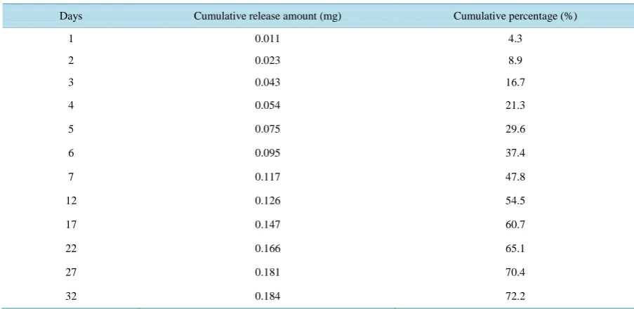
Nanoparticle stability was assessed by quantifying antigen release from the particles over a period of 32 days, with high initial release noted in antigen loaded formulations within the first seven days of incubation (Figure 2). Release amounts during this period were 0.117 mg and reached 47.8% for PLGA-ScpB formulations. Ten days later, there was a moderate continuous cumulative release (Table 1), with a cumulative antigen release of 72.2% noted on the 32nd day.
3.2. Vaccine Efficacy
OALibJ | DOI:10.4236/oalib.1102886 5 August 2016 | Volume 3 | e2886
Figure 1. Scanning electron micrographs of PLGA microspheres incorporating ScpB.
Figure 2. Cumulative release profiles of ScpB from PLGA microspheres.
Table 1. Quantification of antigen release from the particles over a period of 32 days.
Days Cumulative release amount (mg) Cumulative percentage (%)
1 0.011 4.3
2 0.023 8.9
3 0.043 16.7
4 0.054 21.3
5 0.075 29.6
6 0.095 37.4
7 0.117 47.8
12 0.126 54.5
17 0.147 60.7
22 0.166 65.1
27 0.181 70.4
[image:5.595.86.538.500.720.2]OALibJ | DOI:10.4236/oalib.1102886 6 August 2016 | Volume 3 | e2886
agalactiae challenge are shown in Table 2. The first mortalities showed no classical symptoms and were ob-served on day 2 post-challenge, with S.agalactiae successfully isolated from the brain and liver. Three days lat-er, most infected fish showed behavioral changes such as swimming in circles in lateral recumbence, with other clinical signs including opacity of the eyes (unilateral or bilateral) and exophthalmia. The control group (vacci-nated with PBS) had the highest cumulative mortality (up to 55.96%). Group P1 (1 μg/g) had the lowest mortal-ity (12.5%) and the highest RPS (87.66%), while the RPS of groups P3 (3 μg/g) and P5 (5 μg/g) reached 66.80% and 68.01%.
3.3. Lysozyme Activity
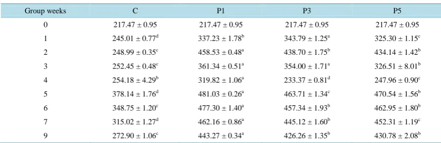
The lysozyme activities from groups P1, P3 and P5 were significant higher than that of control (P < 0.01; Table 3), with relatively higher levels noted 2 weeks post-vaccination followed by a steady decline in subsequent weeks through the fourth week. However, after challenge in the fourth week, the lysozyme activity of groups P1, P3 and P5 rapidly increased to peak in the fifth week. Of these groups, the highest activity levels were noted in group P1 (481.03 ± 0.26; P < 0.01) relative to the other vaccinated groups. In the ninth week, the activity levels for the vaccinated groups were still significantly higher than that of control groups (P < 0.01).
3.4. Superoxide Dismutase Activity
Prior to reaching nine weeks post-vaccination and GBS challenge, SOD activity was examined and found to be significantly higher in the P1, P3, and P5 groups relative to the control groups (P < 0.01; Table 4). In the ninth week, SOD activity was still significantly elevated in groups P1 and P3 relative to the control groups, but not in group P5. The overall trend of SOD activity showed an initial increase followed by a slow decline. In the fourth week post-challenge, the activity initially rose sharply and peaked in the sixth week, with the activity of group P1 being higher than the other groups.
3.5. Enzyme-Linked Immunosorbent Assay (ELISA)
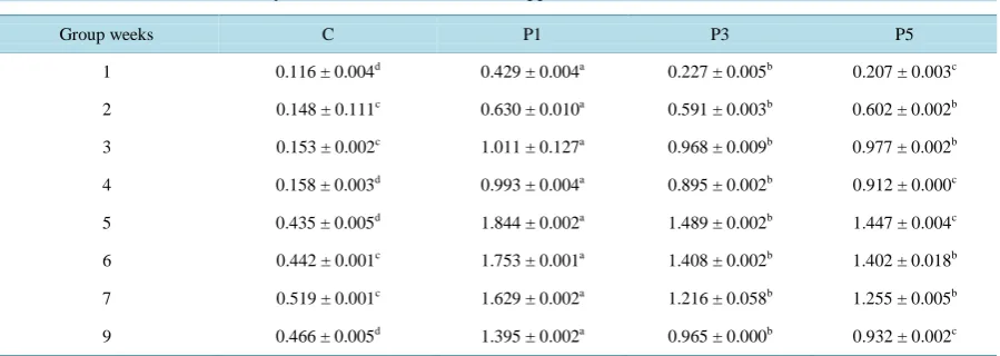
[image:6.595.87.539.453.530.2]After vaccination, the IgM titer levels of group P1, P3 and P5 were significantly elevated relative to the control groups (Table 5). In the third week post-vaccination, the IgM antibody levels for group P1 reached an OD of
Table 2. Immunoprotection of the ScpB vaccine against tilapia Streptococcicosis.
Group Immunizing dose Nos. of fish Nos. of dead fish Mortality (%) RPS (%)
P1 1 μg/g 96 12 12.50 87.66
P3 3 μg/g 113 21 18.58 66.80
P5 5 μg/g 106 19 17.90 68.01
C Control 109 61 55.96 —
Table 3. Kinetics of lysozyme activities in different vaccinated approaches.
Group weeks C P1 P3 P5
0 217.47 ± 0.95 217.47 ± 0.95 217.47 ± 0.95 217.47 ± 0.95
1 245.01 ± 0.77d 337.23 ± 1.78b 343.79 ± 1.25a 325.30 ± 1.15c
2 248.99 ± 0.35c 458.53 ± 0.48a 438.70 ± 1.75b 434.14 ± 1.42b
3 252.45 ± 0.48c 361.34 ± 0.51a 354.00 ± 1.71a 326.51 ± 8.01b
4 254.18 ± 4.29b 319.82 ± 1.06a 233.37 ± 0.81d 247.96 ± 0.90c
5 378.14 ± 1.76d 481.03 ± 0.26a 463.71 ± 1.34c 470.54 ± 1.56b
6 348.75 ± 1.20c 477.30 ± 1.40a 457.34 ± 1.93b 462.95 ± 1.80b
7 315.02 ± 1.27d 462.16 ± 0.86a 445.12 ± 1.60b 452.31 ± 1.19c
9 272.90 ± 1.06c 443.27 ± 0.34a 426.26 ± 1.35b 430.78 ± 2.08b
[image:6.595.87.537.552.698.2]OALibJ | DOI:10.4236/oalib.1102886 7 August 2016 | Volume 3 | e2886
Table 4. Kinetics of SOD activitieses in different vaccinated approaches.
Group weeks C P1 P3 P5
1 29.90 ± 0.11d 61.38 ± 1.39a 57.13 ± 0.20b 54.86 ± 0.70c
2 30.96 ± 0.31d 71.83 ± 0.1a 59.99 ± 0.04b 58.68 ± 0.29c
3 34.70 ± 0.27c 74.82 ± 0.26a 63.88 ± 0.14b 63.38 ± 0.32b
4 36.98 ± 0.09d 64.68 ± 0.39a 55.98 ± 0.41b 54.86 ± 0.49c
5 54.98 ± 0.15d 83.62 ± 0.33a 71.49 ± 0.00b 68.96 ± 0.47c
6 58.29 ± 0.70d 98.16 ± 0.85a 87.56 ± 0.49b 81.60 ± 1.31c
7 46.00 ± 0.33c 63.92 ± 0.08a 58.34 ± 0.31b 59.04 ± 0.65b
9 36.93 ± 0.06c 45.86 ± 0.40a 37.63 ± 0.99b 35.21 ± 0.71c
Notes: The different superscript show extremely significant difference in the same line (P < 0.01), and the same superscript demonstrate insignificant difference (P > 0.05).
Table 5. Kinetics of serum antibody titers in different vaccinated approaches.
Group weeks C P1 P3 P5
1 0.116 ± 0.004d 0.429 ± 0.004a 0.227 ± 0.005b 0.207 ± 0.003c
2 0.148 ± 0.111c 0.630 ± 0.010a 0.591 ± 0.003b 0.602 ± 0.002b
3 0.153 ± 0.002c 1.011 ± 0.127a 0.968 ± 0.009b 0.977 ± 0.002b
4 0.158 ± 0.003d 0.993 ± 0.004a 0.895 ± 0.002b 0.912 ± 0.000c
5 0.435 ± 0.005d 1.844 ± 0.002a 1.489 ± 0.002b 1.447 ± 0.004c
6 0.442 ± 0.001c 1.753 ± 0.001a 1.408 ± 0.002b 1.402 ± 0.018b
7 0.519 ± 0.001c 1.629 ± 0.002a 1.216 ± 0.058b 1.255 ± 0.005b
9 0.466 ± 0.005d 1.395 ± 0.002a 0.965 ± 0.000b 0.932 ± 0.002c
Notes: The different superscript show extremely significant difference in the same line (P < 0.01), and the same superscript demonstrate insignificant difference (P > 0.05).
1.011 ± 0.127, which was significant higher than the other groups (P < 0.05). In the fourth week, the IgM body OD values showed a small decline in all groups. However, in the fifth week post-challenge, the IgM anti-body OD values sharply increased to reach 1.844 ± 0.002. In the next weeks, the values slowly declined, but groups P1, P3 and P5 remained elevated relative to the control groups and group P1 always had a significantly higher value than the other groups.
4. Discussion
ScpB is a surface associated serine protease that plays an important role in the virulence of S.agalactia [10]. It cleaves the chemotactic complement component C5a [20], binds to fibronectin and contributes to cellular inva-sion [21]. While genetic polymorphisms alter the functional activity of C5a peptidase [22], they do not affect its ability to bind fibronectin [23]. ScpB exhibits a conserved nucleotide sequence in several hemolytic streptococ-cal species, is ubiquitously expressed and is lostreptococ-calized to the surface, thus making it a good vaccine candidate for
S.agalactiae and S.pyogenes infections [21] [24].
In this study, the scpB gene was cloned from tilapia S.agalactiae isolates and successfully expressed invivo. ScpB is likely to be membrane-associated or released into extracellular space and thus can serve as a protective immunogen. The obtained subcellular location prediction results were consistent with C5a peptidases located in other sources [12] [25].
[image:7.595.90.540.297.457.2]agalac-OALibJ | DOI:10.4236/oalib.1102886 8 August 2016 | Volume 3 | e2886
tiae is an intracellular pathogen that can survive in phagocytes by subverting bactericidal responses, subunit vaccines should in theory be ineffective gainst a S. agalactiae infection. However, we and other investigators [16] [26] [27] have in several studies observed that some subunit vaccines, when in the form of recombinant proteins purified from E.coli under native conditions and formulated with certain adjuvants, are highly protec-tive against experimental S.agalactiae challenge, with RPS rates around 70% or higher achievable.
PLGA is a degradable polymer microsphere which has been approved by the Food and Drug Administration (FDA), studied extensively for the controlled delivery of therapeutic proteins [28] and has a high potential for gene or protein delivery. While these microspheres are not readily internalized by cells, they are retained in the tissue, thus providing prolonged DNA release [28]. Therefore, the released DNA can transfect the cells at the delivery site with the protein product acting locally or distributed systematically. In this study, recombinant ScpB protein was encapsulated in PLGA and shown to possess good physical performance and sustained release
in vitro. The release amount during the first seven days of incubation was 0.117 mg and reach 47.8% for NP/ScpB formulations. Ten days later, a moderate continuous cumulative release was noted (Table 1), with a cumulative antigen release of 72.2% reached on the 32nd day. This kind of sustained release character for PLGA was also confirmed by other reports [29].
Moreover, ScpB formulated with PLGA afforded a RPS of more than 87% over a period of two months fol-lowing vaccination. Previously, our laboratory showed that Freund’s adjuvant had a strong boosting effect on the specific immunity induced by an S.agalactiae subunit vaccine. However, in order to achieve more than 87% protection when using Freund’s adjuvant, 5 μg/g of recombinant ScpB was necessary. In the present study, only 1 μg/g of recombinant ScpB provided the same level of protection. These findings suggest that PLGA was more effective in providing an improved recombinant ScpB uptake and thus may serve as a more robust adjuvant.
In general, microspheres are too large to enter cells by endocytosis, so instead they can be preferentially taken up by phagocytic cells such as macrophages due to size exclusion [28]. This makes microspheres an ideal DNA carrier for vaccinations or the induction of a cytotoxic T cell response. Furthermore, some microspheres possess bioadhesive properties, making them suitable for mucosal immunizations or oral delivery.
The innate immune defense system is the first line of defense against invading pathogens [30], with lyso-zymes and SODs being an important part of this system. Lysolyso-zymes are catalytic enlyso-zymes able to hydrolyze 1, 4-beta-linkages between N-acetyl-D-glucosamine and N-acetylmuramic acid, which are contained within pepti-dogly can heteropolymers of gram positive bacterial cell walls, and with complement components pertaining to gram negative bacteria also possibly affected [31]-[33]. Lysozymes are abundant in the blood and a number of secretions such as tears, saliva, human milk and mucus. They are also present in cytoplasmic granules of ma-crophages and polymorphonuclear neutrophils (PMNs) [34]. Following the use of a PLGA encapsulated vaccine to inoculate kelp grouper (Epinephelus bruneus), the vaccinated fish sera lysozyme activity was up to 12,000 U/mL, which was significantly higher than levels noted in non-vaccinated or inactivated antigen vaccinated in-dividuals [35]. In the present study, the sera lysozyme activities from the ScpB-PLGA vaccinated groups were significantly higher than those of control groups. These findings suggest that ScpB-PLGA vaccinations can in-crease lysozyme activity and thus antimicrobial activity, and can enhance the ability of tilapia to ward off pa-thogen infections.
SODs are enzymes that alternately catalyze the dismutation of superoxide (O2−) radicals into either ordinary molecular oxygen (O2) or hydrogen peroxide (H2O2). Superoxide is produced as a by-product of oxygen meta-bolism and, if not regulated, causes many types of cell damage [36] [37]. Thus, SODs are an important antioxi-dant defense in nearly all living cells exposed to oxygen and serve as one of the key antioxiantioxi-dant enzymes in fish [38]. While SODs can remove excess free radicals, a dynamic equilibrium may exist between antioxidant levels and the rate of free radical formation. One study found that the SOD activity was closely correlated to immune levels in the body [39] and that it plays an important role in enhancing defense capabilities and immune func-tions in phagocytic cells. In a study that vaccinate grouper (Epinephelusbruneus) with OND-PLGA, serum SOD activity was maintained a high level in 2 - 4 weeks and infections were held at bay [40]. In this study, tilapia vaccinated with ScpB-PLGA showed an increase in serum SOD activity, followed by a later decrease and stabi-lization at the 9th week.
OALibJ | DOI:10.4236/oalib.1102886 9 August 2016 | Volume 3 | e2886 post-immunization, which may correlate to a slow-release as is characteristics of PLGA. The ScpB-PLGA vac-cination provided a 87% survival rate, with a survival rate of only 9% achieved when using ScpB only.
5. Conclusion
In conclusion, the recombinant ScpB encapsulated in PLGA had good immunogenicity against S. agalactiae. These results suggest that PLGA is a more effective than Freund’s adjuvant and enabled a decreased ScpB do-sage for immunization.
Acknowledgements
This work was supported by Key Laboratory of Freshwater Fisheries and Germplasm Resources Utilization Open Project of Ministry of Agriculture (KF201309) and China Agriculture Research System (No. CARS-49).
Conflict of Interest
There is no conflict of interest.References
[1] Chen, M., Li, L.P., Wang, R., Liang, W.W., Huang, Y., Li, J., Lei, A.Y., Huang, W.Y. and Gan, X. (2012) PCR Detec-tion and PFGE Genotype Analyses of Streptococcal Clinical Isolates from Tilapia in China. VeterinaryMicrobiology, 159, 526-530. http://dx.doi.org/10.1016/j.vetmic.2012.04.035
[2] Santillan, D.A., Rai, K.K., Santillan, M.K., Krishnamachari, Y., Salem, A.K. and Hunter, S.K. (2011) Efficacy of Po-lymeric Encapsulated C5a Peptidase-Based Group B Streptococcus Vaccines in a Murine Model. AmericanJournalof ObstetricsandGynecology, 205, 249.e1-249.e8. http://dx.doi.org/10.1016/j.ajog.2011.06.024
[3] Ye, X., Li, J., Lu, M., Deng, G., Jiang, X., Tian, Y., Quan, Y. and Jian, Q. (2011) Identification and Molecular Typing of Streptococcusagalactiae Isolated from Pond-Cultured Tilapia in China. FisheriesScience, 77, 623-632.
http://dx.doi.org/10.1007/s12562-011-0365-4
[4] Evans, J.J., Klesius, P.H. and Shoemaker, C.A. (2004) Efficacy of Streptococcusagalactiae (Group B) Vaccine in Ti-lapia (Oreochromisniloticus) by Intraperitoneal and Bath Immersion Administration. Vaccine, 22, 3769-3773.
http://dx.doi.org/10.1016/j.vaccine.2004.03.012
[5] Pasnik, D.J., Evans, J.J., Panangala, V.S., Klesius, P.H., Shelby, R.A. and Shoemaker, C.A. (2005) Antigenicity of Streptococcusagalactiae Extracellular Products and Vaccine Efficacy. JournalofFishdiseases, 28, 205-212.
http://dx.doi.org/10.1111/j.1365-2761.2005.00619.x
[6] Johri, A.K., Paoletti, L.C., Glaser, P., Dua, M., Sharma, P.K., Grandi, G. and Rappuoli, R. (2006) Group B Strepto-coccus: Global Incidence and Vaccine Development. NatureReviewsMicrobiology, 4, 932-942.
http://dx.doi.org/10.1038/nrmicro1552
[7] Lindahl, G., Stålhammar-Carlemalm, M. and Areschoug, T. (2005) Surface Proteins of Streptococcusagalactiae and Related Proteins in Other Bacterial Pathogens. ClinicalMicrobiologyReviews, 18, 102-127.
http://dx.doi.org/10.1128/CMR.18.1.102-127.2005
[8] Areschoug, T., Stålhammar-Carlemalm, M., Larsson, C. and Lindahl, G. (1999) Group B Streptococcal Surface Pro-teins as Targets for Protective Antibodies: Identification of Two Novel ProPro-teins in Strains of Serotype V. Infectionand Immunity, 67, 6350-6357.
[9] Berner, R., Bender, A., Rensing, C., Forster, J. and Brandis, M. (1999) Low Prevalence of the Immunoglobulin-A- Binding Beta Antigen of the C Protein among Streptococcusagalactiae Isolates Causing Neonatal Sepsis. European JournalofClinicalMicrobiology&InfectiousDiseases, 18, 545-550. http://dx.doi.org/10.1007/s100960050346
[10] Cheng, Q., Debol, S., Lam, H., Eby, R., Edwards, L., Matsuka, Y., Olmsted, S.B. and Cleary, P.P. (2002) Immuniza-tion with C5a Peptidase or Peptidasetype III Polysaccharide Conjugate Vaccines Enhances Clearance of Group B Streptococci from Lungs of Infected Mice. InfectionandImmunity, 70, 6409-6415.
http://dx.doi.org/10.1128/IAI.70.11.6409-6415.2002
[11] Martin, D., Rioux, S., Gagnon, E., Boyer, M., Hamel, J., Charland, N. and Brodeur, B.R. (2002) Protection from Group B Streptococcal Infection in Neonatal Mice by Maternal Immunization with Recombinant Sip Protein. Infectionand Immunity, 70, 4897-4901. http://dx.doi.org/10.1128/IAI.70.9.4897-4901.2002
OALibJ | DOI:10.4236/oalib.1102886 10 August 2016 | Volume 3 | e2886 [13] Heather, C.M., Kelly, S.D. and Victor, N. (2009) Recent Advances in Understanding the Molecular Basis of Group B
Streptococcus Virulence. ExpertReviewsinMolecularMedicine, 3, 1-18.
[14] Dmitriev, A., Tkáciková, L., Suvorov, A., Kantíková, M., Mikula, I. and Totolyan, A. (1999) Comparative Genetic Study of Group B Streptococcal Strains of Human and Bovine Origin. FoliaMicrobiologica, 44, 449-453.
http://dx.doi.org/10.1007/BF02903721
[15] Franken, C., Haase, G., Brandt, C., Weber-Heynemann, J., Martin, S., Lammler, C., Podbielski, A., Lutticken, R. and Spellerberg, B. (2001) Horizontal Gene Transfer and Host Specificity of Beta-Haemolytic Streptococci: The Role of a Putative Composite Transposon Containing scpB and lmb. MolecularMicrobiology, 41, 925-935.
http://dx.doi.org/10.1046/j.1365-2958.2001.02563.x
[16] Li, Q.Y., Ke, X.L., Lu, M.X., Zhu, H.P., Gao, F.Y. and Liu, Z.G. (2014) Prokaryotic Expression and Immunogenicity Analysis of C5a Peptidase (ScpB) of Streptococcusagalactiae Isolated from Tilapia. JournalofFisherySciencesof China, 21, 169-179.
[17] Ogawa, Y., Yamamoto, M., Okada, H., Yashiki, T. and Shimamoto, T. (1988) A New Technique to Efficiently Entrap Leuprolide Acetate into Microcapsules of Polylactic Acid or Copoly (Lactic Glycolic) Acid. Chemical& Pharmaceut-icalBulletin, 36, 1095-1103. http://dx.doi.org/10.1248/cpb.36.1095
[18] Panyam, J., Dali, M.M., Sahoo, S.K., Ma, W., Chakravarthi, S.S., Amidon, G.L., Levy, R.J. and Labhasetwar, V. (2003) Polymer Degradation and inVitro Release of a Model Protein from Poly(D, L-Lactide-co-Glycolide) Nano- and Mi-croparticles. JournalofControlledRelease, 92, 173-187. http://dx.doi.org/10.1016/S0168-3659(03)00328-6
[19] Amend, D.F. (1981) Potency Testing of Fish Vaccines. International Symposium on Fish Biologics: Serodiagnostics and Vaccines. DevelopmentsinBiologicalStandardization, 49, 447-454.
[20] Bohnsack, J.F., Mollison, K.W., Buko, A.M., Ashworth, J.C. and Hill, H.R. (1991) Group B Streptococci Inactivate Complement Component C5a by Enzymic Cleavage at the C-Terminus. BiochemicalJournal, 273, 635-640.
http://dx.doi.org/10.1042/bj2730635
[21] Cheng, Q., Stafslien, D., Purushothaman, S.S. and Cleary, P. (2002) The Group B Streptococcal C5a Peptidase Is Both a Specific Protease and an Invasin. InfectionandImmunity, 70, 2408-2413.
http://dx.doi.org/10.1128/IAI.70.5.2408-2413.2002
[22] Bohnsack, J.F., Takahashi, S., Hammitt, L., Miller, D.V., Aly, A.A. and Adderson, E.E. (2000) Genetic Polymor-phisms of Group B StreptococcusscpB Alter Functional Activity of a Cell-Associated Peptidase That Inactivates C5a. InfectionandImmunity, 68, 5018-5025. http://dx.doi.org/10.1128/IAI.68.9.5018-5025.2000
[23] Tamura, G.S., Hull, J.R., Oberg, M.D. and Castner, D.G. (2006) High Affinity Interaction between Fibronectin and the Group B Streptococcal C5a Peptidase Is Unaffected by a Naturally Occurring Four-Amino-Acid Deletion That Elimi-nates Peptidase Activity. InfectionandImmunity, 74, 5739-5746. http://dx.doi.org/10.1128/IAI.00241-06
[24] Santillan, D.A., Andracki, M.E. and Hunter, S.K. (2008) Protective Immunization in Mice against Group B Strepto-cocci Using Encapsulated C5a Peptidase. AmericanJournalofObstetricsandGynecology, 198, 114.e1-114.e6.
http://dx.doi.org/10.1016/j.ajog.2007.06.003
[25] Cleary, P.P., Matsuka, Y.V., Huynh, T., Lam, H. and Olmsted, S.B. (2004) Immunization with C5a Peptidase from Ei-ther Group A or B Streptococci Enhances Clearance of Group A Streptococci from Intranasally Infected Mice. Vaccine, 22, 4332-4341. http://dx.doi.org/10.1016/j.vaccine.2004.04.030
[26] Li, J., Ye, X., Ke, X.L., Lu, M.X., Chi, Y.Y. and Tian, Y.Y. (2012) Cloning, Expression and Immunogenicity Analysis of Surface Immunogenic Protein (Sip) of Tilapia Streptococcusagalactiae. ActahydrobiologicaSinica, 36, 626-633.
[27] Jia, P., Hu, Y., Heng, C., Sun, B., Yu, W. and Sun, L. (2013) Comparative Study of Four Flagellins of Vibrio anguil-larum: Vaccine Potential and Adjuvanticity. Fish&ShellfishImmunology, 34, 514-520.
http://dx.doi.org/10.1016/j.fsi.2012.11.039
[28] Agarwal, A. and Mallapragada, S.K. (2008) Synthetic Sustained Gene Delivery Systems. CurrentTopicsinMedicinal Chemistry, 8, 311-330. http://dx.doi.org/10.2174/156802608783790965
[29] Stevenson, C.L., Santini, J.T. and Langer, R. (2012) Reservoir-Based Drug Delivery Systems Utilizing Microtechnol-ogy. AdvancedDrugDeliveryReviews, 64, 1590-1602. http://dx.doi.org/10.1016/j.addr.2012.02.005
[30] Magnadóttir, B. (2006) Innate Immunity of Fish (Overview). Fish&ShellfishImmunology, 20, 137-151.
http://dx.doi.org/10.1016/j.fsi.2004.09.006
[31] Yano, T. (1996) The Nonspecific Immune System: Humoral Defense. In: Iwama, G. and Nakanishi, T., Eds., TheFish ImmuneSystem, Academic Press, San Diego, 106-157.
[32] Paulsen, S.M., Engstad, R.E. and Robertsen, B. (2001) Enhanced Lysozyme Production in Atlantic Salmon (Salmosa-lar L.) Macrophages Treated with Yeast Beta-Glucan and Bacterial Lipopolysaccharide. Fish&ShellfishImmunology, 11, 23-37. http://dx.doi.org/10.1006/fsim.2000.0291
OALibJ | DOI:10.4236/oalib.1102886 11 August 2016 | Volume 3 | e2886
http://dx.doi.org/10.1126/science.165.3892.454
[34] Saurabh, S. and Sahoo, P.K. (2008) Lysozyme: An Important Defence Molecule of Fish Innate Immune System. Aq-uacultureResearch, 39, 223-239. http://dx.doi.org/10.1111/j.1365-2109.2007.01883.x
[35] Harikrishnan, R., Balasundaram, C. and Heo, M.S. (2012) Poly D, L-Lactide-co-Glycolic Acid (PLGA)-Encapsulated Vaccine on Immune System in Epinephelusbruneus against Uronemamarinum. ExperimentalParasitology, 131, 325- 332. http://dx.doi.org/10.1016/j.exppara.2012.04.017
[36] Tohru, F. and Masuko, U. (2011) Superoxide Dismutases: Role in Redox Signaling, Vascular Function, and Diseases. Antioxidants&RedoxSignaling, 15, 1583-1606. http://dx.doi.org/10.1089/ars.2011.3999
[37] Garry, R.B. (2011) Superoxide Dismutase in Redox Biology: The Roles of Superoxide and Hydrogen Peroxide. An-ti-CancerAgentsinMedicinalChemistry, 11, 341-346. http://dx.doi.org/10.2174/187152011795677544
[38] Gaëlle, R., Cédric, L.B., Fabienne, G., Christophe, L. and Christine, P. (2015) Immune Responses of Phenoloxidase and Superoxide Dismutase in the Manila Clam Venerupis philippinarum Challenged with Vibrio tapetis—Part II: Combined Effect of Temperature and Two V. Tapetis Strains. Fish&ShellfishImmunology, 44, 79-87.
http://dx.doi.org/10.1016/j.fsi.2014.12.039
[39] Cédric, L.B., Gaëlle, R., Christine, P., Christophe, L., Catherine, S., Olivier, G., Fabrice, P. and Fabienne, G. (2015) Immune Responses of Phenoloxidase and Superoxide Dismutase in the Manila Clam Venerupisphilippinarum Chal-lenged with Vibriotapetis—Part I: Spatio-Temporal Evolution of Enzymes’ Activities Post-Infection. Fish&Shellfish Immunology, 42, 16-24. http://dx.doi.org/10.1016/j.fsi.2014.10.021
[40] Harikrishnan, R., Balasundaram, C. and Heo, M.S. (2012) Poly D, L-Lactide-co-Glycolic Acid-Liposome Encapsu-lated ODN on Innate Immunity in Epinephelusbruneus against Vibrioalginolyticus. VeterinaryImmunologyand Im-munopathology, 147, 77-85. http://dx.doi.org/10.1016/j.vetimm.2012.04.008
Submit or recommend next manuscript to OALib Journal and we will provide best service for you:
Publication frequency: Monthly
9 subject areas of science, technology and medicine Fair and rigorous peer-review system
Fast publication process
Article promotion in various social networking sites (LinkedIn, Facebook, Twitter, etc.) Maximum dissemination of your research work