COGNITIVE LINGUISTIC ABILITIES
Girija, P.C
Department Audiology and
A R T I C L E I N F O
INTRODUCTION
“Cognitive linguistics” is the study of language in its cognitive function, where cognitive refers to the crucial role of intermediate informational structures with our encounters with the world The role of subcortical structures in language function is complex and dependent on language task, with studies increasingly showing subcortical involvement for the production of formulaic language, including recited speech (Arsalidou, Duerden, & Taylor, 2013).
The basal ganglia have traditionally been viewed as m processing nuclei. However, functional neuroimaging evidence has implicated these structures in more complex cognitive and affective processes that are fundamental for a range of human activities. The results of anatomical studies indicated that the basal ganglia participate in multiple circuits or “loops cognitive areas of the cerebral cortex. The activity of neurons within selected portions of the basal ganglia is more related to cognitive or sensory operations than to motor functions. In some instances basal ganglia lesions cause primarily cognitive or sensory disturbances without gross motor impairments (Middleton & Strick, 2000). Extensive evidence now indicates a role for the basal ganglia, in particular the dorsal striatum, in learning and memory (Packard & Knowlton, 2002). The evidence of fMRI which indicated that the components of a left pre-SMA-dorsal caudate nucleus-ventral anterior thalamic loop were active during word generation from rhyming or
International Journal of Current Advanced Research
ISSN: O: 2319-6475, ISSN: P: 2319-6505,
Available Online at www.journalijcar.org
Volume 7; Issue 10(C); October 2018
DOI: http://dx.doi.org/10.24327/ijcar.2018
Copyright©2018 Girija, P.C et al. This is an open access article distributed under the Creative Commons Attribution License, which permits
unrestricted use, distribution, and reproduction in any medium, provided the original work is properly cited.
Article History:
Received 12th July, 2018
Received in revised form 23rd August, 2018
Accepted 7th September, 2018 Published online 28th October, 2018
Key words:
Cognition; Subcortex; Claustrum; Functional Dichotomy
*Corresponding author: Girija, P.C
Department Audiology and Speech Language Pathology, AWH Special College, Calicut, Kerala University of Health Sciences
ISTIC ABILITIES-A COMPARISON BETWEEN RIGHT
AND LEFT SUBCORTEX
Girija, P.C., Sruthy, R and Nayana, N
Audiology and Speech Language Pathology, AWH Special College Kerala University of Health Sciences
A B S T R A C T
The functional dichotomy at the level of subcortex is debatable in terms of cognitive linguistic processes. The current study attempts to resolve these queries through in depth analysis of cognitive linguistic functions by administering CLAP
right subcortical lesion and 15 patients with left subcortical lesion. The results evinced significantly poorer performance by participants with right subcortical lesion. This astounding finding can be accredited to the presence of robust contr
right subcortex to left prefrontal cortex through medial claustrum.
“Cognitive linguistics” is the study of language in its cognitive function, where cognitive refers to the crucial role of intermediate informational structures with our encounters with The role of subcortical structures in language mplex and dependent on language task, with studies increasingly showing subcortical involvement for the production of formulaic language, including recited speech
The basal ganglia have traditionally been viewed as motor processing nuclei. However, functional neuroimaging evidence has implicated these structures in more complex cognitive and affective processes that are fundamental for a range of human activities. The results of anatomical studies indicated that the asal ganglia participate in multiple circuits or “loops‟ with cognitive areas of the cerebral cortex. The activity of neurons within selected portions of the basal ganglia is more related to cognitive or sensory operations than to motor functions. In instances basal ganglia lesions cause primarily cognitive or sensory disturbances without gross motor impairments (Middleton & Strick, 2000). Extensive evidence now indicates a role for the basal ganglia, in particular the dorsal striatum, in memory (Packard & Knowlton, 2002). The evidence of fMRI which indicated that the components of a ventral anterior thalamic loop were active during word generation from rhyming or
category cues (Crosson et. al., 2003). The findings of Tinaz, Schendan, Schon, & Stern, 2006 suggested that circuits involving the frontal lobe and basal ganglia output nuclei are important for picture sequencing and more generally for the sequential ordering of events.
Extensive research has not been yet established regarding the involvement of subcortical structures in language and cognition. The evidence from clinical population with subcortical lesions reveals that even after being diagnosed as non-aphasic, they still exhibit metalinguistic deficits. Hence, it is highly essential to evaluate these skills in depth.
Another grey area is the dichotomy between the right and left subcortical structures in higher cognitive linguistic processing. A noteworthy study was done by Milardi, Bramanti, Milazzo, Finocchio, Arrigo, Santoro, Trimarchi, Quartarone, Anastasi1, and Gaeta, 2015. They used Constrained Spherical Deconvolution (CSD) tractography
connectivity in neurotypical brain. The images displayed ipsilataral as well as contralateral connections between the prefrontal cortex with right and left subcortex through interconnected bundles of medial claustral pathways. This discovery provided a solid base for hypothesizing that, performance of individuals with right subcortical lesion could be poorer in comparison to that of left subcortical lesion, in cognitive linguistic tasks. The current study attempts at throwing light on these unexplored areas.
Aim
To analyze cognitive linguistic functions across right and left subcortical lesion using
CLAP-International Journal of Current Advanced Research
6505, Impact Factor: 6.614
www.journalijcar.org
2018; Page No.15904-15909
//dx.doi.org/10.24327/ijcar.2018.15909.2918
This is an open access article distributed under the Creative Commons Attribution License, which permits unrestricted use, distribution, and reproduction in any medium, provided the original work is properly cited.
Department Audiology and Speech Language Pathology, Kerala University of Health
A COMPARISON BETWEEN RIGHT
Special College, Calicut,
The functional dichotomy at the level of subcortex is debatable in terms of cognitive linguistic processes. The current study attempts to resolve these queries through in depth analysis of cognitive linguistic functions by administering CLAP-M on 15 participants with right subcortical lesion and 15 patients with left subcortical lesion. The results evinced significantly poorer performance by participants with right subcortical lesion. This astounding finding can be accredited to the presence of robust contralateral connections of right subcortex to left prefrontal cortex through medial claustrum.
., 2003). The findings of Tinaz, Schendan, Schon, & Stern, 2006 suggested that circuits involving the frontal lobe and basal ganglia output nuclei are r picture sequencing and more generally for the
Extensive research has not been yet established regarding the involvement of subcortical structures in language and cognition. The evidence from clinical population with tical lesions reveals that even after being diagnosed as aphasic, they still exhibit metalinguistic deficits. Hence, it is highly essential to evaluate these skills in depth.
Another grey area is the dichotomy between the right and left structures in higher cognitive linguistic processing. A noteworthy study was done by Milardi, Bramanti, Milazzo, Finocchio, Arrigo, Santoro, Trimarchi, Quartarone, Anastasi1, and Gaeta, 2015. They used Constrained Spherical Deconvolution (CSD) tractography to evaluate claustral connectivity in neurotypical brain. The images displayed ipsilataral as well as contralateral connections between the prefrontal cortex with right and left subcortex through interconnected bundles of medial claustral pathways. This iscovery provided a solid base for hypothesizing that, performance of individuals with right subcortical lesion could be poorer in comparison to that of left subcortical lesion, in cognitive linguistic tasks. The current study attempts at
these unexplored areas.
To analyze cognitive linguistic functions across right and left -M
Research Article
Method
Participants
30 participants constituted the experimental group, out of which 15 had right subcortical lesion and 15 left subcortical lesion assessed at 6 months to 1 year after the onset of stroke as confirmed by Neurologist and Radiologist
The participants with right (group A) and left (group B) subcortical lesion without cortical involvement assessed at 6 months to 1 year after the onset of stroke as confirmed by Neurologist and Radiologist.
The participants had no history of traumatic brain injury.
The medium of instruction of selected participants was in Malayalam.
Participants were able to speak, read and write Malayalam with a minimum of primary school education.
The participants selected for the study were right-handed.
Participants, who were diagnosed as Non-aphasic by a Speech language pathologist through administering Western Aphasia Battery (Kertesz, 1979), were considered for the study.
Materials
Cognitive Linguistic Assessment Protocol-Malayalam
The cognitive linguistic abilities of both group were assessed using CLAP-M (Cognitive Linguistic Assessment Protocol-Malayalam) developed by Sadia (2009)
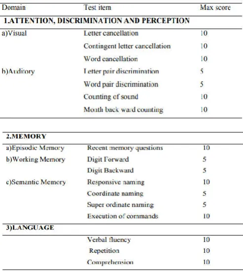
Table 1 indicates the list of domains with its individual test items and their respective scoring as given in CLAP-M.
Table 1Assessment protocol including domains, test items and scoring
Domain 1: Attention, Perception and Discrimination
a) Visual category
1. The cancellation of letter: The cancellation of all pointed letters is the task. This is a task that requires sustained attention.
2. The cancellation of words: It is also sustained attention task .Procedure is same as above.
3. Contingent cancellation: This evaluates the selective cancellation. A pre-requisite contingency before the cancellation is necessary.
4. Scoring: a scoring of one point was given for each correct response.
b) Auditory category
1. Letter pair discrimination:This set is a discrimination predominant task. The discrimination ability of participants for the pair of letters read out by the clinician is assessed. It can be either same/different. 2. Word pair discrimination:This set is a discrimination
predominant task. Procedure is same as discussed above.
3. Auditory sound count: Sustained auditory attention was evaluated by making the participant mentally count how many times a particular letter is repeated in the list.
4. Month backward naming: Patient task was to recite the names of the month in the backward direction (i.e. December to January). This last subtest test requires attention and checks the recall ability.
Scoring: a score of one point was given for each correct response.
Domain 2: Memory
The main processes tested in the domain are:
a) Episodic memory: was tested by asking questions
that tested orientation of self with respect to place, self and time and also few questions of general knowledge.
b) Working memory: was evaluated using digit forward and digit backward repetition task .A maximum of seven digits were included in the list.
Scoring: A score of one was given if all the digits are repeated in correct order.
c) Semantic memory: The following tasks were
included under this category.
1. Responsive naming: The subject was asked to name the target word on which the description has been provided.
2. Co-ordinate naming: The subject was asked to name at least 5 items in a noun class provided.
3. Super ordinate naming: Subject task to identify the class to which the list of items provided belongs. 4. Execution of commands:Two objects like a book and
a pencil were placed in front of the subject. Commands of various levels of complexities which required manipulation of these objects were given.
Scoring: Each item scores one point for the correct answer except execution of commands.
Domain 3: Language
This test include various subtests that evaluate the language functioning.
a. Verbal fluency: This task evaluated the recall ability of the subject and was asked to repeat at least 5 words beginning with a specified letter.
b. Repetition: The repetition subtask included various complex sentences that have to be repeated by the client and the complexity of the sentences was increased. c. Comprehension: Here the client was asked to read the
given passage and answer the questions below that.
Scoring: Each correct answer was provided with two points, except repetition subtask.
Domain 4: Problem solving
The domain tests the reasoning ability that aid in problem solving. The following tests were considered.
a. Sentence formulation: This was a word order unscrambling tasks and the subject was asked to form a grammatically correct sentence.
b. Compare and contrast:The subject task was to identify a similarity and difference between the pair of objects named.
c. Wh-questions: Patient task was to answer the why questions.
d. Sentence disambiguation: Ambiguous sentences were provided to the subject and they were instructed to explain the two interpretations that can be inferred from the sentences.
e. Predicting outcome: Subject was instructed to predict outcome of the described situation.
f. Predicting cause: Task was to predict cause of the described situation.
Scoring: Each task was scored one point for the correct
answer. In sentence disambiguation, each interpretation was scored separately.
Domain 5: Visospatial skills
a. Clock drawing:Subject task was to predict the time in clock, to draw out the time mentioned in the clock and also to draw a clock and mark the given time. This checks the visuospatial ability.
Scoring: All these were timed tasks.1 point for each correct drawing within time.
b. Mazes: To find the way out from the box was the subject task.
Scoring: Scoring was done based on complexity as well as time required for completion.
c. Copying:Subject task was to copy as many pictures as possible from the list provided within the given time.
Scoring: 1 point provided for each picture correctly copied with the time limit.
d. Matching: Consists of two types. In the first one the subject had to match each picture of one side with the target picture on the other side. In the second, subject had to match one picture to two target pictures.
Scoring: One point for each correctly matched picture.
Domain 6: Organization
a. Sequencing events: Formation of stories to check the sequential ability.
Scoring: Each story is separately scored. They are timed tasks. b. Categorization: This check the organization ability of
word class.
c. Analogies: Recognition ability of word concept is measured.
Scoring:one point for each correct answer was given.
Procedure
Data collection
Informed consent
Formal informed consent was obtained from their family members, prior to the testing after explaining to them the purpose and nature of study (Informed Consent -see appendix-3).
Case history
Medical records including objective evaluation reports (MRI) of the experimental group were reviewed and detailed information about each participant was obtained after interviewing the subject and family members.
The participant’s speech and language skills were assessed using Western Aphasic Battery (WAB) and Frenchay Dysarthria Assessment (FDA)
Administration of CLAP-M: The participants (Group A and
B) were made to sit comfortably in a quiet room and were suitably instructed to. An average time taken for administration was nearly 3 ½ hours including 10 to 15 minutes break after each 45 minutes of administration. Scoring was done simultaneously along with the test.
Data analysis
noted down and analyzed. The general pattern of response in the group was also noted.
RESULTS AND DISCUSSION
Domain 1: Attention, Discrimination and Perception
Table 2 shows the mean and standard deviation scores of the domain attention, discrimination and perception in participants with right and left subcortical lesions.
Table 2 Mean and standard deviation scores for right subcortical lesion and left subcortical lesion with CLAP-M
subsection Attention, discrimination and perception
Figure 2 represents the mean scores of participants with right and left subcortical lesion.
Figure 1 Mean score obtained by participants with right and left subcortical
lesion on Domain 1
The mean difference of Attention, discrimination and perception for right subcortical lesion and left subcortical lesion was analyzed using t-test for mean difference. The table 3 indicated the results.
Table 3 shows the t-test results for domain 1 for participants with right and left subcortical lesion
Table 3 t-test results for mean difference of domain 1 for participants with right and left subcortical lesion
The results indicated that right subcortical lesion group performed poorer than left subcortical lesion even though the differences were not statistically significant. The poorer performance of group A could be attributed to the effect of weakened ipsilateral connections of right subcortical structures with their right cortical counterparts. Also adding to this, it can be postulated that the damage to right subcortical regions could impede the activation of right cortex thereby resulting in poorer attention, alertness and motivation. This finding can be correlated with the investigations of Hillis, Newhart, Heidler,
Barker, Herskovits, and Degaonkar, 2005, where they reported the presence of high spatial inattention due to right subcortical lesion.
Domain 2: Memory
Table 4 shows the mean difference and standard deviation for the domain memory for participants with right and left subcortical lesion while figure 2 represents the same aspect graphically
Table 4 Mean and standard deviation for Domain 2 for participants with right and left subcortical lesion
Figure 2 Mean score obtained by participants with right and left subcortical
lesion on Domain 2.
The mean difference of Memory for right subcortical lesion and left subcortical lesion was analyzed using t-test for mean difference. The table 5 indicated the results.
Table 5 t-test results for mean difference of domain 2 for participants with right and left subcortical lesion
Table 5 indicated that t-value is 6.45with p-value <0.01, which indicated there was significant difference between right and left subcortical lesion for Memory.
The present findings revealed that the individuals with right subcortical lesion performed significantly poorer than individuals with left subcortical lesion. The poor performance of the experimental group A (Right subcortical lesion) could be due to the presence of strong contralateral connections of right basal ganglia with left hemisphere. This can be supported with the findings of Milardi D et al (2013) who proved that strong bilateral connections of basal ganglia with contralateral frontal cortex through medial pathways of claustrum.
Domain 3: Language
Table 6 Mean and standard deviation for Domain 3 for participants with right and left subcortical lesion
Figure 3 Mean score obtained by participants with right and left subcortical
lesion on Domain 3.
The mean difference of language for right subcortical lesion and left subcortical lesion was analyzed using t-test for mean difference. The table 7 indicated the results.
Table 7 t-test results for mean difference of domain 3 for participants with right and left subcortical lesion
Results revealed that there was a significant difference between right and left subcortical lesion for Language functions, thereby substantiating the existence of active participation of right subcortical structures. Similar results were obtained by Crosson, B et al (2003)
Domain 4: Problem Solving
Mean and standard deviation scores for right subcortical lesion and left subcortical lesion with CLAP-M subsection Problem Solving are displayed in table 8
Table 8 Mean and standard deviation for Domain 4 for participants with right and left subcortical lesion
Figure 4 represents the mean score for both the group graphically.
Figure 4 Mean score obtained by participants with right and left subcortical
The mean difference of problem solving for right subcortical lesion and left subcortical lesion was analyzed using t-test for mean difference. The table 9 indicates the results.
Table 9 t-test results for mean difference of domain 4 for participants with right and left subcortical lesion.
The significant difference in performance between group A and B ascertain that Right subcortical structures plays an integral role in cognitive functions through their robust contralateral connection to the left prefrontal cortex. The hindrance in this connection could constrain the high level activation required for language related cognitive tasks
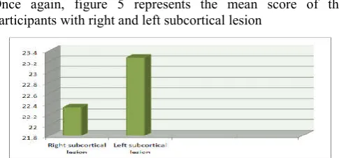
Domain 5: Visuospatial skills
Mean and standard deviation scores for right subcortical lesion and left subcortical lesion with CLAP-M subsection visuospatial skills are displayed in table 10 as well as figure 5.
Table 10 Mean and standard deviation for Domain 5 for participants with right and left subcortical lesion.
Figure 5 Mean score obtained by participants with right and left subcortical
lesion on Domain 5.
The mean difference of visuospatial skills for right subcortical lesion and left subcortical lesion was analyzed using t-test for mean difference. The table 11 indicated the results
Table 11 t-test results for mean difference of domain 5 for participants with right and left subcortical lesion.
Domain 6: Organization
Mean and standard deviation scores for right subcortical lesion and left subcortical lesion with CLAP-M subsection Organization displayed in table 12.
Table 12 Mean and standard deviation for Domain 6 for participants with right and left subcortical lesion.
Once again, figure 5 represents the mean score of the participants with right and left subcortical lesion
Figure 5 Mean score obtained by participants with right and left subcortical
lesion on Domain 5.
The mean difference of organization for right subcortical lesion and left subcortical lesion was analyzed using t-test for mean difference. The table 13 indicated the results.
Table 13 t-test results for mean difference of domain 5 for participants with right and left subcortical lesion.
The task included in the domain Organization required the activation of medial prefrontal cortex, retrosplenial cortex, and angular gyrus, as well as on striatal areas including the caudate nucleus and putamen. The poor performance of the group A once again establishes the strong contralateral connection of right subcortical structures with left prefrontal cortex.
CONCLUSION
Results of the current investigations could conclude that right subcortical structures have prominent role in governing cognitive linguistic processes via contralateral claustral connections to the left prefrontal cortex.
Any lesion at the level of right subcortex can hinder the flow of information to the left prefrontal cortex which will be manifested as poor performance in tasks which require intact cognitive linguistic abilities. Hence the existing notion that, cognitive linguistic functions are exclusively under the dominance of left subcortex, can be challenged through our findings.
Reference
Arsalidou, M., Duerden, E. G., & Taylor, M. J. (2013). The centre of the brain: topographical model of motor, cognitive, affective, and somatosensory functions of the basal ganglia. Human brain mapping, 34(11), 3031-3054.
Crosson, B., Benefield, H., Cato, M. A., Sadek, J. R., Moore, A. B., Wierenga, C. E., & GÖkÇay, D. (2003). Left and right basal ganglia and frontal activity during language generation: contributions to lexical, semantic, and phonological processes. Journal of the International Neuropsychological Society, 9(7), 1061-1077
Middleton, F. A., & Strick, P. L. (2000). Basal ganglia output and cognition: evidence from anatomical, behavioral, and clinical studies. Brain and cognition,
42(2), 183-200.
Milardi, D., Bramanti, P., Milazzo, C., Finocchio, G., Arrigo, A., Santoro, G., ...& Gaeta, M. (2013). Cortical and subcortical connections of the human claustrum revealed in vivo by constrained spherical deconvolution tractography. Cerebral Cortex, 25(2), 406-414.
Packard, M.G., & Knowlton, B. J. (2002). Learning and memory functions of the Basal Ganglia. Annual Review
of Neuroscience, 25, 563-593.
doi:10.1146/annurev.neuro.25.112701.142937
Tinaz, S., Schendan, H. E., Schon, K., & Stern, C. E. (2006). Evidence for the importance of basal ganglia output nuclei in semantic event sequencing: an fMRI study.
Brain research, 1067(1), 239-249.
How to cite this article:
Girija, P.C., Sruthy, R and Nayana, N (2018) 'Cognitive Linguistic Abilities-a Comparison between Right and left subcortex',
International Journal of Current Advanced Research, 07(10), pp. 15904-15909. DOI: http://dx.doi.org/10.24327/ijcar.2018.15909.2918