EFFICIENCY OF TENS IN CONTROLLING PAIN DURING SEPARATION USING TWO
DIFFERENT ORTHODONTIC SEPARATORS
Kala Vani S.V., Jagathesh
Sunil Krishna Kumar
Department of Orthodontics and Dentofacial Orthopedics, Institute of Dental
A R T I C L E I N F O
INTRODUCTION
Pain is perhaps even older than mankind. There is a reason to believe that it is inherent in any life linked with consciousness. Evidence indicates that man has suffered this affliction since his beginning, for one finds testimony to the existence of pain in the chronicles of all races (Fulop Miller) [1]. The International Association of the Study of Pain has defined pain as "an unpleasant sensory and emotional experience with actual or potential tissue damage or described in terms of such damage"[2].Orthodontic tooth movement requires application of force to the tooth which generally causes pain. The general perception of patients is that orthodontic treatment and pain are inseparable and go hand in hand. There are reports that one of the discouraging factors for seeking orthodontic treatment is the individual's fear for the related pain and discomfort [3
It is imperative that pain control during orthodontic treatment should be considered an important aspect
mechanotherapy. The control of pain in orthodontic treatment should include adjusting the forces to a level below the pain threshold. Unfortunately such low forces would have very little if any effect on the tooth movement. To alleviate the and discomfort, clinicians have tried different approaches.
International Journal of Current Advanced Research
ISSN: O: 2319-6475, ISSN: P: 2319-6505,
Available Online at www.journalijcar.org
Volume 8; Issue 04 (A); April 2019; Page No.
DOI: http://dx.doi.org/10.24327/ijcar.201
Copyright©2019 Amber B, Syed Baasit and Kaisar
License, which permits unrestricted use, distribution, and reproduction in any medium, provided the original work is properly
Article History:
Received 15th January, 2019
Received in revised form 7th February, 2019
Accepted 13th March, 2019
Published online 28th April, 2019
Key words:
Transcutaneous electrical nerve stimulation, Separators, Visual analog scale
*Corresponding author: Kala Vani S.V
Department of Orthodontics and Dentofacial Orthopedics, C.K.S. Theja Institute of Dental Sciences and Research, Tirupati
EFFICIENCY OF TENS IN CONTROLLING PAIN DURING SEPARATION USING TWO
DIFFERENT ORTHODONTIC SEPARATORS
Kala Vani S.V., Jagathesh Natarajan, Madhavi O., Hima Bindu S.,
Sunil Krishna Kumar P and Ramesh P
Department of Orthodontics and Dentofacial Orthopedics, C.K.S. Theja Institute of Dental Sciences and Research, Tirupati
A B S T R A C T
Pain is the most commonly cited negative effect of orthodontic treatment and is of major concern to patients as well as clinicians. Pain control during orthodontics is therefore an important aspect of patient compliance. The present study was carried out t
efficiency of transcutaneous electrical nerve stimulation (TENS) in controlling pain during orthodontic separation. Thirty subjects were equally assigned to three groups
group, extraoral group, and control group. In each group, Kesl
mesial and distal to the left maxillary first molar and elastomeric separators on the right side. A scientific medical system physio-multi TENS STIM
electric current. Visual analog scale (VAS) was used to assess the pain. In all the 3 groups left sides (Kesling separator) mean VAS score was more than the right side (elastomeric
separator) means VAS score. There was no statistically significant difference between
intraoral TENS and extraoral TENS group. The TENS sub
pain than the control subjects. The present study suggests that TENS is an effective non pharmacologic method of controlling post- separation orthodontic tooth pain.
Pain is perhaps even older than mankind. There is a reason to believe that it is inherent in any life linked with consciousness. Evidence indicates that man has suffered this affliction since one finds testimony to the existence of pain in the chronicles of all races (Fulop Miller) [1]. The International Association of the Study of Pain has defined pain as "an unpleasant sensory and emotional experience with r described in terms of such [2].Orthodontic tooth movement requires application of force to the tooth which generally causes pain. The general perception of patients is that orthodontic treatment and pain are reports that one of the discouraging factors for seeking orthodontic treatment is the individual's fear for the related pain and discomfort [3-6].
It is imperative that pain control during orthodontic treatment should be considered an important aspect of orthodontic mechanotherapy. The control of pain in orthodontic treatment should include adjusting the forces to a level below the pain threshold. Unfortunately such low forces would have very little if any effect on the tooth movement. To alleviate the pain
clinicians have tried different approaches.
Anti- inflammatory drugs remain the most preferred method but lack of an appropriate protocol for their administration after orthodontic appointments is considered to be a major drawback requiring attention in future research. The non pharmacological approaches include, having p
on something fairly hard for example
analgesic chewing gums, trnascutaneous electrical nerve stimulation (TENS), low-level laser therapy (LLLT), vibratory stimulation and magnetic force field [7
Orthodontic separation forms the first step in fixed orthodontic mechanotherapy wherein space is created mesial and distal to the first molars to accommodate bands. Placement of separators (brass wire, elastomeric, spring type steel separators) results in painful experience in almost all the patients. Although many non
available, one non - invasive, cost effective method of pain control is TENS. Unfortunately, few clinical studies have evaluated the use of electrical stimulation
procedures and some of these have been conducted on an empirical basis. Even though, Acetaminophen is a widely used analgesic during orthodontic treatment as it doesn’t interfere with orthodontic tooth movement, there is now increasing a convincing epidemiological evidence from a range of independent studies implicating paracetamol use in the etiology of asthma and other allergic
putative risk factor not only for the development of asthma due to bronchospasmodic action, but also causes anaphylactic
International Journal of Current Advanced Research
6505, Impact Factor: 6.614
www.journalijcar.org
; Page No.18055-18060
//dx.doi.org/10.24327/ijcar.2019.18060.3442
Amber B, Syed Baasit and Kaisar Ahmad. This is an open access article distributed under the Creative Commons Attribution License, which permits unrestricted use, distribution, and reproduction in any medium, provided the original work is properly
Department of Orthodontics and Dentofacial Orthopedics, C.K.S. Institute of Dental Sciences and Research, Tirupati
EFFICIENCY OF TENS IN CONTROLLING PAIN DURING SEPARATION USING TWO
Natarajan, Madhavi O., Hima Bindu S.,
C.K.S. Theja
of orthodontic treatment and is of major concern to patients as well as clinicians. Pain control during orthodontics is therefore an important aspect of patient compliance. The present study was carried out to assess the efficiency of transcutaneous electrical nerve stimulation (TENS) in controlling pain during orthodontic separation. Thirty subjects were equally assigned to three groups -intraoral group, extraoral group, and control group. In each group, Kesling separators were placed mesial and distal to the left maxillary first molar and elastomeric separators on the right multi TENS STIM -2CH was used to deliver the d to assess the pain. In all the 3 groups left sides (Kesling separator) mean VAS score was more than the right side (elastomeric statistically significant difference between intraoral TENS and extraoral TENS group. The TENS subjects reported significantly less pain than the control subjects. The present study suggests that TENS is an effective
non-separation orthodontic tooth pain.
s remain the most preferred method but lack of an appropriate protocol for their administration after orthodontic appointments is considered to be a major drawback requiring attention in future research. The non-pharmacological approaches include, having patients to chew on something fairly hard for example – a plastic wafer, analgesic chewing gums, trnascutaneous electrical nerve level laser therapy (LLLT), vibratory force field [7-15].
Orthodontic separation forms the first step in fixed orthodontic mechanotherapy wherein space is created mesial and distal to the first molars to accommodate bands. Placement of separators (brass wire, elastomeric, spring type steel painful experience in almost all the patients. Although many non-pharmacological methods are invasive, cost effective method of pain control is TENS. Unfortunately, few clinical studies have evaluated the use of electrical stimulation during orthodontic procedures and some of these have been conducted on an empirical basis. Even though, Acetaminophen is a widely used analgesic during orthodontic treatment as it doesn’t interfere with orthodontic tooth movement, there is now increasing and convincing epidemiological evidence from a range of independent studies implicating paracetamol use in the etiology of asthma and other allergic diseases [16-18]. It is the putative risk factor not only for the development of asthma due dic action, but also causes anaphylactic
Research Article
Efficiency of Tens in Controlling Pain During Separation Using two Different Orthodontic Separators
shock [19]. The purpose of the present investigation was: (1) To evaluate the efficiency of transcutaneous electrical nerve stimulation (TENS), in controlling pain associated with tooth separation using two different separators. (2) To assess the effects of location and duration of TENS therapy during orthodontic separation. (3) To assess the time course of tooth pain following placement of separators.
MATERIALS AND METHODS
Thirty patients selected for comprehensive orthodontic treatment at C.K.S Teja Dental College, Tirupati, were included in this study.
Inclusion Criteria
1. Age group between 18 to 25yrs.
2. Presence of second molars and second bicuspids since separating elastics had to be fixed on the first molars. 3. Subjects with non- extraction treatment plan.
4. Subjects with proper interproximal contacts present mesial and distal to the maxillary 1st molar.
5. Subject with no clinical signs of gingival inflammation.
Exclusion Criteria
1. Use of any medication that could interfere with results before procedure and during the procedure.
2. Patients with cardiac arrhythmias and pacemakers (especially of the demand type).
3. Patients with dermatological lesions e.g. dermatitis, eczema.
4. Patients allergic to electrodes, gels and tapes. 5. Epileptic patients.
Subjects (n = 30) were randomly and equally assigned the following groups using stratified random sampling method: Group 1- TENS intraoral, Group 2- TENS extra-oral, Group 3- control group with equal distribution of males (5) and females (5). In each group, 2 different types of separators were placed. Maxillary left side received Kesling separators and right side, elastomeric separators. Separators were placed mesial and distal to the first permanent maxillary molar. Placebo effects were not taken into consideration in this study as it has been proved by previous studies that there was no significant difference in visual analogue scale (VAS) scores between the placebo and control groups at any time [10,22,23].
Apparatus
A Physio-Multi TENS STIM -2CH (Fig 1) was used to deliver the electric current. Current intensity of this apparatus will be typically in the range of 0-50 mA (milli amperes), though some machines may provide outputs up to 100 mA. Although this is a small current, it is sufficient because the primary target for the therapy is sensory nerves, and so long as sufficient current is passed through the tissues to depolarize these nerves, the modality can be effective. To be clinically effective, it is suggested that the TENS machine should cover the range from about 2-150 pps or Hertz (Hz) pulse frequency. The duration (width) of each pulse was 400 μsec (microseconds). The reason that such short duration pulses were used to achieve these effects is that the targets are the
sufficient. This machine is a dual channel output i.e two pairs of electrodes can be used simultaneously. Widespread and diffuse pain presentations can be usefully treated with a 4 electrode (2 channels) system.
The VAS was used to assess the pain. It consists of horizontal 10 cm scale with description of "no pain" on the left side and "severe pain" on the right side of scale. It is a direct pain scaling method in which the subjects evaluate the level of pain by making a mark on a continuous line. The advantages of using the VAS over observational, self report, behavioral, physiological or verbal rating scales, are the higher sensitivity, reproducibility, and reliability of direct scaling technique [24-28]. The VAS is also a valid and reliable method of measuring discrete pain, being able to discriminate between small changes in pain intensity [29]. These Scales have proven sensitive to both pharmacological and non- pharmacological methods of alleviating pain [30]. It also allows the use of parametric statistical tests [31]. They are less reliable in measuring absolute discomfort at any particular moment than in measuring changes in pain over time, as was done in the present investigation [11].
Methodology of TENS Application
Approval of all the subjects participating in the study was obtained on appropriate consent forms. Subjects receiving TENS therapy were informed that they would assess a pain reduction device that would deliver a mild electric current. Both groups were informed that the current intensity was very small and that they would feel a very slight tingling sensation. Control subjects were informed that they were participating in a study to assess the discomfort experienced during orthodontic treatment.
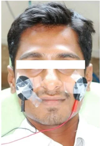
For all subjects Kesling separators made with 0.020” Australian stainless steel wire were placed on the left side maxillary arch mesial and distal to the maxillary first molar. On the right side elastomeric separators were placed mesial and distal to the maxillary first molar (Fig 2). For those subjects in the extraoral groups, electrodes were placed bilaterally over the subjects zygomatic arches (Fig 3). The electrodes were held in position with plasters. The TENS unit was set to deliver a current frequency of 60 Hz with an intensity of 50 mA. The duration of each session was 15 minutes. It was applied for four days (1st, 2nd, 3rd and 4th day). For the intraoral group, current was applied directly to the teeth by placing one pen electrode on the crown of each tooth and the other electrode on the palatal mucosa adjacent to the tooth (Fig 4). The TENS unit was set to a current frequency of 60 Hz with an intensity of 50 mA. The first and second molars and the second premolar on each side of the arch received 6 seconds of treatment current each time. It was repeated on the either side immediately after the placement of the separators for four days.
Subjects in all the three groups were instructed to report every 24 hrs for periodic check up. Any missing separators are replaced immediately and the subjects were instructed not to take any kind of analgesics. The subjects in control group received no treatment procedure.
Analysis of Data
Mean and standard deviation were calculated for each variable. Student ‘t’ test was conducted between the means of the right and left sides VAS score to identify the significant differences and analysis of variance (ANOVA) test was perf
compare the mean between groups and between days. Post hoc test (Tukey) was also performed for multiple comparisons between the groups and the days (p-0.05).
RESULTS
The mean VAS scores for the different groups at the various assessment periods are shown in Table 1.
The pain scores of subjects who received TENS therapy were significantly less than those of controls with both types of separators (p<0.05). The levels of discomfort were found to be significantly different between controls and TENS
all four days. The mean VAS scores with Kesling separators was more than elastomeric separators, but the difference was not statistically significant in TENS groups (p>0.05). Statistically significant difference between the separators was seen in control subjects (p<0.05).
Comparison of VAS scores between groups revealed statistically significant difference between TENS groups and controls on all four days (p<0.01) for both the separators. The difference between intraoral TENS and extra
groups was not statistically significant across the time intervals (p>0.05). Comparison of mean values of VAS scores within the groups using both the separators showed statistically significant difference between first day and the fourth day (p<0.01). The Graph 1 shows the comparison of mean pain scores among the three groups using Kesling separators and Graph 2 shows the comparison with elastomeric separators.
Table 1 Comparison of mean values of VAS scores between groups
Groups Day 1 Day 2
TENS Intraoral Group (n=10) Kesling separator
Mean ± SD Elastomeric separator
Mean ± SD
21.8 ± 8.53 19 ± 4.78
18.9 ± 7.92 15.8± 6.08
14.7 ±5.79 11.8 ±4.75 TENS Extraoral Group
(n=10) Kesling separator
Mean ± SD Elastomeric separator
Mean ± SD
23.3± 8.24
14.6± 9.46 20.3± 7.11 12.5± 7.89
15.7± 4.99 10.9 ±6.15
Control Group (n=10) Kesling separator
Mean ± SD Elastomeric separator
Mean ± SD
46 ±10.4
36.4 ±9.9
40.2± 9.48
31.4± 9.61
33.6± 8.99
27.9± 10.35
e three groups were instructed to report every 24 hrs for periodic check up. Any missing separators are replaced immediately and the subjects were instructed not to take any kind of analgesics. The subjects in control group
Mean and standard deviation were calculated for each variable. ‘t’ test was conducted between the means of the right and left sides VAS score to identify the significant differences and analysis of variance (ANOVA) test was performed to compare the mean between groups and between days. Post hoc test (Tukey) was also performed for multiple comparisons
The mean VAS scores for the different groups at the various
The pain scores of subjects who received TENS therapy were significantly less than those of controls with both types of separators (p<0.05). The levels of discomfort were found to be significantly different between controls and TENS subjects on all four days. The mean VAS scores with Kesling separators was more than elastomeric separators, but the difference was not statistically significant in TENS groups (p>0.05). Statistically significant difference between the separators was
Comparison of VAS scores between groups revealed statistically significant difference between TENS groups and controls on all four days (p<0.01) for both the separators. The difference between intraoral TENS and extra-oral TENS groups was not statistically significant across the time intervals (p>0.05). Comparison of mean values of VAS scores within the groups using both the separators showed statistically significant difference between first day and the fourth day he Graph 1 shows the comparison of mean pain scores among the three groups using Kesling separators and Graph 2 shows the comparison with elastomeric separators.
Comparison of mean values of VAS scores between
Day 3 Day 4
14.7 ±5.79 11.8 ±4.75
9.8 ± 4.63 8.5± 3.92
15.7± 4.99 10.9 ±6.15
11.8± 5.3 7.3 ±4.24
33.6± 8.99
27.9± 10.35
23.4± 7.09
20.7 ±8.97
Fig 1 TENS unit with Electrodes
Fig 2 Intraoral view of Separators
TENS unit with Electrodes
Intraoral view of Separators
Efficiency of Tens in Controlling Pain During Separation Using two Different Orthodontic Separators
Fig 3 Extraoral application of TENS using Electrodes
Fig 4 - Intraoral application of TENS using pen electrode
DISCUSSION
Electrical stimulation has been used as a therapeutic modality from the time of Roman Empire [32]. A comprehensive overview concerning the use of peripheral electrical stimulation for pain relief has been written by Woolf [33].
TENS generates short, low amplitude electrical impulse that travels between two electrodes placed on the skin. The signal from the electrical stimulation of beta fibers (larger nerve fibers for pressure and touch) reaches the central nervous system before the signals from the slower A and C fibers (smaller nerve fibers for pain). Thus, the beta impulse blocks or "closes the gate" to the pain impulses. The electric impulse also stimulates the production of a local analgesic (beta endorphin) and/ or substance "P" in the nerve cells and of serotonin in the brain, raising the patient’s pain tolerance [34]. The first dental application of TENS was for the treatment of myofacial pain dysfunction syndrome [35]. Mumford [36] assessed the effects of TENS on the pain threshold of electrically stimulated tooth pulp and results demonstrated a higher pain perception after TENS application. First controlled experimental design of TENS application was carried out by Hansson and Ekbolm [23] on out patients attending an emergency dental clinic.
Roth and Thrash (1986) [11] assessed for TENS effect on
Abdulhammed et al (1989) [37] assessed the efficiency of TENS in children during placement of a rubber clamp and found significant increase in tooth pain threshold.
Weiss and Carver (1994) [38] reported that TENS reduces the pain associated with debonding and further suggested that it can be used during proximal stripping and incisor recontouring procedures.
Shasikumar and Anup (2009) [39] demonstrated decreased pain thresholds in young patients during initial stages of alignment by extra-oral TENS.
In the present study, no statistically significant difference was found between genders in the subsequent pain responses. Earlier reports by Jones (1984)[ 40], Fernandes et al
(1998)[41], Ngan et al (1989)[42], Bondemark et al (2004)[43] seem to agree with this finding. Few studies claim that females reported more pain/discomfort than males [44,45].
In all the 3 groups, left sides (Kesling separator) mean VAS score was more than the right side (elastomeric separator) means VAS score. Kesling separator was more painful than the elastomeric separators but the difference was statistically non significant (p>0.05).Hoffman WE (1972)[46] and Bondemark L (2004)[43]reported thatboth types of separators caused pain of mild to moderate intensity, with Kesling being more tolerable than elastomeric, but the difference found was not statistically significant. The results show that pain was high in control group than in TENS group. Within the TENS groups, no significant difference was observed with either of separators.
The time of onset was between zero and 1st day/ 24 hours. The pain after placing separator was more on day 1 and day 2. This pain lasted for the 2nd and 3rd day and disappeared between 3rd and 4th day. These findings are similar to the clinical observations reported by Burstone (1962)[47], Hoffman (1972)[46], Roth and Thrash (1986)[11] and by Bondemark L (2004)[43].
The reliability of patients in recording discomfort precisely and at the requested times could not be verified. Nevertheless this study confirms previous findings that the intensity of pain is greatest in 24 hrs. The results also indicate that discomfort was significantly less at every time interval for those who used TENS.
Though our study Supports the use of TENS to Alleviate pain, it Presents few Limitations.
1. As compared to aligning and leveling simulation of orthodontic treatment using separators placement may cause less pain.
2. This design did not consider the response variability inherent in studies using separate treatment and control groups.
3. Influence of personality characteristics such as neuroticism and introversion - extroversion on perception of discomfort by patients was not evaluated.
CONCLUSION
separators. This study provides valuable information concerning the analgesic properties of peripheral electrical stimulation and should help establish a useful baseline for future clinical studies of the efficacy of electrical stimulation during orthodontic procedures. The TENS as a treatment modality to control pain during orthodontic tooth separation has the advantage of being non- invasive, easy to administer and having no adverse tissue reactions. It is worthwhile to look into its potential applications into orthodontics. Future investigations should focus on blinded, randomized control trials, comparing pharmacological and non pharmacological approaches.
References
1. Fulop - Miller R. Triumph over pain. E and C Paul (trans.). New york, Literary Guild of America, 1938;7-8. Alling C, Mahan E. Facial pain. 2nd edition, Philadelphia 1977, Lea and Febiger.
2. Wall P, Melzach R. Text book of pain. p 904, 3rd edition, 1994, Longman Group UK. Limited.
3. Oliver RG, Knapman YM. Attitudes to orthodontic treatment. Br J Orthod 1985; 12:179-88.
4. Jones M, Chan C. Pain in the early stages of orthodontic treatment. J Clin Orthod 1992; 26:311-313.
5. O'Connor PJ. Patient's perceptions before, during, and after orthodontic treatment. J Clin Orthod 2000; 34:591-592.
6. Brown DF, Moerenhout RG. The pain experience and psychological adjustments to orthodontic treatment of preadolescents, adolescents and adults. Am J Orthod Dentofac Orthop 1991; 100: 349-356.
7. Furstman L, Bernick S. Clinical consideration of periodontium. Am J Orthod 1972; 61:138-155.
8. Hwang J, Tee C, Huang A, Taft L. Effectiveness of Thera - Bite wafers in reducing pain. J Clin Orthod 1994; 28:291-292.
9. White LW. Pain and cooperation in orthodontic treatment J Clin Orthod 1984;18:572-575.
10. Roth PM, Trash WJ. Effect of Transcutaneous electrical nerve stimulation for controlling pain associated with orthodontic patients, Am J Orthod 1986; 90:132–138. 11. Lim HM, Lew KK, Tay KL. A clinical investigation of
efficiency of low level laser `therapy in reducing orthodontic postadjustment pain Am J Orthod Dentofac Orthop 1995;108:614-622.
12. Turhani D, Scheriau M, Kapral D, Benesch I, Jonke E, Banthon HP. Pain relief by single low level laser irradiation in orthodontic patients undergoing fixed orthodontic appliance therapy. Am J Orthod Dentofac Orthop 2006;130:371-77.
13. Youssef M, Ashkar S, Hamade E, Gutknecht N, Lampert F, Mir M. The effect of low level laser therapy during orthodontic movement: a preliminary study. Lasers Med Sci 2008;23:27-33.
14. Ste Marie S, Powers M, Sheridan JJ. Vibratory stimulation as a method of reducing pain after orthodontic appliances adjustment J Clin Orthod 2003;37:205-208.
15. Belchman A. Pain - free and mobility - Free orthodontics? Am J Orthod Dentofac Orthop 1998;113:379-83.
16. Etminan M, Sadatsafavi M, Jafari S. Acetaminophen use and the risk of asthma in the children and adults. A
systematic review and meta analysis. Chest 2009;136;1316-1323.
17. Farquhar H, Stewart A, Mitchell E et al. The raole of paracetamol in the pathogenesis of asthma. Clin Exp Allergy 2010;40:32-41
18. Eneli I, Sadri K, Camargo C, Barr RG. Acetaminophen and the risk of asthma Chest 2005;127:604-612.
19. Kumar RK, Savage J. Paracetamol as a cause of anaphylaxis. HK J Paediatr.1998;3:167-8
20. Shaheen SO, Sterne JAC, Songhurst CE, Burney PGJ. Frequent paracetamol use and asthma in adults. Thorax 2000; 55:266-70.
21. Leffers, H, et al (2010). "Intrauterine exposure to mild analgesics is a risk factor for development of male reproductive disorders in human and rat". Human Reproduction 25: 235–244.
22. Kim WS. Clinical study of the management of postoperative pain with transcutaneous electrical nerve stimulation. Pain 1984;20:S68.
23. Hansson P, Ekblom A. Transcutaneous electrical nerve stimulation (TENS) as compared to placebo TENS for the relief of acute oro - facial pain. Pain 1983;15:157-165.
24. Melzack R, Torgenson WS. On the language of pain. Anesthesiology 1971;34:50-59.
25. Mc Grath PA. The measurement of human pain. Endod Dent Traumatol 1986;2:124-29.
26. Duncan GH, Bushnell MC, Lavingne GJ. Comparison of verbal and visual analogue scale for measuring the intensity and unpleasantnessof experimental pain. Pain 1989;37: 295-303.
27. Huskisson EC. Visual analogue scale. In: Melzack R (ed). Pain measurement and assessment, p 33-37. New York 1983, Raven Press.
28. Uskisson E. Measurement of pain. Lancet, 1974; 2:127-131.
29. Seymour R. The use of pain scales in assessing the efficacy of analgesics in post operative dental pain. Eurp Clin Pharmacol 1982; 23: 441- 444.
30. Bergius M, Kiliaridis, and Berggren U. Pain in orthodontics. A review and discussion of the literature, J. Orofac. Orthop 2000;61:125-137.
31. Bhat M. Statistical analysis and design characteristics of studies on dentinal sensitivity. Endod Dent Traumatol 1986;2:165-171.
32. Wu CH. Electrical fish and the discovery of animal electricity. Am Scient 1984; 72:598.
33. Woolf CJ. Trancutaneous and implanted nerve stimulation in: Wall PD, Melzack R (ed): Textbook of pain, p 679, Edinburgh, Churchill Livingstone, 1984. 34. Esposito, C.J.; Shay, J.S.; and Morgan, B. Electronic
dental anesthesia: A pilot study, Quintess. Int 1993;24:167-170.
35. Denholtz M. Treatment of the myofascial pain dysfunction syndrome with transcutaneous electrical nerve stimulation. In Ersek RA (editor): Pain control with T.E.N.S., principles and practice. St. Louis, 1981, Warren H. Green, Inc.
Efficiency of Tens in Controlling Pain During Separation Using two Different Orthodontic Separators
37. Abdulhameed SM, Feigal RJ, Rudney JD, Kajander KC. Effect of peripheral electrical stimulation on measures of tooth pain threshold and oral soft tissue comfort in children. Anesth Prog 1989;36:52-57. 38. Weiss.D, Carver. D. Transcutaneous electrical neural
stimulation for pain. J Clin Orthod 1994;28:670-671. 39. Shashikumar and Anup B. Transcutaneous electrical
nerve stimulation - A non-invasive, simple, chairside, pain control technique in orthodontics. Orthodontic Cyber Journal, Nov 2009.
40. Jones M L. An investigation into the initial discomfort caused by placement of an archwire. Eur J Orthod 1984;6:48–54.
41. Fernandes L M , Øgaard B , Skoglund L. Pain and discomfort experienced after placement of a conventional or super elastic NiTi aligning archwire. A randomized clinical trial. J Orofac Orthop 1998;59:33 – 339.
42. Ngan p, Kess B, Wilson S. Perception of discomfort by patients undergoing orthodontic treatment. Am J Orthod Dentofac Orthop 1989;96:47-53.
43. Bondemark.L, Fredricksson.K, Susanna.I. Separation effect and perception of pain and discomfort from two types of orthodontic separators. World J Orthod 2004;5:172-176.
44. Kvam E, Bondevik O, Gjerdet N R. Traumatic ulcers and pain in adults during orthodontic treatment. Community Dentistry and Oral Epidemiology 1989;17:154–157.
45. Scheurer P A, Firestone A R, Bürgin W B. Perception of pain as a result of orthodontic treatment with fixed appliances. Eur J Orthod 1996;18:349 – 357.
46. Hoffman WE. A study of four types of orthodontic separators. Am J Orthod 1972;62:67-73.
47. Burstone C J. The biomechanics of tooth movement. In: Kraus B S, Riedel R A (eds). Vistas in orthodontics, pp. 197 – 213, Lea & Febiger , Philadelphia 1962.
How to cite this article:
Kala Vani S.V et al (2019) 'Efficiency of Tens in Controlling Pain During Separation Using two Different Orthodontic Separators', International Journal of Current Advanced Research, 08(04), pp. 18055-18060.
DOI: http://dx.doi.org/10.24327/ijcar.2019.18060.3442