Original Research Article
Serum 25-hydroxy vitamin D in asthmatic children and its
relation to disease severity
Ihab Hafez El Sawy
1, Passant Al-Said Moaz
2, Ghada Mohamed Farouk El Deriny
1*,
Mohamed Sami Abd El Moniem El Kholy
1INTRODUCTION
Asthma, one of the most substantial health problem affecting children worldwide, is a chronic respiratory disease distinguished by airway hyper-responsiveness, intensive airway, inflammation and airflow obstruction in response to particular triggers.1
Asthma prevalence among Egyptian children aged 3-15 years was estimated to be 8.2%.2,3 This high prevalence of asthma worldwide may be affected by changing environmental factors associated with westernized lifestyles.
The exact mechanisms involved in occurrence of asthma are somewhat not well understood, in part due to the
ABSTRACT
Background: Asthma is a chronic immunological disorder of the lungs. Vitamin D has several effects on the innate and adaptive immune systems. Little is known about vitamin D level and its impact on severity of asthma in children. This study aimed to determine vitamin D levels in asthmatics versus control children; studying the relation if any between these levels and asthma severity.
Methods: This cross-sectional study was conducted on 60 asthmatic children and 20 apparently healthy children as controls. Asthma patients were divided into 3 groups (mild, moderate, severe; 20 each). Asthma severity was based on GINA criteria. Vitamin D level was measured to all study group.
Results: The difference between the mean values of vitamin D level in control and asthmatic patients was statistically significant (p<0.001). This difference between control group and each asthma subgroup and between asthma subgroups versus each other were statistically significant being highest in control and lowest in patients with severe asthma (p<0.001). Differences in vitamin D status in control and all asthmatic patients were statistically significant (p<0.001). The difference between control group and each asthma subgroup according to vitamin D status were statistically significant (p<0.001). Concerning asthma subgroups the difference in vitamin D status between severe versus mild and moderate asthma were statistically significant (p<0.001), while between mild and moderate asthma it was not.
Conclusions: Significantly lower vitamin D level in asthmatic children compared to controls and a differential decrease in vitamin D levels in asthmatic children being lowest in severe asthma was confirmed.
Keywords: Asthma, Vitamin D, Asthma severity, Children
1
Department of Paediatrics, 2Department of Clinical and Chemical Pathology, Faculty of Medicine, University of Alexandria, Egypt
Received: 09 December 2018
Revised: 22 December 2018
Accepted: 26 December 2018
*Correspondence:
Dr. Ghada Mohamed Farouk El Deriny, E-mail: [email protected]
Copyright: © the author(s), publisher and licensee Medip Academy. This is an open-access article distributed under the terms of the Creative Commons Attribution Non-Commercial License, which permits unrestricted non-commercial use, distribution, and reproduction in any medium, provided the original work is properly cited.
marked heterogeneity of the disorder in both adults and childrenand multiple aberrated immune responses which are associated with this disarray.1,4,5,6
Countless dietary hypotheses have been introduced for the prevention and treatment of asthma, among the nutrients and antioxidants included in this theories, vitamin D is of distinct interest.7-9 Increasing evidence demonstrates that vitamin D deficiency plays a role in chronic diseases including asthma, presumably through the immunomodulatory effects of vitamin D.10-12 Vitamin D has been shown to have a prominent role in both innate and adaptive immunity by promoting phagocytosis and modulating the effects of Th1, Th2 and regulatory T cells.13-15 Vitamin D deficiency has been associated with increased airway hyper-responsiveness (AWH),decreased pulmonary function, bad asthma control and increased steroid resistance.16,17 Cellular studies revealed that vitamin D harmonize the activity of multiple defence and immune cells including monocytes, lymphocytes, macrophages, and epithelial cells. Vitamin D was proved to promote the production of Treg cells, IL-10–secreting Tregs which are of specific importance, as IL-10 is an influential anti-inflammatory cytokine and it inhibits TH1 and TH2 immune responses, which has led to considerable interest in its role in allergic inflammation.18
Airway epithelial cell express enzymes of the vitamin D metabolism and are competent to transform the precursor 25(OH)D3 into the active 1,25(OH)2D3 form.19,20 This
activated form of vitamin D is able to modify the inflammatory cascade after a viral infection by blocking the poly (I:C) induced cytokine and chemokine production while preserving the antiviral activity.21 Since the epithelial cells are primary distension of respiratory microorganisms and since Vitamin D induce the production cathelicidin which has antiviral and antibacterial activity, a seasonal decrease of vitamin D dependent epithelial host defence could be reason for the increased frequency of lower respiratory tract infection during winter.19,20
Respiratory viral infections are important triggers of asthma exacerbations. Recent data suggest that vitamin D reduces the risk of respiratory viral infection by damping of inflammation which is a sequence of infections. In a randomized study it was found that those receiving double the daily requirements of vitamin D had a 60% decrease in the incidence of seasonal influenza or common cold. However, those taking 2,000 IU/d had a 90% reduction.22 Thus, deficiency of vitamin D might have been responsible for the increased incidence of respiratory tract infections, and accordingly, more asthma worsening.23
The focus of the present work was to determine 25- hydroxy vitamin D levels in asthmatics versus control children and to detect the relation if any between these levels and asthma severity.
METHODS
This cross-sectional study was conducted on 60 patients with proven diagnosis of persistent bronchial asthma and 20 apparently healthy age and sex matched children served as control. The patients and controls were recruited from Asthma Clinic and Outpatient departments of El-Shatby Alexandria University Children’s Hospital from October 2014 to January 2015 Asthma patients were divided into 3 groups; 20 with mild persistent asthma, 20 with moderate persistent asthma and 20 with severe persistent asthma based on GINA criteria.24
Inclusion criteria
Inclusion criteria were patients with persistent asthma aged >2 years to exclude occasional wheezes due to viral respiratory tract infections.
Exclusion criteria
Patients were excluded if they were mild intermittent asthma; receiving drugs that may affect vitamin D serum level as phenytoin, phenobarbital, rifampicin; having other chronic illness as congenital heart disease, cystic fibrosis, etc; receiving vitamin D supplementation.
Vitamin D serum level was measured using the 25(OH) Vitamin D Total Assay. It is a direct competitive chemiluminescence immunoassay for human serum or plasma intended for use on the DiaSorin LIAISON automated analyzer.25 vitamin D status was considered to be deficient if serum level <10 ng/ml, insufficient from 10-30 ng/ml and sufficient from 30-70ng/ml.26
RESULTS
Data were fed to the computer and analyzed using IBM SPSS software package version 20.0.
Qualitative data were described using number and percent. Quantitative data were described using mean and standard deviation for normally distributed data while abnormally distributed data was expressed using median, minimum and maximum.
Comparison between different groups regarding categorical variables was tested using Chi-square test. When more than 20% of the cells have expected count less than 5, correction for chi-square was conducted using Fisher’s Exact test or Monte Carlo correction.
For normally distributed data, comparison between two independent population were done using independent t-test while more than two population were analyzed F-t-test (ANOVA) to be used and Post Hoc test (Scheffe).
Significance test results are quoted as two-tailed probabilities. Significance of the obtained results was judged at the 5% level.
Concerning age, the mean values of control and patients with mild, moderate and severe persistent asthma were (6.61±2.46), (6.58±1.52), (5.63±1.93) and (5.99±1.96) respectively. There were no statistical significant differences between control group and asthmatic subgroups or between the asthmatic subgroups versus each other (p=0.338). As for sex, the asthmatic subgroups and control were also matched as (p=0.419) (Table 1).
Table 1: Demographic data in studied groups.
Control Mild asthma Moderate asthma Severe asthma
P value
No % No % No % No %
Sex
Male 12 60.0 7 35.0 8 40.0 9 45.0 F
p= 0.419
Female 8 40.0 13 65.0 12 60.0 11 55.0
Age
Min.–Max. 2.80–11.0 4.0–9.20 3.30–11.0 3.50–11.0
F
p = 0.338
Mean±SD 6.61±2.46 6.58±1.52 5.63±1.93 5.99±1.96
Median 5.80 6.15 4.54 6.0
P value for comparing between the studied groups; 2: Chi square test; F: F test (ANOVA).
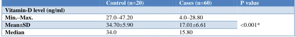
Tables 2: Comparison between mean serum level of vitamin D in control and all asthma patients.
Control (n=20) Cases (n=60) P value Vitamin-D level (ng/ml)
Min.–Max. 27.0–47.20 4.0–28.80
<0.001*
Mean±SD 34.70±5.90 17.01±6.61
Median 34.0 15.80
P valuefor Student t-test for comparing between the two studied group; *: Statistically significant at p≤0.05.
Table 3: Comparison between control and asthma subgroups according to vit D level.
Control Mild asthma Moderate Severe P value Vitamin-D level (ng/ml)
Min.–Max. 27.0–47.20 18.90–28.80 10.80– 23.20 4.0–17.70
<0.001*
Mean±SD 34.70±5.90 24.11±3.17 16.48±3.67 10.44± 3.66
Median 34.0 24.80 14.90 10.85
p1 <0.001* <0.001* <0.001*
p2 <0.001
*
<0.001*
p3 <0.001*
P value=Post Hoc Test (Scheffe) was used for comparing between the two studied groups; p1: value for comparing between control with each other group; p2: value for comparing between mild asthma with moderate and severe; p3: value for comparing between moderate and severe; *: Statistically significant at p≤0.05.
Table 4: Comparison between the two studied groups according to vitamin-D status.
Control (n=20) Cases (n=60)
P value
No % No %
Vitamin-D level (ng/ml)
Deficiency (<10) 0 0.0 7 11.7
<0.001*
Insufficiency(10 - <30) 5 25.0 53 88.3
Sufficient (≥30) 15 75.0 0 0.0
Total 20 100.0 60 100.0
Table 5: Vitamin-D status in different asthma subgroups and control.
Control Mild asthma Moderate Severe
P value
No % No % No % No %
Vitamin-D level (ng/ml)
Deficiency (<10) 0 0.0 0 0.0 0 0.0 7 35.0
<0.001*
Insufficiency(10 - <30) 5 25.0 20 100.0 20 100.0 13 65.0
Sufficient (≥30) 15 75.0 0 0.0 0 0.0 0 0.0
Total 20 100.0 20 100.0 20 100.0 20 100.0
FE
p1 <0.001* <0.001* <0.001*
FE
p2 - 0.008*
FE
p3 0.008*
P value for Monte Carlo test for comparing between the studied group 2: Chi square test FE: Fisher Exact test p1: value for comparing between control with each other group; p2: value for comparing between mild asthma with moderate and severe; p3: value for comparing between moderate and severe*: Statistically significant at p≤0.05.
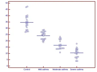
Figure 1: Scattered plot for the different studied groups according to vitamin-D level.
The mean values of vitamin D serum level in control and all asthmatic patients were 34.70±5.90 ng/ml and 17.01±6.61 ng/ml respectively, this difference was statistically significant (p<0.001) (Table 2).
Concerning the mean values of vitamin D serum level in control group and asthma patients subgroups (mild, moderate and severe persistent asthma) were 34.70±5.90 ng/ml, 24.11±3.17 ng/ml, 16.48±3.67 ng/ml and 10.44±3.66ng/ml respectively. The difference between control group and each asthma subgroup and between asthma subgroups versus each other were statistically significant being highest in control group and lowest in patients with severe persistent asthma (p<0.001) (Table 3), (Figure 1).
As for the vitamin D status it was (0.0) deficient, 5 (25%) insufficient, 15 (75%) sufficient level in the controls and
7 (11.7%) deficient, 53 (88.3%) insufficient, and (0.0) sufficient in the asthmatic patients, this difference in vit D status was statistically significant (p<0.001) (Table 4).
Regarding the vitamin D status in control and asthma patients subgroups (mild, moderate and severe persistent asthma) (Table 5). 5 (25%) of control had insufficiency, 15 (75%) had sufficient level while insufficient level was found in 20 (100%) of both mild and moderate subgroups. In patients with severe persistent asthma, 13 (65%) were insufficient and 7 (35%) were deficient. The difference between control group and each asthma subgroup according to vitamin D status were statistically significant (p<0.001). Concerning asthma subgroups the difference between severe versus mild and moderate persistent asthma were statistically significant (p<0.001), with no statistical difference detected between mild and moderate persistent asthma subgroups (Table 5).
0 5 10 15 20 25 30 35 40 45 50
V
it
a
m
in
-D
l
e
v
e
l (n
g
/m
l)
DISCUSSION
Although it is well known that positive atopic status, exposure and sensitization to environmental allergens and/or familial history of allergic disease are significant risks factors associated with the development of asthma, a lot of evidence suggests that vitamin D deficiency may also predispose to allergic phenotype.27,28 Vitamin D is considered to be a potent modulator of the immune system and is involved in regulating cell proliferation and differentiation. Epidemiological evidence suggests that there is a worldwide epidemic of vitamin D deficiency, and lack of vitamin D has been linked to increased incidence of asthma and increased severity of asthma in children.29,30
In the present study, mean serum vitamin D levels in all patients with persistent bronchial asthma was significantly lower than that of healthy controls. With respect to disease severity, the differences in mean serum vitamin D between asthma subgroups versus each other and versus the control group were highly statistically significant (p<0.001) being lowest in severe asthma subgroup and highest in control group.
Furthermore the results showed significant difference concerning vitamin D status, where 11.7% of all asthmatic patients were vitamin D deficient and 88.3% had vitamin D insufficient while none of the control group had vitamin D deficiency, 25% had insufficiency and 75% had sufficient vitamin level. These difference were statistically significant (p<0.001). Comparing between each asthma subgroups concerning vitamin D status 65% of severe cases had vitamin D insufficiency and 35% had vitamin D deficiency, while in moderate and mild cases 100% were vitamin D insufficient.
Bener et al found that a high proportion of Qatari children were deficient in vitamin D.31 This deficiency was more frequent in children suffering from asthma compared to non-asthmatic controls. Also, Litonjua and Weiss studies had revealed 35% of American asthmatic children were vitamin D deficient and these children were at a greater risk of severe asthma attacks.29 These epidemiological studies suggest a possible association between vitamin D deficiency and asthma and support our current observations.
Furthermore Ginde et al have presented evidence to show that low vitamin D levels were associated with higher frequency of respiratory tract infections in asthmatic patients and with increased asthma severity.32 Adequate vitamin D status may improve handling of respiratory infections early in life by up regulating the production of antimicrobial proteins, such as cathelicidin and beta defensins.20
In another study Freishtat et al documented that 86% of African- American asthmatic children living in
Washington, D.C. were vitamin D deficient while 19% of non-asthmatics were having the deficiency.33 Similarly El-banna et al demonstrated an association between vitamin D and the antimicrobial peptides (cathelicdin) in asthmatic patients where they found a highly significant decrease in 25(OH)D in asthmatics versus control group (p<0.001).34
In this respect but in a different track Brehm et al in a cross sectional study on mild to severe persistent asthma, they stated that increased 25(OH)D was associated with reduced hospitalization, reduced anti-inflammatory medication use and reduced airway hyper-responsiveness.35 Chinellato et al in a cross-sectional study regarding well and poor asthma control, they stated that hypovitaminosis D is frequent in children with asthma living in a Italy.36 In those children, lower levels of vitamin D were associated with reduced asthma control. Alyasin et al results showed that serum 25-hydroxy vitamin D levels were inversely associated with asthma severity, and there was a direct and significant relationship between vitamin D levels and pulmonary function test outcomes in asthmatic children.37 Also Merve et al in a cross-sectional trial detected that the frequency of vitamin D deficiency and insufficiency was higher in children with asthma, compared to the controls.38 Lower levels of vitamin D were associated with poor asthma control and increased asthma severity.
In contrast to the result of previous studies and to the present study Menon et al in a case-controlled study in 2012 conducted on 263 subjects of ages 2-19 years with asthma who were compared to 284 non-asthmatic controls of similar ages, Serum 25(OH)D was measured in all subjects, they found no differences in mean 25(OH) D levels between asthmatic patients and controls, and they stated that asthma severity had no relationship to mean 25(OH)D levels.39
It is difficult to ascertain from cross-sectional studies whether vitamin D deficiency is responsible for reduced asthma control or whether uncontrolled asthma associated lifestyles, such as decreased exposure to sunlight, less outdoor exercise are responsible for lower serum levels of vitamin D. Interventional trials aimed at detecting the effect of vitamin D supplementation on asthma exacerbations and double-blind, placebo-controlled trials in vitamin D–deficient children with asthma are needed to explore whether there is a causal relationship or a simple association between the two events. There is no evidence to suggest that asthmatic patients should be screened for vitamin D deficiency or insufficiency. However, the high-risk group must be screened for this deficiency.
CONCLUSION
The present study confirmed a significantly lower serum vitamin D level in asthmatic children compared to controls and a differential decrease in serum vitamin D levels in asthmatic children being lowest in severe persistent asthma. These findings support the growing body of evidence that vitamin D may have a role in pathophysiological events behind bronchial asthma.
ACKNOWLEDGEMENTS
We are grateful to all the patients for their helpful Co-operation.
Funding: No funding sources Conflict of interest: None declared
Ethical approval: The study was approved by the Institutional Ethics Committee
REFERENCES
1. Moore WC, Meyers DA, Wenzel SE, Teague WG, Li H, Li X, et al. National heart, lung, and blood institute’s severe asthma research program, identification of asthma phenotypes using cluster analysis in the severe asthma research program. Am J Respir Crit Care Med. 2010;181:315-23.
2. Zedan M, Settin A, Farag M, Ezz-Elregal M, Osman E, Fouda A. Prevalence of bronchial asthma among Egyptian school children. Egypt J Bronchol 2009;3(2):124-30.
3. Hassan AA, Sabah Abdou Hagrass. Prevalence of Bronchial Asthma in Primary School Children. Am J Med Med Sci. 2017;7(2):67-73.
4. Fitzpatrick AM, Teague WG, Meyers DA, Peters SP, Li X, Li H, et al. National Institutes of Health/National Heart, Lung, and Blood Institute Severe Asthma Research Program. Heterogeneity of severe asthma in childhood: confirmation by cluster analysis of children in the National Institutes of Health/National Heart, Lung, and Blood Institute Severe Asthma Research Program. J Allergy Clin Immunol. 2011;127:382-9.
5. Holt PG, Strickland DH. Interactions between innate and adaptive immunity in asthma pathogenesis:new perspectives from studies on acute exacerbations. J Allergy Clin Immunol. 2010;125:963-72.
6. National Hospital Discharge Survey, Mortality Component of the National Vital Statistics System, National Center for Health Statistics. CDC, 2011. 7. Allan K, Devereux G. Diet and asthma:nutrition
implications from prevention to treatment. J Am Diet Assoc. 2011;111:258-68.
8. Nurmatov U, Devereux G, Sheikh A. Nutrients and foods for the primary prevention of asthma and allergy: systematic review and meta-analysis. J Allergy Clin Immunol. 2011;127:724-33.
9. Mason RS, Sequeira VB, Gordon-Thomson C. Vitamin D:the light side of sunshine. Eur J Clin Nutr. 2011;65:986-93.
10. Reed CE. The natural history of asthma. J Allergy Clin Immunol. 2006;118(3):543-8.
11. Antonella LoMauro A, Aliverti A. Sex differences in respiratory function. Breathe (Sheff). 2018;14(2):131–40.
12. Niloufer S Ali, Kashmira Nanji. A Review on the Role of Vitamin D in Asthma. Cureus. 2017;9(5):e1288.
13. Gombart AF, Borregaard N, Koeffler HP. Human cathelicidin antimicrobial peptide (CAMP) gene is a direct target of the vitamin D receptor and is strongly up-regulated in myeloid cells by 1,25-dihydroxyvitamin D3. FASEB J. 2005;19:1067-77. 14. Matheu V, Bäck O, Mondoc E, Issazadeh-Navikas
S. Dual effects of vitamin D–induced alteration of TH1/TH2 cytokine expression:enhancing IgE production and decreasing airway eosinophilia in murine allergic airway disease. J Allergy Clin Immunol. 2003;112:585-92.
15. Sandhu MS, Casale TB. The role of vitamin D in asthma. Ann Allergy Asthma Immunol. 2010;105:191-9.
16. Penna G, Roncari A, Amuchastegui S. Expression of the inhibitory receptor ILT3 on dendritic cells is dispensable for induction of CD4_Foxp3_ regulatory T cells by 1, 25-dihydroxyvitamin D3. Blood. 2005;106:3490–7.
17. Ghoreishi M, Bach P, Obst J, Kmoba M, Fleet JC, Dutz JP. Expansion of antigen-specific regulatory T cells with the topical vitamin D analog calcipotriol. J Immunol. 2009;182:6071–8.
18. McGlade JP, Gorman S, Zosky GR. Suppression of the asthmatic phenotype by ultraviolet B-induced, antigen-specific regulatory cells. Clin Exp Allergy. 2007;37:1267–76.
19. Hansdottir S, Monick MM, Hinde SL, Lovan N, Look DC, Hunninghake GW. Respiratory epithelial cells convert inactive vitamin D to its active form: potential effects on host defense. J Immunol. 2008;181:7090-9.
20. Hansdottir S, Monick MM, Lovan N, Powers L, Gerke A, Hunninghake GW. Vitamin D decreases respiratory syncytial virus induction of NF-kappa Blinked chemokines and cytokines in airway epithelium while maintaining the antiviral state. J Immunol. 2010;184:965-74.
21. Litonjua AA, Weiss ST. Is vitamin D deficiency to blame for the asthma epidemic? J Allergy Clin Immunol. 2007;120:1031-5.
22. Aloia JF, Li-Ng M. Re:epidemic influenza and vitamin D. Epidemiol Infect. 2007;135:1095–8. 23. Grant WB. Variations in vitamin D production
could possibly explain the seasonality of childhood respiratory infections in Hawaii. Pediatr Infect Dis J. 2008;27:853-9.
education guidelines for the prevention program expert panel report 3, guideline for the diagnosis and management of asthma. 2007.
25. Ersfeld DL, Rao DS, Body JJ, Sackrison Jr JL, Miller AB, Parikh N, et al. Analytical and clinical validation of the 25 OH vitamin D assay for the LIAISON automated analyzer. Clin Biochem. 2004;37:867–74.
26. Hollis BW, Wagner CL, Drezner MK, Binkley NC. Circulating vitamin D3 and 25-hydroxyvitamin D in humans:an important tool to define adequate nutritional vitamin D status. J Steroid Biochem Mol Biol. 2007;103(3-5):631-4.
27. Bouzigon E, Nadif R, Le Moual N, Dizier MH, Aschard H, Boudier A, et al. Genetic and environmental factors of asthma and allergy:Results of the EGEA study. Rev Mal Respir. 2015;32(8):822-40.
28. Chang JH, Cha HR, Lee DS. 25-Dihydroxyvitamin D3 Inhibits the Differentiation and Migration of TH17 Cells to Protect against Experimental Autoimmune Encephalomyelitis. PLoS One. 2010;5(9):e12925.
29. Weiss ST, Litonjua AA. Maternal diet versus lack of exposure to sunlight as the cause of the epidemic of asthma, allergies and other autoimmune diseases. Thorax. 2007;62:746–8.
30. Brehm JM, Celedon JC, Soto-Quiros ME, Avila L, Hunninghake GM, Forno E, et al. Serum vitamin D levels and markers of severity of childhood asthma in Costa Rica. Am J Respir Crit Care Med. 2009;179:765–71.
31. Bener A, Al-Ali M, Hoffmann GF. High prevalence of vitamin D deficiency in young children in a highly sunny humid country:a global health problem. Minerva Pediatr. 2009;61:15–22.
32. Ginde AA, Mansback JM, Camargo CA Jr. Association between serum-25-hydroxyvitamin D level and upper RTI in the 3rd National Health and
Nutrition Examination Survey. Arch Intern Med. 2009;39:875–82.
33. Freishtat RJ, Iqbal SF, Pillai DK, Klein CJ, Ryan LM, Benton AS, et al. High prevalence of vitamin D deficiency among inner-city African American youth with asthma in Washington, DC. J Pediatr. 2010;156:948-52.
34. El-banna EAM, Salah KM, Ahmed HS. Effects of vitamin D and the antimicrobial peptide in asthma, Egypt J Pediatr Allergy Immunol. 2012;10(2):101-7.
35. Brehm JM, Celedon JC, Soto-Quiros ME, Avila L, Hunninghake GM, Forno E, et al. Serum vitamin D levels and markers of severity of childhood asthma in Costa Rica. Am J Respir Crit Care Med. 2009;179:765–71.
36. Chinellato I, Piazza M, Sandri M, Peroni D, Piacentini G, Boner AL. Vitamin D serum levels and markers of asthma control in Italian children. J Pediatr 2011;158:437-4.
37. Alyasin S, Momen T, Kashef S, Alipour A, Amin R. The relationship between serum 25 hydroxy vitamin D Levels and asthma in children. Allergy Asthma Immunol Res. 2011;3(4):251-5.
38. Merve H, Cem HR, Ayse D, Ali OK, Nesibe A. Effects of 25 hydroxy vitamin D levels on the severity and asthma control in school age asthma Arch Argent Pediatr. 2017;115(4):336-42.
39. Menon J, Maranda L, Nwosu BU. Serum 25-hydroxyvitamin D levels do not correlate with asthma severity in a case-controlled study of children and adolescents. J Pediatr Endocrinol Metab. 2012;25(7-8).