Original Research Article
Analysis of cervical liquid-based cytology results in Eskişehir, Turkey:
correlation of cytology results with histology, immunocytocemical HPV
Ab and HPV DNA results of 18404 women
Bahattin Erdoğan
1*, Cengiz Bal
2, Binnur Önal
3INTRODUCTION
Cervical cancer is an important health problem 530 000 cases seen worldwide every year and 275 000 people die consequently. It is the third most common cancer after breast and large intestine cancer in women.1 According to data from the Ministry of health–Turkey public health department, cervical cancer is ranked at the 9th among
women’s cancer cases for all age groups in Turkey with a rate of 2.4%.2
The Bethesta System (TBS) used in cytological interpretation is based on internationally accepted terminology and morphological evidence. The system is linked to the biology and management of the disease and is concise and practical. It is flexible enough to be adopted in different geographical and laboratory
ABSTRACT
Background: To analyze retrospectively the results of HPV DNA, immunocytochemical HPV antibody staining of
gynecologic smear samples evaluated in a public hospital and to observe their compatibility with histologic diagnosis. At the same time, the contribution of ICC HPV Ab staining experience results to the morphological evaluation is discussed in this paper.
Methods: In this study, liquid-based cytology test results of patients who applied to the gynecology between 2014
and 2017 were analyzed. The Ultravision Quanto Detection System was modified for immunocytochemical staining. HPV DNA tests were performed with the Qiagen Hybrid Capture test.
Results: The 18404 test result was included in the research. The percentage of smear that epithelial cell atypia is seen
was 3.4%, the rate of ASC/SIL was 1.89%. Compared to the first 3 years of the study, the increase in the rate of LSIL is seen with a partial decrease in ASCUS rate in year 2017 (p<0.05). The atypical positive test rate with histologic confirmation was 73.61%. Among 138 HPV Ab results, 58.7% of them were negative and 41.3% of them were positive. Sensitivity and specificity rates were determined 76.19% and 52.17% for SIL. Among 53 HPV DNA results (53% negative and 46.3% positive); sensitivity and specificity rates were determined 92.86% and 50% for SIL.
Conclusions: İmmunocytochemical HPV Ab staining provided statistically significant contribution to LSIL (p<0.05).
It is thought that it also will provide additional evidence for morphological findings while cytological evaluation and may help the clinician in managing the conditions for disease.
Keywords: Liquid-based cytology, HPV DNA, Immunocytochemistry, Turkey
1
Department of Pathology, Eskişehir City Hospital, Eskişehir, Turkey
2
Department of Biostatistics, ESOGU Medical Faculty, Eskişehir, Turkey
3
Ankara, Turkey
Received: 14 December 2018
Revised: 16 January 2019
Accepted: 17 January 2019
*Correspondence:
Dr. Bahattin Erdoğan,
E-mail: [email protected]
Copyright: © the author(s), publisher and licensee Medip Academy. This is an open-access article distributed under
the terms of the Creative Commons Attribution Non-Commercial License, which permits unrestricted non-commercial use, distribution, and reproduction in any medium, provided the original work is properly cited.
environments. It is suitable to change and improve with the increase of knowledge and experience.3 In spite of all these features, the inhibition continues due to the sensitivity especially for some diagnostic categories, and the intra-observer repeatable and inter observer compliance is less than ideal.4,5
Any decline in sensitivity and specificity depends on human performance and can be avoided by taking appropriate preventive measures. As clinical cytologists, attention has been drawn to the risk of falling in susceptibility to PAP testing due to unfamiliarity with abnormal cells and to the decreased specificity where the fear of missing important diseases may lead to overloading of benign abnormalities.6
The discovery of a strong association between human papilloma virus (HPV) infection and cervical cancer has changed perspectives on cervical cancer prevention by accelerating the development of protective vaccines with HPV DNA and RNA tests. Specifically developed DNA-based HPV molecular tests do not distinguish between transient and persistent infection. It alone is not as specific as the primary screening method for the detection of significant lesions. At the same time, HPV testing is ordinarily expensive and is therefore not easily accessible to anyone who can benefit from it. Therefore, additional markers are needed to improve routine clinical practice.7 Immunocytochemical (ICC) detection of molecular changes caused by HPV in host cells can be used additionally in cytological interpretation by improving sensitivity (SE) without compromising potentially specificity (SP). For this purpose, the cyclin-dependent kinase inhibitor p16 (p16INK4a) and Ki-67 proliferation marker (MIB-1) have been investigated as additional molecular markers for cervical lesions and have been reported to be useful as an adjunct to increase the specific sensitivity of HPV testing as well as cytological screening. However, clear methodological standards are required for optimal performance in the ICC in the clinical setting.7
Clinical guidelines recommend a routine HPV test (co-test) in conjunction with cytology for cervical cancer screening program in women over 30 years of age. It is stated that screening interval is safe every 3 years for women with normal cellular morphology and negative for HPV DNA test. It is also indicated that to estimate the feasibility and reliability of test guidelines, routine clinical practice is required.8, 9
In our study, we analyzed the results of liquid-based cytology (LBC) of patients who applied to Eskisehir State Hospital (EDH) gynecology clinic for four years retrospectively. We compared the results of SE, SP values and histologic diagnosis cytological, HPV DNA and ICC HPV Ab results. We discussed the contribution of the ICC HPV Ab results to the cytological evaluation.
METHODS
This study included LBC test results from patients admitted to the gynecology clinic of the hospital between 2014 and 2017. HPV DNA results of the patients and pathological diagnoses of the cervical biopsy specimens were obtained from the electronic records of EDH and Eskişehir Osmangazi University (ESOGÜ) Medical Faculty Hospital Pathology Departments. The Ultra Vision Quanto Detection System was modified for ICC staining and HPVAb3 (clone K1H8) was used as primer antibody. In cases with epithelial cell atypia, a new smear was made from the remaining LBC specimen of cellular evaluation and was air-dried. Deparaffinization, hydration prior to staining and antigen retrieval steps in the manufacturer's procedure were not applied. It kept in Lam Tris buffer saline (TBS) for 5 minutes. The slide was shaken and the liquid on it was thrown out, spreading is limited by the limitation pen. It washed in in order of Hydrogen peroxidase (3% Aqueous): 10 minutes, Phosphate buffer saline (PBS) pH: 7.4: 3 minutes. (2 times), Ultra B Block: 5 minutes, Primary antibody (HPV Ab-3): 45 minutes, PBS: 3 minutes (2 times), Amplifier: 20 minutes, PBS: 3 minutes (2 times), HRP Polymer: 30 minutes, PBS: 3 minutes. (2 times), AEC chromogen: 15 minutes and running tap water for 30 second. It is colored 1 minute in hematoxylin, 1 minute in running tap water. After washing, it was rinsed in distilled water for 30 sec and sealed with Aqueous mounting medium. Brown staining in the epithelial cell nucleus was interpreted as a positive reaction (Figure 1). HPV DNA tests that could be detected in patient records were performed with Qiagen Hybrid Capture test. The data is analyzed in the IBM SPSS 24 program. Biopsy results are used as reference values in the calculation of sensitivity and specificity.
RESULTS
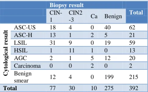
(HSIL) and 2.6% were diagnosed as carcinoma. The cross-distribution of cytological results and biopsy diagnoses is shown in Table 2. There was a significant correlation between cytology and histology results
(p<0.05). Sensitivity and specificity rates were obtained as 72.09%-91.28% for LSIL and 73.33-100% for HSIL, respectively.
Table 1: Cytological result vs. year cross tabulation.
Year
Total
2014 2015 2016 2017
Cyto
logic
al re
sult ASC-US
Count 77 51 108 108 344
% Within year 1.7 1.1 2.3 2.4 1.9
ASC-H Count 8 5 12 9 34
% Within year 0.2 0.1 0.3 0.2 0.2
LSIL Count 33 25 36 79 173
% Within year 0.7 0.5 0.8 1.8 0.9
HSIL Count 2 7 6 6 21
% Within year 0.0 0.2 0.1 0.1 0.1
AGC Count 0 8 23 22 53
% Within year 0 0.2 0.5 0.5 0.3
Carcinoma Count 1 0 0 1 2
% Within year 0 0 0 0 0
Benign smear Count 4494 4301 4499 4130 17424
% Within year 96.8 94.4 94.5 93.0 94.7
Nondiagnostik Count 28 161 76 88 353
% Within year 0.6 3.5 1.6 2.0 1.9
Total Count 4643 4558 4760 4443 18404
% Within year 100 100 100 100,0 100
(ASC-US: atypical squamous cells of undetermined significance, ASC-H: atypical squamous cells- cannot exclude HSIL, LSIL: low grade squamous ıntraepithelial lesion, HSIL: hıgh grade squamous ıntraepithelial lesion. AGC: atypical glanular cell).
Table 2: Cytological result vs. biopsy diagnosis cross tabulation.
Biopsy result
Total
CIN-1
CIN2
-3 Ca Benign
C
ytol
og
ic
al result
ASC-US 18 4 0 40 62
ASC-H 13 1 2 5 21
LSIL 31 9 0 19 59
HSIL 1 11 1 0 13
AGC 2 1 5 12 20
Carcinoma 0 0 2 0 2 Benign
smear 12 4 0 199 215
Total 77 30 10 275 392
Among HPV-DNA results of 55 patients, 52.7% were negative, and 47.3% were positive. Biopsy diagnosis of 28 of these patients was attained (14 benign, 10 LSIL and 4 HSIL) (Table 3). The correlation between HPV DNA and biopsy results was statistically significant (K=0.068). According to biopsy results, SE and SP values are for LSIL (90-50%), HSIL (100-50%) and for total SIL (92.86- 50%).
In our series, 51 of the ICC HPV Ab studied cases were resulted as HPV DNA (Table 4). The correlation between the two results was statistically significant (K=0.404).
When the cases between 2014 and 2016 were compared with the case results evaluated in 2017, decrease in ASCUS results and increase in LSIL results were significant (P<0.05).
ICC HPV Ab. was applied in 138 patients, 58.7% were negative and 41.3% were positive. There were 45 biopsy specimens (23 benign, 15 LSIL, 6 HSIL and 1 carcinoma) (Table 3). The correlation between ICC HPV Ab results and biopsy results is significant (K=0.032). According to biopsy results, SE and SP values are for LSIL (86.67-52.17%), for HSIL (50-52.17%) and for total SIL (76.19-52.17%).
Fıgure 1: Immunohistochemical HPV antibody staining; dark brown positive staining is observed in
Table 3: Biopsy diagnosis vs. ICC HPV Ab and HPV DNA results cross tabulation.
ICC HPV Ab
Total HPV DNA Total
Negative Positive Negative Positive
Bi
op
sy dia
gno
sis
Benign Count 12 11 23 7 7 14
% within biopsy diagnosis 52.2 47.8 50 50 CIN I
(LSIL)
Count 2 13 15 1 9 10
% within biopsy diagnosis 13.3 86.7 10 90 CIN II-III
(HSIL)
Count 3 3 6 0 4 4
% within biopsy diagnosis 50 50 0 100
Malign Count 1 0 1 0 0 0
% within biopsy diagnosis 100 0 0 0
Total Count 18 27 45 8 20 28
% within biopsy diagnosis 40 60 28.6 71.4
(CIN: Cervical ıntraepithelial lesion
Table 4: ICC hrHPV Ab vs. HPV DNA crosstabulation.
HR HPV DNA
Total
Negative Positive
ICC HPV Ab. Negative
Count 21 8 29
% within ICC hrHPV Ab 72.4 27.6
Positive Count 7 15 22
% within ICC hrHPV Ab 31.8 68.2
Total count 28 23 51
DISCUSSION
Pap smear test is an important screening test for investigating cervical-derived neoplastic lesions and that effective for reducing the incidence of cervical cancer. It is a noninvasive, inexpensive, simple and effective method described by Papanicolaou in 1942. Although this test contributes to the reduction of cervical cancer mortality rates, problems with consistency got ahead of the benefits of it. Due to problems with sampling, scanning and interpretation, new technologies have been put into practice to reduce false negative results. Techniques of preparing thin film spreads using a liquid medium such as LBC have been developed to overcome these problems. Although these has a higher rate of satisfaction than conventional cytology in studies and the possibility of detecting HPV DNA from remaining samples, the preference of them should be discussed due to their higher cost.10
The aim of quality in gynecological cytology is to improve the performance of the test by trying to minimize false positive and false negative results. Objective data is needed to evaluate the test performance. Sample adequacy (proportion of unsatisfactory samples), workload records, the report scales of the main categories of diagnoses for each cytopathologist and comparison with national standards, positivity rate, ASC ratio, ASC/SIL, return time, and cytology-histology compliance ratio (PPV: positive test percentage with
histological confirmation of cervical dysplasia) are some of the data that can be used for this purpose.11-13
Workload is a determinant that affects quality parameters in gynecological cytology. The limit for the maximum number of slides that can be scanned by the scanning staff is 100 slides per workday, independent of the other tasks that can be performed by staffing.12 In addition to the daily work load, when 7 slides/day as "ECA-adjusted workload" that uses a parameter with a higher correlation is evaluated, it means 70 slides/day with 10% ECA.14,15 In the study conducted by Türkmen İÇ and his colleagues, no correlation was established between the ASC / SIL rates and the workload of staff.15 The average workload in our study is 22 slides per day.
can be representative throughout Turkey, ratios that was reported by the participating centers ranged from 0.3 to 16.6%. According to the type of participant centers, average values are 2.81% in State Hospitals, 3.16% in University Hospitals and 12.32% in Private Hospitals.14 The positivity rate in our study is 3.4% (ASC-US: 1.9%, ASC-H: 0.2%, LSIL: 0.9%, HSIL: 0.1%, AGS: 0.3%, Ca: 0.01%) (Table1). The positivity rates are affected by the geographical, socio-cultural variability of the studied patient populations and the volume of cases studied; it is also expected that the values produced by the laboratory will be between the statistical intervals recommended for that region. When compared with the first 3 years in our study, the increase in LSIL rate with the partial decrease in ASCUS ratio in 2017 was statistically significant (p<0.05). We believe that this difference has emerged as a result of the ICC HPV Ab. staining which has been used for cyto-virological confirmation within the past year. ASCUS and ASC-H represent the uncertainty status. The ASCUS diagnosis has more usage potency in the study. For this reason, the ASC / SIL ratio is another parameter recommended for usage in quality control. This ratio has been reported as 1.73%-2.05% for cytologists and 0.87%- 4.5% for cytotechnologists.25 The CAP Laboratory Accreditation Program values the rates between 0.4% and 5.1% within acceptable limits. These rates have been reported by various studies published in Turkey between 2.25% and 12.6%.15, 17-24 In two separate studies that may reflect the Turkey-wide situation, the ratios are reported as 1.8% and 2.87%.14,17 The ASC / SIL ratio in our study is 1.89% and it is in acceptable limits. Comparisons of gynecological cytology results with biopsy diagnoses have been a common activity in the laboratories and are required by the Clinical Laboratory Improvement Amendment (CLIA). Cytology-biopsy correlation represents a summary of many activities in the cytological detection, diagnosis and validation of cervical anomalies. No correlation can reflect; cytological screening and diagnosis problems, problems related to histological diagnosis, or clinical sampling problems occurring repeatedly as a primary cause in cytology and biopsy specimens. This monitor should not be viewed as an assessment of the performance of the cytopathology laboratory. This is an actual "system" monitor. Statistics represent the performance of all staff and processes in the acquisition, processing, and evaluation of cytology and biopsy specimens.12 Over the last 30 years, there have been many studies on susceptibility problems in cervical cytology. Rates of SE: 61%-95% and SP: 78%-100% ratios have been reported in LBC samples.1,23,27 In our study, the values of SE: 72.09%, SP: 91.28%, PPV: 62% for LSIL, the values of SE: 73.33%, SP: 100%, PPV: 100% for HSIL were obtained.
Using the HPV test as a reflex test or cytology scan can be a tool to optimize cyto-virological correlation/ screening accuracy. A high-risk HPV DNA study is generally accepted for the triage of women diagnosed with ASC-US. However, there is no consensus on the
optimal management method of LSIL.28 The HPV test should only be performed in women over 30 years of age because of the prevalence of transient infection and the low prevalence of underlying high-grade lesions. It is stated that HPV DNA testing in women under 30 may cause unnecessary evaluation and excessive treatment. In studies comparing cytology and HPV testing in Europe and North America, the sensitivity of cytology ranges from 40 to 80%, while the sensitivity of clinical-based HPV testing is consistently above 85%. The specificity of both tests shows an increase with time, but on average, cytology (96%) is more specific than the HPV test (91%).29
In two studies conducted in Turkey, HPV positivity in patients with abnormal cytology was reported as 57% and 90.8%, respectively. This rate varies between 29%-61% in the literature.30 In our study, HPV DNA positivity rate is 47.3%. The rate of HPV Ab detected by immunocytochemical method is 41.3%. There are no previous studies found in reference to ISC HPV Ab staining in LBC specimens. Our work reflects the first experience. The correlation between sensitivity and specificity values calculated by accepting biopsy results detected as reference values and ISC HPV Ab and HPV DNA results is statistically significant, and the contribution to morphological evaluation is promising. Considering that cytopathologists are expert morphologists and use immunological dyes for interpretation, an immunomodulator applied in a preparation derived from LBC may be a tool for the detection of possible lesions of progression to high grade squamous intraepithelial lesions. ICC is also fast, simple and relatively inexpensive and provides information related to cytomorphology. Moreover, immunocyto-chemical confirmation of the molecular changes that HPV causes in host cells can be used additionally in cytological interpretation, potentially improving SE without compromising SP.
CONCLUSION
ACKNOWLEDGEMENTS
We thank the faculty members of the Department of Pathology of Eskişehir Osmangazi University Faculty of Medicine for the support they have given to our work.
Funding: No funding sources Conflict of interest: None declared
Ethical approval: The study was approved by the Institutional Ethics Committee
REFERENCES
1. Arbynm M, Castellsague X, de-Sanjose S, Bruni L, Saraiya M, Bray F,et al. Worldwide burden of cervical cancer in 2008. Annals Oncol. 2011;22:2675-86.
2. Directorate General of Public Health, Department of Cancer. Türkiye kanser istatistikleri, 2014. Avaible at: http://kanser.gov.tr/Dosya/ca_istatistik/2014-RAPOR._uzuuun.pdf. Accessed on 13 November 2018.
3. Nayar R, Wilbur DC. The Bethesda System for Reporting Cervical Cytology: Hystorical Perspective. Acta Cytologyca. 2017;61:659-72. 4. Stoler MH, Schiffman M. İnterobserver
reproducibility of cervical cytologyc and histologc interpretations: realistic estimates from the ASCUS-LSIL Trage Study. JAMA. 2001;285(11):1500-5. 5. Edebal Z, Akıncıoğlu E, Kiseli M, Doğan H, Gürsoy
A Y, Ataoğlu Ö. ASC-US Tanılı Hastalarda tekrarlanabilirlik, Tanı Kriterlerinin Sevikal Biyopsiye Yansıması ve Servikal Biyopsilerde P16 Antikoru Araştırması. Acta Oncol tur. 2017;50(1):50-5.
6. Syrjänen K, Di Bonito L, Gonçalves L, Murjal L, Santamaria M, Mahovlic V, et al. Cervical cancer screening in Mediterranean countries: implications for the future. Cytopathology. 2010;21(6):359-67. 7. Pinto AP, Degen M, Villa LL, Cibas ES.
Immunomarkers in Gynecologyc Cytology: Te Search for the Ideal “Biomolecular Papanicolaou Test”. Acta Cytologyca. 2012;56:109-21.
8. Katki HA, Wacholder S, Solomon D, Castle PE, Schiffman M. Risk estimation for the next generation of prevention programmes for cervical cancer. Lancet Oncol. 2009;10(11):1022–3.
9. Katki H A, Kinney W K, Fetterman B, Lorey T, Poitras NE, Cheung L ant at. Cervical cancer Risk for 330000 women Undergoing Concurrent HPV Testing and Cervical Cytology in Routine Clinical Practice at a Large Managed Care Organization. Lancet Oncol. 2011;12(7):663-72.
10. İlter E, Midi A, Haliloğlu B, Çelik A, Yener AN, Ulu İ, Bozkurt HS, Özekici Ü. Comparison of conventional and liquid-base cutology: do the diagnostic benefits outweigh the financial aspect? Tur J med Sci 2012;42(1):1200-1206.
11. Branca M, Longatto-Fiho A. Recommendadtions on Quality Control and Quality Assurance in
Cervical Cytology. Acta Cytologyca. 2015;59:361-9.
12. Mario-Lucio C. Araujo Jr, Santana DA, Almeida LB, Shirley BS. Quintana, et al. Quality in cytopathology: an analysis of the internal quality monitoring indicators of the Instituto Nacional de Câncer. J Bras Patol Med Lab. 2015(51);102-7. 13. Smith B, Matisic J, Hayes M. Quality
Assurance/Quality Control – Cervical Cytology and Histopathology. BC Cancer Agency. Vancouver, BC, Canada: 2016.
14. Renshaw AA, Elsheikh TM. Predicting screening sensitivity from workload in gynecologyc cytology: a review. Diagn Cytopathol. 2011;39(11):832-6. 15. Türkmen İÇ, Usubütün A, Çakır A, Aydın Ö, Bolat
FA, Akbulut M. What does the Data of 354725 Patients from Turkey Tell Us About Cevical Smear Epithelial Cell Abnormalities? Turk patoloji Derg. 2017;33:134-43.
16. Arbyn M, Anttila A, Jordan J, Gonco G, Schenk U, Segnan N, et al. European guidelines for quality assurance in cervical cancer screening - Second Edition. 2008: 84.
17. Turkish Cervical Cancer and Cervical Cytology Research Group. Prevalence of cervical cytological abnormalities in Turkey. J gynecol Obs. 2009;106(3):206-9.
18. Keskin HL, Seçen Eİ, Taş EE, Kaya S, Avşar AF.Servikal Smear Sitoljisi ile Kolposkopi Eşliğinde Servikal Biyopsi Korelasyonu. Türk Jinekoljik Onkoloji Dergisi. 2011;3:71-5.
19. Türkmen IÇ, Başsüllü N, Korkmaz P, Günenç B, Baykal CM, Güdücü N, et al. Patients with epithelial cell abnormality in PAP smears: Correlation of results with follow-up smears and cervical biopsies. Turk Patoloji Derg. 2013;29:179-84.
20. Mehmetoğlu HÇ, Sadikoğlu G, Özçakır A, Bilgel N. Pap smear screaning in the primary health care setting: A study from Turkey. N Am J med Sci. 2010;2(10):467-72.
21. Coşkun A, Köskü B, Kıran G, Arıkan DC, Analan A. Pap smear Screening Results in Kahramanmaraş Gynecol Obstet Reprod Med. 2008;14(3):182-5. 22. Desticioğlu R, Yıldırım M, Süngü N, Akyol M,
Yavuz AF. The Evaluation of Cervical cytology results in a tertiary Health centre between 2006-2015. Ankara med J. 2007;(4):267-74.
23. Nermin KOC. Evaluation of 50,465 Cervical Smear results in Zeynep Kamil Maternity and Pediatric research and Training Hospital. Zeynep kamil Tıp Bülteni. 2016;47:3.
25. Nascimento AF, Cibas ES. The ASC/SIL ratio forcytopatologists as a quality control measure. A follow-up study. Am J Clin Pathol. 2007;128:653-6. 26. Yeşil C, Önder S, Boynukalın K, Ergül Ö, Fırat P, Kuzey GM, Usubütün A. Corelation Between Cytological and Histological Diagnosis in Premalignant Lesions of the Cervix. Turk Patoloji Dergisi. 2010;26(1):38-43.
27. Nanda K, McCrory DC, Myers E. Accuracy of the Papanicolaou test in screening for and follow-up of cervical cytologic abnormalities: A systematic review. Ann Intern Med. 2000;132:810–9.
28. Verdoodt F, Szarewski A, Halfon P, Cuschieri K, Arbyn M. Triage of women with minor abnormal cervical cytology: meta-analysis of the accuracy of an assay targeting messenger ribonucleic acid of 5 high-risk human papillomavirus types. Cancer Cytopathol. 2013;121(12):675-87.
29. Lorincz A, Castanon1 A, Lim WWA, Sasieni P. New strategies for human papillomavirus-based cervical screening. Women's Health. 2013;9(5):443–52.
30. Dursun P, Ayhan A, Mutlu L, Çağlar M, Haberal A, Güngör T, et al. HPV Types in Turkey: Multicenter Hospital based Evaluation of 6388 patients in Turkish Gynecologic Oncology group Centers. Türk Patoloji Derg. 2013;29:210-6.
Cite this article as: Erdoğan B, Bal C, Önal B.