University of Pennsylvania
ScholarlyCommons
Publicly Accessible Penn Dissertations
2017
Normal And Epilepsy-Associated Pathologic
Function Of The Dentate Gyrus
Christopher Dengler
University of Pennsylvania, [email protected]
Follow this and additional works at:
https://repository.upenn.edu/edissertations
Part of the
Neuroscience and Neurobiology Commons
This paper is posted at ScholarlyCommons.https://repository.upenn.edu/edissertations/2254 For more information, please [email protected].
Recommended Citation
Dengler, Christopher, "Normal And Epilepsy-Associated Pathologic Function Of The Dentate Gyrus" (2017).Publicly Accessible Penn Dissertations. 2254.
Normal And Epilepsy-Associated Pathologic Function Of The Dentate
Gyrus
Abstract
The dentate gyrus plays critical roles both in cognitive processing and in regulating propagation of
pathological, synchronous activity through the limbic system. The cellular and circuit mechanisms underlying these diverse functions overlap extensively. At the cellular level, the intrinsic properties of dentate granule cells combine to make these neurons fundamentally reluctant to activate, one of their hallmark traits. At the circuit level, the dentate gyrus is one of the more heavily inhibited regions of the brain, with powerful feedforward and feedback GABAergic inhibition dominating responses to afferent activation. In pathologic states such as epilepsy, disease-associated alterations within the dentate gyrus combine to compromise this circuit’s regulatory properties, culminating in a collapse of its normal function. Through the use of dynamic circuit imaging and electrophysiological brain slice recordings, pharmacology, immunohistochemistry, and a pilocarpine model of epilepsy, I characterize the emergence of dentate granule cell firing properties during brain development and then examine how the circuit’s normal activation properties become corrupted as epilepsy develops. I find that, in the perinatal brain, dentate granule cells activate in large numbers. As animals mature, these cells become less excitable and activate in extremely sparse populations in a precise, repeatable, frequency-dependent manner. This sparse activation is mediated by local circuit inhibition and not by alterations in afferent innervation of granule cells. Later, in a pilocarpine model of epilepsy, I demonstrate that normally sparse granule cell activation is massively enhanced during both epilepsy development and
expression. This augmentation in excitability is mediated primarily by local disinhibition, and the mechanistic cause of this compromised inhibitory function varies over time following epileptogenic injury. My results implicate a reduction in chloride ion extrusion as a mechanism compromising inhibitory function and contributing to granule cell hyperactivation specifically during early epilepsy development. In contrast, we demonstrate that sparse dentate granule cell activation in chronically epileptic mice is rescued by glutamine application, implicating compromised GABA synthesis as a mechanism of disinhibition in chronic epilepsy. We conclude that compromised feedforward inhibition within the local circuit is the predominant mediator of the massive dentate gyrus circuit hyperactivation evident in animals during and following epilepsy
development.
Degree Type
Dissertation
Degree Name
Doctor of Philosophy (PhD)
Graduate Group
Neuroscience
First Advisor
Douglas A. Coulter
Keywords
dentate granule cell, dentate gyrus, epilepsy, epileptogenesis
Subject Categories
Neuroscience and Neurobiology
NORMAL AND EPILEPSY-ASSOCIATED PATHOLOGIC FUNCTION OF
THE DENTATE GYRUS
Christopher G. Dengler
A DISSERTATION
in
Neuroscience
Presented to the Faculties of the University of Pennsylvania
in
Partial Fulfillment of the Requirements for the
Degree of Doctor of Philosophy
2017
Supervisor of Dissertation
_______________ Douglas A. Coulter, PhD
Professor of Pediatrics
Graduate Group Chairperson
_________________
Joshua I. Gold, PhD
Professor of Neuroscience
Dissertation Committee:
Diego Contreras, MD, PhD, Professor of Neuroscience Michael P. Nusbaum, PhD, Professor of Neuroscience Eric D. Marsh, MD, PhD, Assistant Professor of Neurology
NORMAL AND EPILEPSY-ASSOCIATED PATHOLOGIC FUNCTION OF
THE DENTATE GYRUS
COPYRIGHT
2017
Christopher Gail Dengler
This work is licensed under the Creative Commons Attribution- NonCommercial-ShareAlike 3.0 License
To view a copy of this license, visit
iii
DEDICATION
This thesis is dedicated to my parents, Gail and Susanne Dengler,
iv
ACKOWLEDGMENT
I would like thank to my mentor, Douglas A. Coulter for his tireless support, advice, and
guidance in generating this body of work. Further, I would like to acknowledge all
previous and current members of the Coulter Lab for their technical and experimental
support, especially Dr. Hajime Takano for lending incomparable support to imaging
experiments and analysis, Dr. Cuiyong Yue for aid in physiology experiments, Alicia
White and Shareen Nelson for assistance with animal husbandry and epilepsy model
implementation,
and Dr. Ethan Goldberg for thoughtful discussions and further
experimental support.
Further thanks go to my current and former thesis committeemembers, Diego Contreras, Michael Nusbaum, Eric Marsh, Viji Santhakumar, and
v
ABSTRACT
NORMAL AND EPILEPSY-ASSOCIATED PATHOLOGIC FUNCTION OF
THE DENTATE GYRUS.
Christopher G. Dengler
Douglas A. Coulter
The dentate gyrus plays critical roles both in cognitive processing and in
regulating propagation of pathological, synchronous activity through the limbic system.
The cellular and circuit mechanisms underlying these diverse functions overlap
extensively. At the cellular level, the intrinsic properties of dentate granule cells combine
to make these neurons fundamentally reluctant to activate, one of their hallmark traits.
At the circuit level, the dentate gyrus is one of the more heavily inhibited regions of the
brain, with powerful feedforward and feedback GABAergic inhibition dominating
responses to afferent activation. In pathologic states such as epilepsy, disease-associated
alterations within the dentate gyrus combine to compromise this circuit’s regulatory
properties, culminating in a collapse of its normal function. Through the use of dynamic
circuit imaging and electrophysiological brain slice recordings, pharmacology,
immunohistochemistry, and a pilocarpine model of epilepsy, I characterize the
emergence of dentate granule cell firing properties during brain development and then
examine how the circuit’s normal activation properties become corrupted as epilepsy
develops. I find that, in the perinatal brain, dentate granule cells activate in large
numbers. As animals mature, these cells become less excitable and activate in extremely
sparse populations in a precise, repeatable, frequency-dependent manner. This sparse
activation is mediated by local circuit inhibition and not by alterations in afferent
vi
normally sparse granule cell activation is massively enhanced during both epilepsy
development and expression. This augmentation in excitability is mediated primarily by
local disinhibition, and the mechanistic cause of this compromised inhibitory function
varies over time following epileptogenic injury. My results implicate a reduction in
chloride ion extrusion as a mechanism compromising inhibitory function and
contributing to granule cell hyperactivation specifically during early epilepsy
development. In contrast, we demonstrate that sparse dentate granule cell activation in
chronically epileptic mice is rescued by glutamine application, implicating compromised
GABA synthesis as a mechanism of disinhibition in chronic epilepsy. We conclude that
compromised feedforward inhibition within the local circuit is the predominant
mediator of the massive dentate gyrus circuit hyperactivation evident in animals during
vii
TABLE OF CONTENTS
DEDICATION ... III
ACKOWLEDGMENT ... IV
ABSTRACT ... V
TABLE OF CONTENTS ... VII
LIST OF ILLUSTRATIONS ... X
CHAPTER 1: INTRODUCTION ... 1
Temporal lobe epilepsy and the hippocampus ... 2
The dentate gyrus: function and structure ... 6
Objectives and Organization of the Proposed Studies ... 14
Chapter 1 Bibliography ... 16
CHAPTER 2: NORMAL AND EPILEPSY-ASSOCIATED PATHOLOGIC
FUNCTION OF THE DENTATE GYRUS. ... 21
Abstract ... 21
Introduction ... 21
Activity in the DG is sparse ... 23
What functions are served by sparsely activating DGCs? ... 28
Dentate gating: a secondary consequence of the DG’s sparse code ... 29
Which mechanisms contribute to the DG gate breakdown in epilepsy? ... 44
Conclusions ... 51
Chapter 2 Bibliography ... 52
viii
Abstract ... 60
Introduction ... 61
Materials and Methods ... 63
Results ... 72
Development of DG gating function–VSDI recordings ... 72
MCI of DGC activation to afferent stimulation ... 77
Repeatability of the active versus silent DGC response ... 90
Selective recruitment of DGC responses by θ and γ frequency stimulation ... 95
Regional distinction in proportion DGC activation ... 98
Discussion ... 102
Chapter 3 Bibliography ... 108
CHAPTER 4: MASSIVELY AUGMENTED HIPPOCAMPAL DENTATE
GRANULE CELL ACTIVATION ACCOMPANIES EPILEPSY
DEVELOPMENT. ... 112
Abstract ... 112
Introduction ... 113
Results ... 115
Changes in DGC activation during epileptogenesis ... 115
Alterations in amplitudes of evoked Ca2+ transients during epileptogenesis ... 123
Location of responsive DGCs within the granule cell layer ... 128
Juxtacellular recordings of DGCs during epileptogenesis. ... 132
Alterations in GABAergic efficacy during epileptogenesis ... 137
Disruption in inhibitory function degrades sparse DGC activation ... 142
Metabolic rescue of circuit collapse in the chronically epileptic DG ... 146
Discussion ... 151
Acknowledgments ... 156
Methods ... 157
Chapter 4 Bibliography ... 165
CHAPTER 5: FUTURE DIRECTIONS AND CONCLUSIONS ... 170
Understanding DGC activation in vivo ... 170
Chronic modulation of DGC excitability ... 172
DGC excitability as a biomarker ... 174
ix
x
LIST OF ILLUSTRATIONS
Figure 1.1. Basic anatomical position and organization of the DG. ... 8
Figure 1.2. Intact EC-DG-Hilar-CA3 excitatory projections in horizontal
hippocampal-entorhinal cortex slices. ... 13
Figure 2.1. “Gatekeeper” function of the DG is maintained by GABAergic inhibition. .... 35
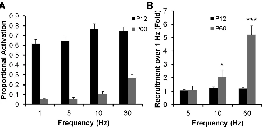
Figure 2.2. Postnatal development of DG gating behavior ... 38
Figure 2.3. Decreased DGC activation during postnatal development ... 41
Table 3.1. Specifications for VSD and calcium imaging probes ... 68
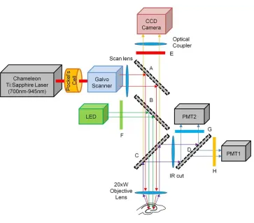
Figure 3.1. System diagram for the sequential VSD and calcium imaging microscope .... 69
Figure 3.2. Postnatal development of DG gating behavior ... 76
Figure 3.3. Relationship of AP activation to calcium transient amplitude in DGCs ... 80
Figure 3.4. Decreased DGC activation during postnatal development ... 84
Figure 3.5. DGC activation timing precision increases during postnatal development ... 89
Figure 3.6. Repeatability of activate/remain silent DGC response ... 94
Figure 3.7. Increased θ and γ frequency recruitment of DGCs during postnatal
development ... 97
Figure 3.8. Enhanced proportional activation of DGCs in the infrapyramidal blade of the
DG ...101
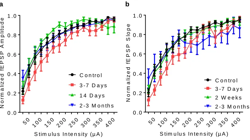
Figure 4.1. Input-output relationships of perforant path stimulation during
epileptogenesis ... 117
Figure 4.2. EEG-instrumentation (headcaps) and sub convulsive pilocarpine treatment
do not alter DGC responsiveness ... 118
Figure 4.3. Meier Kaplan survival curve depicting epilepsy onset in pilocarpine treated
xi
Figure 4.4. Changes in DGC activation during epileptogenesis ... 122
Figure 4.5. Alterations in evoked Ca2+ transient amplitudes during epileptogenesis ... 127
Figure 4.6. Location of responsive DGCs within the GCL ... 131
Figure 4.7. Juxtacellular recordings from active DGCs during epileptogenesis ... 136
Figure 4.8. IPSC alterations during epileptogenesis ... 140
Figure 4.9, m- and s-IPSCs during epileptogenesis ... 141
Figure 4.10. Disruption in inhibitory function degrades sparse DGC activation ... 145
Figure 4.11. Metabolic rescue of circuit collapse in chronically epileptic DG ... 149
Figure 4.12. Pharmacologic blockade of recurrent mossy fiber synapses ... 150
xii
AUTHOR CONTRIBUTIONS
Chapter 1 was written by C.G.D., and edited by C.G.D. and D.A.C.
Chapter 2 was originally published as:
Dengler CG, Coulter DA (2016) Normal and epilepsy-associated pathologic function of the dentate gyrus. Prog Brain Res 226:155–178.
It was written by C.G.D. following discussions between C.G.D. and D.A.C., and edited by C.G.D. and D.A.C.
Chapter 3 was originally published as:
Yu EP, Dengler CG, Frausto SF, Putt ME, Yue C, Takano H, Coulter DA (2013)
Protracted postnatal development of sparse, specific dentate granule cell activation
in the mouse hippocampus. J Neurosci 33:2947–2960.
E.P.Y. and D.A.C. designed research; E.P.Y., C.G.D., S.F.F., C.Y., and H.T. performed research; E.P.Y., C.G.D., M.E.P., and H.T. analyzed data; E.P.Y. and D.A.C. wrote the paper. Specifically, C.G.D. contributed figures 3.3, 3.5, and 3.7 as second author.
Chapter 4 was originally published as:
Dengler CG, Yue C, Takano H, Coulter DA (2017) Massively augmented hippocampal
dentate granule cell activation accompanies epilepsy development. Sci Rep 7:42090.
C.G.D. and D.A.C. designed research; C.G.D. and C.Y. performed research; all data and figures generated by CGD with exception of figures 4.8 and 4.9, in which data were collected and initial analysis conducted by C.Y.; C.G.D. and H.T. contributed new analytic tools; C.G.D., C.Y. and H.T. analyzed data; and C.G.D. and D.A.C. wrote the paper.
1
CHAPTER 1: Introduction
The research in this dissertation aims to understand the mechanisms by which
the principal cells of the dentate gyrus (DG), dentate granule cells (DGCs), are activated
by afferent input and how these activation properties may become corrupted as a result
of circuit changes occurring during epilepsy development. The DG plays critical roles
both in cognitive processing and in regulating propagation of pathological seizure
activity in epilepsy. Understanding how DGCs activate within their local circuit is
fundamental to understanding how the DG accomplishes its cognitive function of pattern
separation, the process whereby one discriminates between similar experiences. Studies
have demonstrated that DGCs exhibit firing in extremely small subpopulations during
execution of cognitive tasks, consistent with theories that DGCs transform incoming
information into a sparse neural code (Jung and McNaughton, 1993; Acsády and Káli,
2007). This sparse DGC activation has also been hypothesized to be particularly
important with regard to epilepsy. The DG is the primary gateway regulating cortical
input to the hippocampus and a possible contributor to cortical-hippocampal
interactions underlying seizures in temporal lobe epilepsy (TLE). The DG acts as a
regulated “gate” or “filter,” capable of protecting downstream hippocampal areas from
runaway excitation and epileptiform activity (Lothman et al., 1992; reviewed in Dengler
and Coulter, 2016). In animal models of - and patients with - TLE, the DG undergoes
marked circuit perturbations including cell loss, alterations in local inhibitory circuits,
axonal and dendritic sprouting, disruptions in ionic homeostasis, and
increased/dysregulated neurogenesis. It is unclear how these epilepsy-associated circuit
2
compromised, which circuit changes are responsible. We know little about the
mechanisms mediating the sparse network activation properties of the DG and the
emergence of this property during brain development. We know even less about how
epilepsy development may erode these properties which are critical to normal limbic
system function. This dissertation aims to address these gaps in our understanding of
this watershed circuit. To provide a general context for our ensuing studies, I begin by
introducing relevant background on epilepsy as well as the hippocampus and DG’s
relevance to both normal and pathological brain function in epilepsy.
Temporal lobe epilepsy and the hippocampus
Epilepsy is defined as the condition of having two or more recurrent, unprovoked
seizures in period of longer than 24 hours (I.L.A.E., 1993). With more than 40 clinical
variants, epilepsy is estimated to affect over 65 million people worldwide (Ngugi et al.,
2010; Thurman et al., 2011). Current estimates suggest that one in 26 people will develop
epilepsy in their lifetime (Hesdorffer et al., 2011), yet approximately 1/3 of epileptic
patients receiving anticonvulsant medications have inadequate seizure control or
experience intolerable side effects of this therapy (Jacobs et al., 2001). In addition to its
primary clinical presentation - the occurrence of seizures – epilepsy has many additional
consequential impacts on patients and families. Seizures themselves can alter and
damage the brain to produce significant cognitive comorbidities (Jokeit and Ebner,
2002) and, further, the sedative and dissociative side-effects of anticonvulsant
medications can negatively impact cognitive ability. Beyond pathological changes in the
brain function, epilepsy restricts patients’ ability to live independently, drive a motorized
3
prevalence, lack of responsiveness to current therapies, treatment-related side-effects,
and its myriad of comorbidities are all critical reasons to study this disorder.
Among all brain regions, the temporal lobe is the most epileptogenic (likely to
generate seizures; Tatum, 2012). TLE is a chronic disorder that involves structural
damage, dysregulated function and hyperexcitability in temporal lobe structures,
including the amygdala, entorhinal cortex, parahippocampal cortex, and most
predominately, the hippocampus (Thom et al., 2009; Tatum, 2012). TLE represents the
prevalent epilepsy variant in adults and is often associated with a brain injury in a
patient’s history, such as status epilepticus, traumatic brain injury, central nervous
system infection, stroke, and febrile seizures (French et al., 1993; Christensen et al.,
2009; Ferguson et al., 2010). As such, TLE is often designated an “acquired epilepsy,”
meaning that the initial injury may have incited the development of spontaneous
recurrent seizures in a previously healthy brain. This process has been termed
epileptogenesis and is classically thought to occur in three phases (Goldberg and Coulter,
2013; Maguire, 2016). The first phase of epileptogenesis is the initial precipitating injury,
in which some insult to the brain occurs. The second phase is a so-called “latent period”
in which the numerous circuit and cellular changes caused by the preceding insult
transform a healthy brain into one which can spontaneously generate seizures. This
latent period is often characterized as the epileptogenic period, a time in which no
seizures have yet occurred, despite the brain’s increasing propensity for generating
pathological discharges. Finally, the third phase is the emergence of epilepsy, a point at
which the alterations occurring in the brain during the latent period foment into
4
TLE can be effectively modeled by generating a controlled epileptogenic brain
insult in an experimental animal, most typically rodents. This injury can take many
forms. Among the most common experimental models of epilepsy is one induced by an
episode of status epilepticus (a prolonged, unremitting seizure), initiated by either local
or systemic administration of convulsant agents such a pilocarpine or kainate (Curia et
al., 2008; Ben-Ari, 2010; e.g. this model is implemented in Chapter 4). Pilocarpine is an
acetylcholine (ACh) agonist at muscarinic-type ACh receptors, while kainate is a
glutamate agonist at ionotropic glutamate receptors. In these models, animals undergo a
pharmacologically-induced, prolonged seizure which is then quelled with
anticonvulsants. After a short latent period (typically days to weeks), spontaneous,
recurrent seizures emerge, and the animal becomes epileptic. During epilepsy
development and presentation, alterations occurring in the brain can be assessed
experimentally. Other common models of acquired epilepsy include repetitive electrical
stimulation (kindling) of temporal lobe structures, physical injury (e.g. fluid percussive
injury), as well as early hypoxia-induced ischemic injuries and hyperthermic models of
febrile seizures (Bender et al., 2004; Pitkänen et al., 2006; Grone and Baraban, 2015).
After TLE becomes established in patients, it is one of the most intractable
epilepsies, with 60-75% of patients experiencing inadequate seizure control with
pharmacological treatments (Spencer, 2002). Often, surgical resection of the ictogenic
(seizure-initiating) brain region, typically the hippocampus and parahippocampal
structures, is the only effective treatment for these uncontrolled epilepsies. Surgical
resection eliminates seizures completely in 50-70% of patients, and can significantly
5
Since TLE seizures are often first recorded in the hippocampus (Toyoda et al.,
2013), there is a stereotypic pattern of pathology evident in this structure in patients
with epilepsy, and because its surgical removal can prevent seizures, the
epilepsy-associated alterations to this brain structure have been studied extensively. The changes
occurring in the hippocampi of TLE patients and animal models are numerous and share
clear patterns of hippocampal damage on several levels. At the gross structural level,
hippocampal sclerosis, a combination of structural atrophy and gliosis, is observed in
both TLE patients and animal models. (Margerison and Corsellis, 1966; Nadler et al.,
1978; Ben-Ari, 1985). At the circuit and cellular levels, there is evidence of numerous
epilepsy-associated changes in the hippocampus, among them: synaptic receptor
re-organization (Gibbs et al., 1997; Brooks-Kayal et al., 1998; Bernard, 2012; Nadler, 2012),
axonal sprouting (Tauck and Nadler, 1985; Zhang et al., 2009; Peng et al., 2013),
establishment of pathological recurrent excitatory pathways (Tauck and Nadler, 1985;
Buckmaster, 2010), strengthening of existing excitatory pathways (Ang et al., 2006), cell
death in a variety of neuronal populations (Margerison and Corsellis, 1966; Sloviter,
1987; Blümcke et al., 2000; Niquet et al., 2012), and aberrant generation of new neurons
(Parent et al., 1997; Hattiangady et al., 2004; Parent and Kron, 2012). Collectively, these
circuit changes within the hippocampus are thought to transform the hippocampus into
a structure capable of generating and propagating seizures. Further, since these same
disrupted limbic circuits are important in cognitive processing, these changes are likely
to alter normal circuit activation and function within the hippocampus and lead to
deficits in hippocampal-dependent cognitive functions (Liu et al., 2003; Kleen et al.,
6
The dentate gyrus: function and structure
Within the hippocampal formation, the DG has long been implicated as a key
structure for seizure initiation and propagation because of several early observations.
DGCs are among the few cell types of the hippocampus that survive in significant
numbers in epileptic patients (Margerison and Corsellis, 1966), Further, the combined
loss of DG inhibitory interneurons and sprouting of recurrent, auto-excitatory mossy
fiber synapses in the epileptic DG might allow for pathological circuit activation (Tauck
and Nadler, 1985; Sloviter, 1987). Later, many additional aberrant properties of the DG’s
circuitry were discovered to follow epileptogenic injuries, establishing the ever
increasing inquiry into this structure’s ability to both generate seizures and/or allow for
their propagation (reviewed in detail in Dengler and Coulter, 2016 [vide infra, Chapter 2]).
These alterations to the basic DG circuit have received particularly strong
attention owing to its position as the gateway to the hippocampus: the site of the first
synapse in the canonical trisynaptic loop (Figure 1.1b). The DG is situated between the
entorhinal cortex and the hippocampus proper (Figs. 1.1a,b), where it processes
incoming multimodal, entorhinal-cortical spatial information (Sargolini et al., 2006;
Fyhn et al., 2008). The DG transforms these cortical inputs into a sparse neural code in a
process termed “pattern separation,” before relaying its output onto area CA3 via the
mossy fiber pathway (Leutgeb et al., 2007; Moser et al., 2008; de Almeida et al., 2009;
Fig 1.1b). These mossy fiber outputs to CA3 are the second synapse of the trisynaptic
circuit. In this location, and owing to its relatively inexcitable activation properties, the
7
downstream CA structures (Coulter and Carlson, 2007; Hsu, 2007; Dengler and Coulter,
2016). The DG’s output is then processed locally in area CA3 in a process termed
“pattern completion” (Leutgeb and Leutgeb, 2007; Bakker et al., 2008; Neunuebel and
Knierim, 2014). This transformed information is then transferred to area CA1 via the
Schaffer collaterals in the third synapse of the loop (Figure 1.1b). CA1 then further
processes this DG-CA3-input and integrates this information together with direct
cortical inputs from the temporo-ammonic pathway (Ang et al., 2005; Acsády and Káli,
2007), before relaying the hippocampus’s final output back to the subiculum and
entorhinal cortex, thus completing the hippocampal loop (Figure 1.1b). As a whole, the
hippocampal trisynaptic loop structure has been implicated as a key circuit involved in
many aspects of spatial and declarative memories (Moser et al., 2008). At same time,
this excitatory, interconnected “circular” structure has also raised concerns that
pathological excitation may reverberate through the hippocampal loop during seizures
(Stringer and Lothman, 1992; Rafiq et al., 1993; Iijima et al., 1996).
Before reviewing the functional roles and properties of the DG and their
relevance to both cognitive function and epilepsy in Chapter 2, I will first review the
relevant neuroanatomy and cells of the DG circuit. The DG is organized into repeating
units called lamellae which are oriented transverse to the long axis of the hippocampus
(Figs. 1.1a,b). Each lamella of the DG can be divided into three basic areas. The
outermost layer of the DG, the molecular layer, contains both the primary excitatory
afferent fibers innervating the DG as well as the dendrites of the DG’ principle cells,
DGCs. The next area of the DG is the granule cell layer, which primarily contains DGC
somata. Finally, the hilus (or polymorph layer) harbors a variety of inhibitory
8
a
b
c
9
Figure 1.1. Basic anatomical position and organization of the DG.(a) A sagittal view of the
mouse brain highlighting the location of the entorhinal cortex and hippocampus. Red lines
indicate the hippocampus’ tranverse, lamellar axis. One lamella is shown in (b). (b) A single
hippocampal lamella is illustrated to show the classical “trisynaptic circuit”, consisting of the PP
axons of entorhinal cortical layer II neurons; mossy fiber (MF) axons of DGCs to CA3; and finally,
Schaffer collateral axons (SC) of CA3 to CA1. Red lines indicate area shown in (c). (c) Schematic
illustration of layers in a DG lamella depicting basic connectivity of DG neurons: Black, the DG’s
principle excitatory cells, DGCs; Grey, inner molecular layer projecting, excitatory mossy cells;
red, somatically-targeting inhibitory basket cells with axons in granule cell layer; and purple, dendritically-targeting inhibitory hilar interneurons with axonal projections throughout the
10
The primary excitatory afferent input to the DG is the perforant path (PP)
projection. The PP originates from layer II stellate cells of the entorhinal cortex and
terminates through excitatory, en passant glutamatergic synapses onto DGC dendrites. The PP is composed of two sub-pathways, the lateral and medial perforant paths. The
lateral perforant path (LPP) originates in the lateral portion of the entorhinal cortex and
projects onto the outer dendrites of DGCs, defining the outer molecular layer, whereas
the medial perforant path (MPP) originates in the medial entorhinal cortex and projects
onto the middle dendrites of DGCs comprising the middle molecular layer. Additional,
minor external inputs to the DG include cholinergic inputs and supramammilary fibers
terminating in the inner molecular layer. Finally, the DG receives internal excitatory
drive from mossy cells in the hilus, whose axons project onto the hilar neurons and inner
molecular layer, synapsing on proximal DGC dendrites. Additionally, the hilus of the DG
receives multiple neuromodulatory inputs including cholinergic, noradrenergic and
serotonergic fibers from extrahippocampal nuclei. The principle output of the DG is the
mossy fiber pathway. This pathway terminates onto excitatory mossy cells and various
inhibitory interneurons in the hilus and onto inhibitory interneurons and pyramidal cells
in CA3 (See Witter, 1993, for detailed review).
The majority of DGCs comprise a relatively homogenous cell population with two
exceptions: adult-born granule cells (abDGCs) and semilunar granule cells. Small
numbers of abDGCs are generated in the DG throughout an animal’s(and human’s) life
(Altman and Das, 1965; Eriksson et al., 1998). During their maturation, these neurons
display some unique electrophysiological properties, until maturing and become
functionally indistinguishable from mature DGCs (van Praag et al., 2002). Semilunar
11
morphology present in the inner molecular layer. Their unique plateau potentials enable
them to fire continuously during phasic bursts onto their postsynaptic targets which
include hilar interneurons and mossy cells (Williams et al., 2007).
The GABAergic neurons of the DG have somata present in all layers of the DG,
and different classes of these cells have layer-specific synaptic targets. One of the most
common subtypes is the basket cell, a class of inhibitory interneurons typically
characterized by fast-spiking membrane behavior and uniformly possessing axonal
arbors with exclusively perisomatic-targeting via basket-like plexuses surrounding DGC
somata and axon initial segments (Freund and Buzsáki, 1996; Fig. 1.1b). Basket cells
typically have a pyramidal cell body located at the periphery of the GC layer. These
interneurons provide somatic GABA-ergic inhibition to DGCs via both feedforward
(afferents from EC; Seress and Pokorny, 1981) and feedback (innervation from DGCs;
Ribak and Peterson, 1991) pathways (Freund and Buzsáki, 1996). Basket cells commonly
express the calcium-binding protein parvalbumin (Ribak et al., 1990), but can also
express other markers such as neuropeptide Y (NPY), cholecystokinin (CCK), and
vasoactive-intestinal peptide (VIP), or exhibit no clear expression of known-markers
(Gulyás et al., 1991; Acsády et al., 1996).
The second major class of inhibitory interneurons in the DG is the hilar
interneurons. These neurons commonly contain the neuropeptides somatostatin and/or
neuropeptide Y and their axonal terminals primarily target DGC dendrites in the middle
and outer molecular layers (Bakst et al., 1986; Chan-Palay et al., 1986; Köhler et al.,
1987; Amaral et al., 1988). These neurons are commonly ascribed to operate in a
feedback capacity, receiving the majority of their excitatory input from DGCs (but also
12
limit the efficacy of entorhinal cortical afferents onto DGCs (Freund and Buzsáki, 1996;
Houser, 2007).
Two less common subtypes of interneurons that are present in the DG are HICAP
cells and neurogliaform cells. HICAP cells (hilar interneurons projecting to the
commissural/associational pathway) have somata localized in the hilus and axons that
innervate the inner molecular layer (Han et al., 1993). Finally, neurogliaform cells have
somata located in the molecular layer of the DG with dense local axonal plexuses in the
molecular layer and are thought to provide inhibition through “bulk-transmission” of
GABA through extrasynaptic receptors (Freund and Buzsáki, 1996; Armstrong et al.,
2012).
Owing to the repeating, lamellar structure of the DG and hippocampus, intact,
well-connected ex vivo brain slices can be prepared by slicing the brain along the transverse axis of the hippocampus. Figure 1.2 demonstrates this within-slice
connectivity via retro-anterograde labeling using the lipophilic dye, DiI, in a standard
hippocampal-entorhinal cortical slice. This labeling demonstrates that horizontally
oriented hippocampal-entorhinal cortical slices contain PP axons originating in the EC
that project onto and around the molecular layer of the DG. These PP path axons are
functionally intact and allow for direct afferent stimulation of downstream DG
structures. Further, DG efferent connectivity is maintained to both the hilus and CA3. In
both of the primary research of chapters of this dissertation (3 and 4), I study functional
properties of the DG using these brain slice preparations utilizing both imaging and
13
Figure 1.2. Intact EC-DG-Hilar-CA3 excitatory projections in horizontal
hippocampal-entorhinal cortex slices.A 350 μm thick, hippocampal-entorhinal cortical brain slice,
demonstrating intact projections from the EC to the DG, and from the DG to CA3. Grey,
transmitted light micrograph. Red, DiI, a lipophilic fluorescent dye, used to trace cell membranes.
, placement of a small DiI crystal in the DG molecular layer retrogradely traces DG afferent PP
fibers originating in the EC, and anterogradely labels DG MF axonal projections to the hilus and
CA3.
EC
DG
PP
MF
CA3
14
Objectives and Organization of the Proposed Studies
The two primary aims of this dissertation are to (1) characterize the development
of network firing properties in the healthy DG and (2) determine the nature of DGC
activation within the DG network in animals with epilepsy. This thesis is, in large part, a
compilation of 3 self-contained, published manuscripts, accompanied by additional
background, future directions, and final conclusions.
Earlier in the present chapter, I introduced relevant background on epilepsy, the
hippocampus, DG neuroanatomy, and the general aims of this thesis as to provide
overall context to this dissertation’s research.
Chapter 2 provides a detailed literature review of the DG’s sparse activation
properties, its relevance to cognitive processing, and its secondary role in functioning as
a regulated “gate” or “filter” of cortical afferents. Additionally, this chapter discusses
many of the epilepsy-associated circuit changes known to occur within the DG and their
possible roles in altering this circuits’ normal function.
Chapter 3 details the results of a study focused on characterizing the emergence
of sparse DGC activation during brain development and how this property accompanies
the associated development of an animal’s cognitive functions (Aim 1). We demonstrate a
protracted, progressive sparsification of DGC responses during brain development,
accompanied by increased temporal precision and frequency dependence of activation.
Chapter 4 characterizes the collapse of normal sparse DGC activation in a mouse
model of TLE, as well as the mechanisms of this circuit failure (Aim 2). This study
reveals that epilepsy emergence is accompanied by massive enhancement of the
15
local network, though the mechanistic cause of this compromised inhibitory function
varied over time following epileptogenic injury.
Finally, Chapter 5 presents the future directions of this research program and
16
Chapter 1 Bibliography
Acsády L, Arabadzisz D, Freund TF (1996) Correlated morphological and neurochemical features identify different subsets of vasoactive intestinal
polypeptide-immunoreactive interneurons in rat hippocampus. Neuroscience 73:299–315. Acsády L, Káli S (2007) Models, structure, function: the transformation of cortical
signals in the dentate gyrus. Prog Brain Res 163:577–599.
Altman J, Das GD (1965) Autoradiographic and histological evidence of postnatal hippocampal neurogenesis in rats. J Comp Neurol 124:319–335.
Amaral DG, Insausti R, Campbell MJ (1988) Distribution of somatostatin immunoreactivity in the human dentate gyrus. J Neurosci 8:3306–3316.
Ang CW, Carlson GC, Coulter DA (2005) Hippocampal CA1 circuitry dynamically gates direct cortical inputs preferentially at theta frequencies. J Neurosci 25:9567–9580. Ang CW, Carlson GC, Coulter DA (2006) Massive and specific dysregulation of direct
cortical input to the hippocampus in temporal lobe epilepsy. J Neurosci 26:11850–
11856.
Armstrong C, Krook-Magnuson E, Soltesz I (2012) Neurogliaform and Ivy Cells: A Major Family of nNOS Expressing GABAergic Neurons. Front Neural Circuits 6:23.
Bakker A, Kirwan CB, Miller M, Stark CEL (2008) Pattern separation in the human hippocampal CA3 and dentate gyrus. Science 319:1640–1642.
Bakst I, Avendano C, Morrison JH, Amaral DG (1986) An experimental analysis of the origins of somatostatin-like immunoreactivity in the dentate gyrus of the rat. J Neurosci 6:1452–1462.
Ben-Ari Y (1985) Limbic seizure and brain damage produced by kainic acid: Mechanisms and relevance to human temporal lobe epilepsy. Neuroscience 14:375–403.
Ben-Ari Y (2010) Kainate and temporal lobe epilepsies: Three decades of progress. Epilepsia 51:40.
Bender RA, Dubé C, Baram TZ (2004) Febrile seizures and mechanisms of
epileptogenesis: insights from an animal model. Adv Exp Med Biol 548:213–225. Bernard C (2012) Alterations in synaptic function in epilepsy.
Blümcke I, Suter B, Behle K, Kuhn R, Schramm J, Elger CE, Wiestler OD (2000) Loss of
hilar mossy cells in Ammon’s horn sclerosis. Epilepsia 41 Suppl 6:S174-80.
Brooks-Kayal AR, Shumate MD, Jin H, Rikhter TY, Coulter DA (1998) Selective changes in single cell GABA(A) receptor subunit expression and function in temporal lobe epilepsy. Nat Med 4:1166–1172.
Buckmaster PS (2010) Mossy fiber sprouting in the dentate gyrus. Epilepsia 51:39–39. Chan-Palay V, Köhler C, Haesler U, Lang W, Yasargil G (1986) Distribution of neurons
17
human hippocampus. J Comp Neurol 248:360–375.
Christensen J, Pedersen MG, Pedersen CB, Sidenius P, Olsen J, Vestergaard M (2009) Long-term risk of epilepsy after traumatic brain injury in children and young adults: a population-based cohort study. Lancet (London, England) 373:1105–1110. Coulter DA, Carlson GC (2007) Functional regulation of the dentate gyrus by
GABA-mediated inhibition. Prog Brain Res 163:235–243.
Curia G, Longo D, Biagini G, Jones RSG, Avoli M (2008) The pilocarpine model of temporal lobe epilepsy. J Neurosci Methods 172:143–157.
de Almeida L, Idiart M, Lisman JE (2009) The input-output transformation of the hippocampal granule cells: from grid cells to place fields. J Neurosci 29:7504–7512. Dengler CG, Coulter DA (2016) Normal and epilepsy-associated pathologic function of
the dentate gyrus. Prog Brain Res 226:155–178.
England MJ, Liverman CT, Schultz AM, Strawbridge LM (2012) Epilepsy across the spectrum: Promoting health and understanding. A summary of the Institute of Medicine report. Epilepsy Behav 25:266–276.
Eriksson PS, Perfilieva E, Björk-Eriksson T, Alborn AM, Nordborg C, Peterson DA, Gage FH (1998) Neurogenesis in the adult human hippocampus. Nat Med 4:1313–1317. Ferguson PL, Smith GM, Wannamaker BB, Thurman DJ, Pickelsimer EE, Selassie AW
(2010) A population-based study of risk of epilepsy after hospitalization for traumatic brain injury. Epilepsia 51:891–898.
French JA, Williamson PD, Thadani VM, Darcey TM, Mattson RH, Spencer SS, Spencer DD (1993) Characteristics of medial temporal lobe epilepsy: I. Results of history and physical examination. Ann Neurol 34:774–780.
Freund TF, Buzsáki G (1996) Interneurons of the hippocampus. Hippocampus 6:347–
470.
Fyhn M, Hafting T, Witter MP, Moser EI, Moser M-B (2008) Grid cells in mice. Hippocampus 18:1230–1238.
Gibbs JW, Shumate MD, Coulter DA (1997) Differential epilepsy-associated alterations in postsynaptic GABA(A) receptor function in dentate granule and CA1 neurons. J Neurophysiol 77:1924–1938.
Goldberg EM, Coulter DA (2013) Mechanisms of epileptogenesis: a convergence on neural circuit dysfunction. Nat Rev Neurosci 14:337–349.
Grone BP, Baraban SC (2015) Animal models in epilepsy research: legacies and new directions. Nat Neurosci 18:339–343.
Gulyás AI, Tóth K, Dános P, Freund TF (1991) Subpopulations of GABAergic neurons containing parvalbumin, calbindin D28k, and cholecystokinin in the rat
hippocampus. J Comp Neurol 312:371–378.
18
the dentate gyrus of the rat hippocampus. Eur J Neurosci 5:395–410.
Hattiangady B, Rao MS, Shetty AK (2004) Chronic temporal lobe epilepsy is associated with severely declined dentate neurogenesis in the adult hippocampus. Neurobiol Dis 17:473–490.
Hesdorffer DC, Logroscino G, Benn EKT, Katri N, Cascino G, Hauser WA (2011) Estimating risk for developing epilepsy. Neurology 76:23–27.
Houser CR (2007) Interneurons of the dentate gyrus: an overview of cell types, terminal fields and neurochemical identity. Prog Brain Res 163:217–232.
Hsu D (2007) The dentate gyrus as a filter or gate: a look back and a look ahead. Prog Brain Res 163:601–613.
I.L.A.E. (1993) Guidelines for epidemiologic studies on epilepsy. Commission on Epidemiology and Prognosis, International League Against Epilepsy. Epilepsia 34:592–596.
Iijima T, Witter MP, Ichikawa M, Tominaga T, Kajiwara R, Matsumoto G (1996) Entorhinal-Hippocampal Interactions Revealed by Real-Time Imaging. Science (80- ) 272:1176–1179.
Jacobs MP, Fischbach GD, Davis MR, Dichter MA, Dingledine R, Lowenstein DH, Morrell MJ, Noebels JL, Rogawski MA, Spencer SS, Theodore WH (2001) Future directions for epilepsy research. Neurology 57:1536–1542.
Jokeit H, Ebner A (2002) Effects of chronic epilepsy on intellectual functions. Prog Brain Res 135:455–463.
Jung MW, McNaughton BL (1993) Spatial selectivity of unit activity in the hippocampal granular layer. Hippocampus 3:165–182.
Kleen JK, Scott RC, Holmes GL, Lenck-Santini PP (2010) Hippocampal interictal spikes disrupt cognition in rats. Ann Neurol 67:250–257.
Köhler C, Eriksson LG, Davies S, Chan-Palay V (1987) Co-localization of neuropeptide tyrosine and somatostatin immunoreactivity in neurons of individual subfields of the rat hippocampal region. Neurosci Lett 78:1–6.
Leutgeb JK, Leutgeb S, Moser M-B, Moser EI (2007) Pattern separation in the dentate gyrus and CA3 of the hippocampus. Science 315:961–966.
Leutgeb S, Leutgeb JK (2007) Pattern separation, pattern completion, and new neuronal codes within a continuous CA3 map. Learn Mem 14:745–757.
Liu X, Muller RU, Huang L-T, Kubie JL, Rotenberg A, Rivard B, Cilio MR, Holmes GL (2003) Seizure-induced changes in place cell physiology: relationship to spatial memory. J Neurosci 23:11505–11515.
Lothman EW, Stringer JL, Bertram EH (1992) The dentate gyrus as a control point for seizures in the hippocampus and beyond. Epilepsy Res Suppl 7:301–313.
Maguire J (2016) Epileptogenesis: More than just the latent period. Epilepsy Curr 16:31–
19
Margerison JH, Corsellis JAN (1966) Epilepsy and the temporal lobes: A clinical, electroencephalographic and neuropathological study of the brain in epilepsy, with particular reference to the temporal lobes. Brain 89:499–530.
Moser EI, Kropff E, Moser M-B (2008) Place cells, grid cells, and the brain’s spatial
representation system. Annu Rev Neurosci 31:69–89.
Nadler J (2012) Plasticity of Glutamate Synaptic Mechanisms. In Jasper’s Basic Mechanisms of the Epilepsies, 4th edition pp 1–17.
Nadler JV, Perry BW, Cotman CW (1978) Intraventricular kainic acid preferentially destroys hippocampal pyramidal cells. Nature 271:676–677.
Neunuebel JP, Knierim JJ (2014) CA3 retrieves coherent representations from degraded input: Direct evidence for CA3 pattern completion and dentate gyrus pattern
separation. Neuron 81:416–427.
Ngugi AK, Bottomley C, Kleinschmidt I, Sander JW, Newton CR (2010) Estimation of the burden of active and life-time epilepsy: A meta-analytic approach. Epilepsia 51:883–890.
Niquet J, Lopez-Meraz M-L, Wasterlain CG (2012) Programmed Necrosis After Status Epilepticus.
Parent JM, Kron MM (2012) Neurogenesis and Epilepsy. In Jasper’s Basic Mechanisms of the Epilepsies, 4th edition.
Parent JM, Yu TW, Leibowitz RT, Geschwind DH, Sloviter RS, Lowenstein DH (1997) Dentate granule cell neurogenesis is increased by seizures and contributes to aberrant network reorganization in the adult rat hippocampus. J Neurosci 17:3727–
3738.
Peng Z, Zhang N, Wei W, Huang CS, Cetina Y, Otis TS, Houser CR (2013) A reorganized GABAergic circuit in a model of epilepsy: evidence from optogenetic labeling and stimulation of somatostatin interneurons. J Neurosci 33:14392–14405.
Pitkänen A, Schwartzkroin P, Moshe SL (2006) Models of Seizures and Epilepsy. Elsevier Academic Press.
Rafiq A, DeLorenzo RJ, Coulter DA (1993) Generation and propagation of epileptiform discharges in a combined entorhinal cortex/hippocampal slice. J Neurophysiol 70:1962–1974.
Ribak CE, Nitsch R, Seress L (1990) Proportion of parvalbumin-positive basket cells in the GABAergic innervation of pyramidal and granule cells of the rat hippocampal formation. J Comp Neurol 300:449–461.
Ribak CE, Peterson GM (1991) Intragranular mossy fibers in rats and gerbils form synapses with the somata and proximal dendrites of basket cells in the dentate gyrus. Hippocampus 1:355–364.
20
Seress L, Pokorny J (1981) Structure of the granular layer of the rat dentate gyrus. A light microscopic and Golgi study. J Anat 133:181–195.
Sloviter RS (1987) Decreased hippocampal inhibition and a selective loss of interneurons in experimental epilepsy. Science 235:73–76.
Spencer SS (2002) When should temporal-lobe epilepsy be treated surgically? Lancet Neurol 1:375–382.
Stringer JL, Lothman EW (1992) Reverberatory seizure discharges in hippocampal-parahippocampal circuits. Exp Neurol 116:198–203.
Tatum WO (2012) Mesial temporal lobe epilepsy. J Clin Neurophysiol 29:356–365. Tauck DL, Nadler J V (1985) Evidence of functional mossy fiber sprouting in
hippocampal formation of kainic acid-treated rats. J Neurosci 5:1016–1022. Thom M, Eriksson S, Martinian L, Caboclo LO, McEvoy AW, Duncan JS, Sisodiya SM
(2009) Temporal Lobe Sclerosis Associated With Hippocampal Sclerosis in Temporal Lobe Epilepsy: Neuropathological Features. J Neuropathol Exp Neurol 68:928–938.
Thurman DJ et al. (2011) Standards for epidemiologic studies and surveillance of epilepsy. Epilepsia 52:2–26.
Toyoda I, Bower MR, Leyva F, Buckmaster PS (2013) Early activation of ventral hippocampus and subiculum during spontaneous seizures in a rat model of temporal lobe epilepsy. J Neurosci 33:11100–11115.
van Praag H, Schinder AF, Christie BR, Toni N, Palmer TD, Gage FH (2002) Functional neurogenesis in the adult hippocampus. Nature 415:1030–1034.
Witter MP (1993) Organization of the entorhinal-hippocampal system: a review of current anatomical data. Hippocampus 3:33–44.
21
CHAPTER 2:
Normal and epilepsy-associated pathologic
function of the dentate gyrus.
Abstract
The dentate gyrus plays critical roles both in cognitive processing and in the
regulation of the induction and propagation of pathological activity. The cellular and
circuit mechanisms underlying these diverse functions overlap extensively. At the
cellular level, the intrinsic properties of dentate granule cells combine to endow these
neurons with a fundamental reluctance to activate, one of their hallmark traits. At the
circuit level, the dentate gyrus constitutes one of the more heavily inhibited regions of
the brain, with strong, fast feedforward and feedback GABAergic inhibition dominating
responses to afferent activation. In pathologic states such as epilepsy, a number of
alterations within the dentate gyrus combine to compromise the regulatory properties of
this circuit, culminating in a collapse of its normal function. This epilepsy-associated
transformation in the fundamental properties of this critical regulatory hippocampal
circuit may contribute both to seizure propensity, and cognitive and emotional
comorbidities characteristic of this disease state.
Introduction
Situated as the initial component of the canonical trisynaptic circuit, the dentate
gyrus (DG) is a critical entry point to the hippocampus, functioning as a key regulator of
cortical input to the limbic system. The DG is involved in the performance of
hippocampal-dependent tasks and is postulated to accomplish these cognitive functions
through a transformation of highly active and multimodal afferent cortical inputs into a
22
discuss how the sparse, selective activation properties of the DG’s principal cells, dentate
granule cells (DGCs), are critical to hippocampal cognitive function and how this
characteristic firing property plays a key role in the regulation of seizure activity in
diseases such as epilepsy. Finally, we will consider mechanisms that may erode
appropriate DG circuit activation and play a pivotal role in epileptogenesis, seizure
propensity in epilepsy, as well as the cognitive comorbidities associated with the disease.
In vivo recording studies have demonstrated that DGCs exhibit spatially selective firing in extremely small populations (Jung and McNaughton, 1993a; Chawla et al.,
2005). This characteristic sparse activation is thought to enable DGCs to participate in
the execution of cognitive functions such as pattern separation and novelty detection
(Leutgeb et al., 2007). The propensity for DGCs to generate action potentials (APs) is
normally tightly constrained by a combination of cell-intrinsic properties and powerful
local inhibitory control, culminating in a population of neurons that are extremely
reluctant to activate. However, when DGCs do activate, they exert strong excitatory
influence on their downstream targets. A secondary consequence of the DG’s low
excitability is its ability to restrict relay of pathological, synchronous cortical activity into
the hippocampus and regulate seizure activity in diseases such as epilepsy: a
phenomenon termed “dentate gating” (Lothman et al., 1992; Stringer and Lothman,
1992). However, if this regulatory ability becomes compromised, the DG’s powerful
excitatory influence on its downstream targets in the hippocampus can allow it to relay
and amplify synchronous pathological activity through the limbic system, potentially
fomenting seizure activity.
These characteristic DG circuit properties are significantly disrupted both in
humans with epilepsy and in animal models of the disorder. A large aggregation of
23
sprouting of pathologic, recurrent excitatory networks, molecular and cellular alterations
of local inhibitory circuits, aberrant neurogenesis, astrocytic gliosis, and changes in the
intrinsic properties of DGCs. These pathology-associated alterations have generated a
prevalent hypothesis that the DG’s normal gating function is compromised during
epilepsy and its development and contributes to increased seizure propensity. However,
this DG gate failure hypothesis has yet to receive adequate experimental support, and if
found to exist, the exact mechanisms mediating such a circuit collapse are likely
mechanistically complex.
Activity in the DG is sparse
Among the most unusual properties of the DG is the uniquely low level of
neuronal activity among its principal cell population. Assays measuring DGC activity
over the last several decades have consistently described DGC activation as unusually
low compared to many other brain areas. In vivo unit recordings during spatial navigation tasks have demonstrated exceedingly low levels of DGC activity; the vast
majority of recorded DGCs display extremely low mean firing rates (typically <0.5 Hz)
and activate only in tightly restricted spatial and directional receptive fields (Jung and
McNaughton, 1993a; Leutgeb et al., 2007; Neunuebel and Knierim, 2012). Moreover,
studies examining neuronal expression of activity-dependent immediate-early genes
such as c-Fos, Zif286, and Arc as surrogate measures of DGC AP firing have found
exceptionally sparse labeling in DGCs, with only 2–5% of DGCs expressing these genes,
even after exposure to multiple environments or spatial cognitive tasks (Chawla et al.,
2005; Tashiro et al., 2007; Alme et al., 2010). In comparison, similar spatial navigation
24
cells in downstream CA subfields (Guzowski et al., 1999), highlighting the comparative
paucity of DGC activation.
The extremely sparse activation of DGCs is particularly puzzling given the
anatomy of their primary afferents from the entorhinal cortex (EC). DGCs receive
massively convergent and divergent inputs from the medial and lateral entorhinal
cortices. Singly-labeled EC stellate cells projecting to the DG can project to as much as
1/3 of the rodent DG, making en passant synapses with DGCs throughout this extensive
axonal arbor (Tamamaki and Nojyo, 1993). Conversely, retrograde tracing studies have
shown that discrete regions of the DG can receive inputs from substantial portions of the
EC, with a single DGC receiving inputs from 3600 to 5600 neurons widely distributed
across the EC (Patton and McNaughton, 1995; van Groen et al., 2003). In addition to the
massive convergence and divergence of DG afferents, the originating EC stellate cells of
this pathway, particularly “grid cells” in the medial EC, are promiscuously active. These
neurons exhibit large-scale activation and demonstrate tessellated grid-like receptive
fields across spatial environments (Fyhn et al., 2004, 2007). Given the repeating nature
of grid cell activity throughout spatial environments, it appears that a significant portion
of DG afferents are almost always active, given that the vertices of grid cells receptive
fields comprise approximately 20% of the entire environment (Lisman, 2011). Thus, the
convergence, divergence, and nearly continuous activity of the EC perforant path
projection provide substantial excitatory input to the DG.
Given this robust and constant excitatory drive impinging onto DGCs from the
EC, why are so few of these cells active during behavioral tasks? It is likely that
cell-intrinsic biophysical properties of DGCs contribute in part to their relative inexcitability.
DGCs exhibit hyperpolarized membrane potentials compared to most hippocampal
25
endows them with a long time constant for integrating synaptic inputs. Further, they lack
regenerative calcium conductances to permit phasic or “burst mode” firing and they
exhibit remarkable spike frequency adaptation during sustained depolarizations (Fricke
and Prince, 1984; Spruston and Johnston, 1992; Staley et al., 1992). Finally, DGC
dendrites display significant attenuation of synaptic input owing to the lack of active
conductances that would allow dendritic spiking or input amplification (Krueppel et al.,
2011). This combination of properties endows DGCs with an innate reluctance to fire
APs.
In addition to these relatively hypoexcitable cell-intrinsic properties, DGCs reside
in a strongly inhibitory local network, which provides rich feedforward, feedback, and
tonic inhibitory input. Feedforward inhibitory drive onto DGCs is particularly strong due
to a number of factors. First, several classes of feedforward inhibitory interneurons,
including both somatically targeting fast-spiking interneurons on the granule cell layer’s
periphery as well as dendritically targeting hilar interneurons, are far more easily
recruited by perforant path activation than are DGCs (Scharfman, 1991; Ewell and Jones,
2010). Second, the fast-spiking basket cell interneurons of the DG are particularly
specialized to integrate afferent inputs, generate APs, and powerfully inhibit DGCs upon
perforant path activation with extraordinary rapidity. Among the many specializations of
these basket cells are high K+–Na+ conductance ratios in their dendrites which allow for
rapid and temporally precise integration and activation (Hu et al., 2010), nonuniform
cable properties that accelerate the time course of fast somatic synaptic potentials and
elevate the efficacy of slower distal inputs (Nörenberg et al., 2010), and highly efficient
calcium-buffering abilities that allow them to rapidly and repeatedly couple synchronous
transmitter release with AP firing (Aponte et al., 2008; see Hu et al., 2014; for review of
26
constrain DGC firing, and limit firing rates of DGCs when spiking occurs, because this
inhibitory input primarily targets DGC somata and axon initial segments, the cellular
domains of synaptic integration of excitatory and inhibitory inputs.
Third, feedback inhibition in the DG also appears particularly specialized. DGC
axons are normally not present in the molecular layer or granule cell layer, but are
instead restricted exclusively to the hilus (Claiborne et al., 1986), where 50% of their
target cell population is composed of GABAergic interneurons (Houser and Esclapez,
1994; Houser, 2007), suggesting that many of the DGCs’ postsynaptic targets may be
inhibitory neurons. DGCs are unique among cortical principal cells in that they are
endowed with a repertoire of differing synaptic terminal types in their axons. These
axons give rise to 7–12 mossy fiber boutons which synapse with excitatory hilar mossy
cells and onto CA3 pyramidal cells, but they have a far larger number (100–150) of
smaller terminals, including filopodial extensions and en passant synapses, which
primarily target interneurons (Acsády et al., 1998). Due to this target selectivity, the vast
majority of DGCs’ postsynaptic targets may be inhibitory interneurons. These targets
include both hilar feedback interneurons (a majority of which are somatostatin positive)
that provide both dendritic feedback inhibition onto DGCs, which limits the strength of
cortical afferent inputs, as well as fast-spiking interneurons with somatically targeted
inhibition (typically also performing feedforward functions). This biases synaptic
integration toward restriction of DGC activation (Freund and Buzsáki, 1996). This
feedback inhibition is hypothesized to contribute to making the DG a competitive
network, or a “winner-take-all” scheme, in which the activation of a sparse population of
DGCs effectively silences the remaining majority of the population (de Almeida et al.,
27
Given the high levels of activity of its upstream cortical afferents in relation to the
surprisingly low activity levels of its principal cells, the information transformation
within the DG has been described as a “sparsification” operation or as a form of “sparse
coding” in which the DG’s output becomes a more sparse representation of its incoming
neuronal activity pattern, both in terms of number of DGCs activating, as well as their
limited firing rates (O’Reilly and McClelland, 1994; Treves and Rolls, 1994; Acsády and
Káli, 2007). The sheer paucity of DGC activation raises an important question with
regard to cognitive processing. If so few DGCs are active, how can their extremely sparse
activity contribute in any significant way toward neural processing of
hippocampal-dependent cognitive tasks? In short, the DG could only effectively transmit information
through an amplifying mechanism by which DGCs could strongly excite their
downstream targets in the hilus, and perhaps more critically, the pyramidal cells of CA3,
without relying on synaptic convergence (since DGCs only target a few CA3 pyramidal
cells). Without such a mechanism, the sparse coding in the DG would be a synaptic “dead
end” and little, if any, information content could be relayed to downstream hippocampal
structures. In vivo recordings have shown that single DGCs are indeed capable of reliably activating CA3 pyramidal cells (Henze et al., 2002). Unique properties of DGC axon
terminals appear to be specialized to achieve this end. Mossy fiber boutons synapse with
excitatory hilar mossy cells and CA3 pyramidal cells. These synapses are unique in the
mammalian brain in that they are exceptionally large, contain multiple release sites, and
display extraordinarily robust frequency facilitation, even at very low frequencies (Salin
et al., 1996). Furthermore, even though the majority of DGCs’ synaptic targets are
feedback (onto DGCs) and feedforward (onto CA3) inhibitory interneurons, these
connections are mediated by filopodial synapses, which, in contrast to mossy fiber
28
specialized synaptic properties allow DGCs to act as conditional “detonators”
(McNaughton and Morris, 1987) of their postsynaptic CA3 targets during periods of
elevated firing rates.
What functions are served by sparsely activating DGCs?
What are the implications of a sparsely coding network of cells that rarely
activate, yet when they do, exert massive excitatory influence on their postsynaptic
targets? The answer varies depending on whether one is considering the DG’s role in
cognition and memory, or epilepsy. The DG’s position at the entrance to the
hippocampus combined with the sparse activation of DGCs led to the theory that DG
circuitry transforms promiscuous and highly active cortical input into a sparse neural
code suitable for representing novel aspects of episodic memories (Treves and Rolls,
1994; Treves et al., 2008). This DG-mediated transformation is critical to many aspects
of hippocampal-dependent neural processing, most notably pattern separation and
pattern completion, which are fundamental components of episodic memory encoding
(Leutgeb et al., 2007; McHugh et al., 2007; Nakashiba et al., 2012; Neunuebel and
Knierim, 2014). Behaviorally, pattern separation is the ability to discriminate subtle
differences between similar episodes (e.g. “where one parked one’s car today is not
necessarily the same place as yesterday”; O’Reilly and McClelland, 1994). From a
computational perspective, this translates to the "process of transforming similar inputs
into more dissimilar outputs" (Piatti et al., 2013), thereby detecting novelty and isolating
salient differences between inputs. Computational models suggest that the DG could
separate similar patterns through the output of different, small subsets of DGCs with
distinct DGC ensembles activating to each unique stimulus (O’Reilly and McClelland,
29
reporter studies have both reported that the same sparse population of DGCs may
activate in multiple environments (Leutgeb et al., 2007; Alme et al., 2010) and that the
rest of the population may remain essentially dormant during a given behavioral epoch.
This conflicts with the expectation of sparse firing of multiple, distinct DGC ensembles in
different environments (Jung and McNaughton, 1993b; Chawla et al., 2005) predicted by
computational models (O’Reilly and McClelland, 1994; de Almeida et al., 2009b). In this
case, rate coding within the active population of cells could instead differentiate inputs.
Other models hypothesize that newborn and mature DGC’s contribute differentially to
the active population of DGCs involved in pattern separation (Aimone et al., 2011;
Lisman, 2011; see Piatti et al., 2013 for review of neurogenesis and sparse coding). It is
not yet known which of these coding schemes is utilized by the DG in execution of
DG-dependent tasks such as pattern separation. What remains abundantly clear, however, is
that information coding is achieved through sparse activation of principal cells within
the DG and that this is critical to normal execution of DG-dependent cognitive function.
Dentate gating: a secondary consequence of the DG’s sparse code
A secondary consequence of the DG’s sparse activation and low excitability has
received considerable attention with regard to epilepsy: the DG is capable of preventing
and restricting the relay of pathological, synchronous activity in the EC into the
hippocampus and limbic system, regulating seizure activity in diseases such as epilepsy.
This phenomenon has been termed “dentate gating” (Heinemann et al., 1992; Lothman
et al., 1991). The term, “dentate gating,” and others, such as “the dentate gate,”
“gatekeeper,” “regulatory checkpoint,” “critical checkpoint,” and “filter function,” have
all been used extensively by epilepsy researchers to describe the phenomena in which the
30
hippocampus proper. These terms do not semantically imply that this behavior is the
function of the DG, but rather a role that emerges as a consequence of the DG’s
transformation of cortical inputs into a sparse, neural code during execution of its
cognitive functions.
Some of the first evidence suggesting a “gatekeeper” role for the DG appeared in
early in vivo field recordings from the DG molecular layer in rabbits during electrical stimulation of the EC and perforant path. In these studies, the DG was capable of
filtering out higher frequency inputs (>10 Hz) via EPSP habituation due to large, slow
post-stimulus IPSPs as long as the stimulus train was brief; higher frequency stimulation
for longer durations (several seconds) appeared to break this filter down and facilitate
DG EPSPs (Andersen et al., 1966). Later, Collins et al., (1983) more directly
demonstrated the DG’s role in regulating seizure propagation in behaving rats. Seizure
-like activity was initiated in the EC by focal application of chemoconvulsants, and then
activation of hippocampal structures was measured post hoc using a metabolic
deoxyglucose autoradiography assay in sectioned hippocampal slices. When convulsant
injection failed to induce epileptic activity, 10 or fewer spike events were observed per
minute in the EC and there were no apparent changes in deoxyglucose autoradiography
or behavior. When mild convulsive activity was initiated (10–30 interictal spikes per
minute), animals exhibited slight to no signs of behavioral seizures, which included
intermittent staring and sniffing. In these animals, deoxyglucose uptake was restricted to
the EC and DG molecular layer and there was no indication of propagation of seizure
activity into the DG granule cell layer or CA fields of the hippocampus proper. However,
when greater than 40 interictal spikes per minute were induced (by either convulsants
alone or in combination with electrical stimulation), animals displayed clear behavioral