Rethinking an Algorithm: The Utility of the Techinicium
99 m Labeled Red Cell (RBC) Scanning for Lower
Gastrointestinal Bleeding (LGIB)
Shaffer R. S. Mok1, Chijioke Ojiako2, Ankur Kalra1, Mithil Gajera3, Sri Sujanthy Rajaram3
1
Division of Internal Medicine, Department of Internal Medicine, Cooper University Hospital, Robert Wood Johnson Medical School, University of Medicine and Dentistry of New Jersey, Camden, USA; 2Mulica Hill Medical Associates, Mullica Hill, USA; 3Division of Critical Care Medicine, Department of Internal Medicine, Cooper University Hospital, Robert Wood Johnson Medical School, University of Medicine and Dentistry of New Jersey, Camden, USA.
Email: [email protected]
Received December 9th, 2011; revised January 18th, 2012; accepted February 26th, 2012
ABSTRACT
Purpose: Technetium 99 m (99 m-Tc) labeled scan is often doneto localize bleeding to facilitate treatment. No level 1 or 2 data supports this approach. The aim of this study was todetermine the correlation between site of bleeding by nu-clearscan and findings at surgery, angiogram or colonoscopy. Methods: Records of patients admitted to Cooper Uni-versity Hospital from January 2001-December 2005 with LGIBwho had 99mTc scan were analyzed. Results: 164 of 170 patients were eligible to be evaluated. There were 45 positive (27.5%) and 119 negative scans (72.5%). 21 of 45 patients with positive scans had angiography. 7 patients (33.3%) had positive and 14 (66.6%) negative angiograms. In 6 patients (85.7%) with (+) angiograms, therewas correlation on the area of bleed as seen on the 99 m-Tc scan (p = 0.125). 20 patients, in the positive scan group, required surgery.In 15 (75%) the findings at surgery correlated with the scanresult (p = 0.04). 31 patients (68.8%) with positive scan hadcolonoscopy. There was correlation in 27 patients (87.0%) (p < 0.001). The patients with (+) scan received a total of 372 (8.2 per patient)transfusions of packed red blood cells (PRBC) compared to 333 (2.7 per patients) transfusions in patients with (–) scans.Surgeons documented in 7 pa-tients that the result of scan influencedsurgery. Patients with (+) and (–) scans had similar rates ofcolonoscopy (73.35% vs 76.4%), hospital length of stay (14.3 vs 12.10 days), while mortality rate was (8.8% vs 6.72%) respectively, Conclu-sion: 99 m-Tc scan has low yield in the evaluation of LGIB. However when positive, they tend to correlate with find-ingsat angiogram, surgery and colonoscopy.
Keywords: Technicium 99 m; Lower Gastrointestinal Bleed; LGIB; Tc-99 m
1. Introduction
Gastrointestinal Bleeding (GIB) is a major source of mor- bidity and mortality in the Unites States, encompassing 800,000 discharge diagnoses yearly. Of these known diagnoses lower GIB (LGIB), as defined as bleeding distal to the ligament of Trietz, included 20 to 27 cases per 100,000 admissions in the United States [1]. To better understand this condition, a diagnostic algorithm was created to proceed with an accurate and cost-effective method for diagnosis [2]. Patients that are hemodynamically unstable undergo upper endoscopy followed by additional diagnostic means if no source of bleeding is located. One such diagnostic modality is the 99 m-Technitium (99 m- Tc) labeled RBC scan, however no level 1 or 2 data exists to support this diagnostic modality in the setting of acute LGIB.
When examining the existing data accuracy rates were
scopy and angiography, influence of the scan on surgical intervention, blood transfusion requirements, mortality and hospital length of stay (LOS).
2. Methods
2.1. Study Design
This retrospective case series study was approved by the IRB at Cooper University Hospital and protocol was consistent with all ethical guideline set forth by the dec- laration of Helsinki. Eligible subject data was screened by admission diagnosis of acute lower gastrointestinal bleed (LGIB) and underwent 99 m-Technitium (99 m-Tc) Scanning. All subjects were admitted to Cooper University Hospital/The University of Medicine and Dentistry of New Jersey-Robert Wood Johnson Medical School-Cam- den from January 2001 to December 2005.
This study population was examined first for age and sex. After this distinction was made, the sample was then analyzed for the following: 1) Accuracy Rate of 99 m-Tc scan; 2) Correlation between site of bleeding as detected by 99 m-Tc scan and findings at the time of surgery; 3) The influence of the 99 m-Tc scan on the type of surgery; 4) Correlation between findings at the time of scan and findings at time of angiogram and colonoscopy; 5) Blood transfusion requirements (units of packed red blood cells (PRBC)); 6) Mortality and hospital length of stay (days).
2.2. Study Measurements
The sample population underwent 99 m-Technetium tag- ged RBC scanning and followed all guidelines set forth by The Society of Nuclear Medicine [9]. Intravenous (IV) access was first obtained utilizing standard techniques. Subjects were then injected with 20 to 30 mCi (750 to 1100 MBq) of an isotope containing 99 m-Technetium RBCs. After injection, the technician waited 15 to 20 min- utes for the isotope to circulate. At this time, the subject population underwent a series of gamma-camera nuclear scan guided imaging at 1 min image duration, taken at 5 minute intervals in cine mode for 90 minutes to localize extravagation of the isotope. Both anterior-posterior and oblique imaging was obtained to differentiate imaging form the bladder. The rectal glove was also imaged at this time to localize sources of rectal bleeding. If no tracer was extravagated by 90 minutes, the study was terminated. All imaging was scrutinized by board certified Radiolo- gists accustomed to analyzing nuclear tagged RBC scans.
Angiographic techniques involved the department of Surgery in conjunction with Interventional Radiology and were in accordance with guidelines set forth by the Ameri- can College of Cardiology/American Heart Association [10]. Finally exploratory laparotomy was performed by
the Department of Surgery, specific guideline could not be elicited.
2.3. Statistical Methods
Several different analyses were utilized to determine the results of this study. A sample size of 150 was determined to obtain adequate power for this study. Ascertaining 40 from the positive group and 110 from the negative group achieve 86% power for detecting a difference with 95% confidence interval. Categorical data (e.g. mortality) was compared using Fischer’s exact test. A chi-square was completed for each of these variables, with the p value, 95% CI, being calculated by standard means. Continuous variables (e.g. Length of hospital stay) were compared using the two-tailed student’s t-test. Multivariate analy-ses were used to examine the elements of outcome.
3. Results
3.1. Sample Population
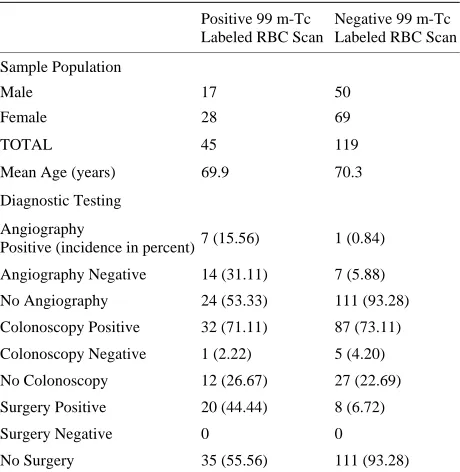
[image:2.595.308.538.499.735.2]Of the total sample population of 170 subjects, 164 sub- jects were included in the study. The exclusion of 6 sub- jects occurred due to incomplete medical records. Of the 164 subjects (n = 164), 67 (40.9%) were male, 97 (59.1%) were female. The mean age of the sample population was determined as 70.18 years of age (+/– 12.36, standard deviation (SD)). The mean age of the positive RBC scan subgroup showed no statistically significant difference when compared to the negative subgroup (p = 0.85) (Ta- ble 1).
Table 1. Incidence of gender, age and diagnostic test results in sample population.
Positive 99 m-Tc Labeled RBC Scan
Negative 99 m-Tc Labeled RBC Scan
Sample Population
Male 17 50
Female 28 69
TOTAL 45 119
Mean Age (years) 69.9 70.3
Diagnostic Testing
Angiography
Positive (incidence in percent)7 (15.56) 1 (0.84)
Angiography Negative 14 (31.11) 7 (5.88)
No Angiography 24 (53.33) 111 (93.28)
Colonoscopy Positive 32 (71.11) 87 (73.11)
Colonoscopy Negative 1 (2.22) 5 (4.20)
No Colonoscopy 12 (26.67) 27 (22.69)
Surgery Positive 20 (44.44) 8 (6.72)
Surgery Negative 0 0
3.2. Subgroup Analysis
Of the 164 subjects, 45 (27.5%) of the sample population had a positive test, while 119 (72.5%) of the total sample size had negative 99 m-Tc labeled RBC scans. Within the 45 subjects who had positive 99 m-Tc Scanning, 21 (46.67%) underwent angiography, 20 (44.44%) subjects required surgery and 31 (68.89%) subjects required co- lonoscopy. The rates of colonoscopy were similar be-tween the positive RBC scan group (73.35%) when com- pared with the negative group (76.4%). Among the 21 subjects who underwent angiography, 7 (33.33%) sub-jects had positive angiograms and 14 (66.67%) subsub-jects had negative angiograms (Table 1).
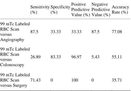
The sensitivities, specificities, positive predictive val- ues (PPV) and negative predictive values of the 99 m-Tc Scan when compared to angiography results, colonoscopy results and surgical results can be summarized in Table 3. The accuracy rate of the tagged RBC scan was reported as 35.71% when compared to surgery, 55.11% versus colonoscopy and 77.08% when compared to angiography.
In 6 of the 7 subjects, who had positive 99 m-Tc la- beled RBC scans and positive angiography, there was correlation found between the angiographic findings and the results of the tagged RBC scan. These findings were not statistically significant (p = 0.125). Within the surgi- cal subgroup 15 of the 20 subjects, that required surgery and had positive 99 m-Tc labeled RBC scans, had results that correlated between the scan and surgical findings (p = 0.04). Attending surgeons in 7 (46.67%) of subjects who underwent surgery reported that results of the 99 m-Tc scan “influenced” their surgery. The colonoscopy sub- group showed correlation in 27 subjects (87%, p < 0.001), also statistically significant.
When examining the transfusion requirements of the groups with positive 99 m-Tc labeled RBC scanning with the negative group, the positive group was transfused 372 units of PRBC (8.3 per subject), compared to 333 units of PRBC in the negative group (2.8 units per subject) (p < 0.001). Subjects in the 99 m-Tc labeled RBC scan positive group demonstrated a mean hospital LOS of 14.4 days when compared with 10.3 days of the negative subgroup (p = 0.09). Mortality rates were reported as 8.8% and 6.7% among the positive and negative sub-groups respectively (p = 0.85) (Table 2).
4. Discussion
[image:3.595.307.538.120.267.2]The diagnostic approach to LGIB has been defined in an algorithmic approach [2]. Within this approach, is the subdivision of subjects that are hemodynamically unsta- ble, defined by hypotension, tachycardia or postural changes positive in blood pressure. The approach to these patients required upper endoscopy as the initial diagnostic step. If
Table 2. Transfusion requirements, hospital length of stay and mortality of sample population.
Positive 99 m-Tc Labeled RBC Scan
Negative 99 m-Tc Labeled RBC Scan p Value
Transfusion Requirements
Total Number of
PRBCs (units) 372 333 <0.001
PRBC/subject
(units/subject) 8.3 2.8 <0.001
Hospital Length of
Stay (days) 14.4 10.3 0.09
Mortality (%) 8.8 6.7 0.85
Table 3. Diagnostic Parameters of 99 m-Technitium Labeled RBC Scan when compared with various diagnostic modali-ties.
Sensitivity (%)
Specificity (%)
Positive Predictive Value (%)
Negative Predictive Value (%)
Accuracy Rate (%)
99 mTc Labeled RBC Scan versus Angiography
87.5 33.33 33.33 87.5 77.08
99 mTc Labeled RBC Scan versus Colonoscopy
26.89 83.33 96.97 5.43 55.11
99 mTc Labeled RBC Scan versus Surgery
71.43 0 100 0 35.71
bleeding persisted despite endoscopic intervention, the recommendation is to proceed additional diagnostic mo- dalities one of which is the 99 m-Tc labeled RBC scan. Though included in the diagnostic approach for LGIB, variable data existed in our literature search to support utilization of the 99 m-Tc RBC scan.
The accuracy of the scan is limited by the method that 99 m-Tecnitium is instilled into the patient. By utilizing the sulfur colloid approach (SC), studies suggested that sources of bleeding could be identified for patients bleed- ing as low as 0.05 to 0.1 milliliter/minute (mL/min) [11]. Future studies determined bleeding rates as low as 0.04 mL/min by utilizing instillation of 99 m-Tc tagged RBC directly into the patient [12]. Indeed this modality ap-peared promising, however when put to the test, results were less hopeful.
[image:3.595.308.539.316.476.2]Additionally, the accuracy rate of the 99 m-Tc scan ranged from 35.71% to 77.08% when compared with various other investigative techniques. These results are likely due to the varying diagnostic parameters of the other, more ac- curate, diagnostic tests.
The 99 m-Tc RBC scan likely was found to have a higher accuracy rate when compared to angiography, as the reported sensitivity of angiography ranged from 30% - 47% [13-15]. Similarly, the accuracy rate of the RBC scan when compared to colonoscopy was likely due to the ability of endoscopy to detect 74% - 82% of source of GIB, which is higher than that of angiography [16]. Fi- nally, the accuracy rate of the 99 m-Tc scan was lowest when compared to surgery, as surgery allows direct visu- alization of the patient in questions anatomy and is there- fore the most accurate diagnostic approach.
A large review of the data on radiolabeled RBC scan- ning, by Zucker et al., yielded the percentages of “cor- rect” scans from zero to 96% [6]. The confirmation of the 99 m-Tc labeled RBC scan’s results occurred through endoscopic, angiographic and surgical means. Our study utilized similar means to correlate the labeled RBC scans. In terms of correlation with angiography, the results of our study determined that one third of the subjects with a “positive” tagged RBC scan also had angiographic evi- dence of bleeding. Furthermore, six of those seven had correlation with the location of bleeding, though not sta- tistically significant (p = 0.125). These results are likely explained by the variability of both diagnostic tests for determining location of GIB, as stated above.
In contrast, statistical significance was found when the 99 m-Tc scans were utilized to localize bleeding in com- parison to surgery (p = 0.04) and colonoscopy (p < 0.001) (Table 1). In comparison to data from Hunter et al., the localization error was comparable when 99 m-Tc scan- ning was weighed against surgical means in approximately 25% of cases [7]. The danger of assuming correlation be- tween the 99 m-Tc scan and surgery is the potential to perform surgery when unnecessary. Orecchia, et al., found a correlation of 94% among his sample population, while Voeller demonstrated failure of localization in 85% of his sample population [8,17]. The data is conflicting, however our data does show statistical correlation between 99 m-Tc scanning and surgery/colonoscopy. Despite this signify- cance, the surgeons of seven subjects were “influenced” by 99 m-Tc RBC scans. This small number alluded to the need for imaging more advanced than the RBC scan, when pursuing surgical intervention in LGIB. Additionally, if the diagnostic yield is comparable, perhaps RBC scan is an unnecessary and costly step in the diagnostic work up of LGIB. Arguments for the contrary would offer con- tention to the invasiveness of angiography, endoscopy and surgical means.
Another interesting result of this study is the relation- ship between the subjects with a positive 99 m-Tc scan and those with a “negative” scan. As defined in Table 2, the subjects who had a positive 99 m-Tc labeled RBC scan, required a statistically significant amount of PRBC per subject when compared with the subjects with a nega- tive scan. When examining hospital LOS, the positive RBC scan subgroup had an additional 4.1 days when compared with the negative subgroup, though not statistically sig- nificant (p = 0.09). Finally, there was no significant dif- ference calculated between the all cause mortalities of the scan positive and negative subgroups (p = 0.85). To our knowledge, no literature existed to correlate transfusion demands, LOS or all cause mortality with 99 m-Tc labeled RBC scanning. The results of this study appeared to de- monstrate the transfusions demands increased with a posi- tive test, but no statistically significant difference existed in LOS or mortality between the two subgroups.
Potential limiting factors included the retrospective na- ture of this study, which could be prevented with a future prospective study. Additionally, the sample size of sub- jects who underwent further diagnostic investigation was small and may have limited data extrapolation. The pres- ence of small sample size was consistent with data found during our literature search. Future study with a larger cohort may expand knowledge of the true clinical utility of this test for LGIB. Yet despite these potential limita- tions, the results of this study call into question the diag- nostic utility of 99-Tc scanning when confirmatory test- ing exists with greater accuracy. Though less invasive, the potential for localization error of 25% may lead to a high number of unnecessary surgeries in patients with LGIB.
5. Conclusion
The 99 m-Tc labeled RBC scan is a diagnostic study for LGIB with relatively low diagnostic accuracy rates. How- ever when the study was positive, the location of LGIB appeared to correlate with the results of angiography, colonoscopy and surgery. There was a statistically sig- nificant increase in the units of PRBC per subject in the subgroup with a positive RBC scan when compared to the subgroup with a negative scan. Finally, there was no sta- tistical significance between the hospital LOS and mor- tality of the positive versus negative subgroups.
6. Acknowledgements
REFERENCES
[1] J. J. Farrell and L. S. Friedman, “Gastrointestinal Bleeding in the Elderly,” Gastroenterology Clinics of North America, Vol. 30, No. 2, 2001, pp. 377-407.
doi:10.1016/S0889-8553(05)70187-4
[2] A. S. Fauci, E. Braunwald, D. L. Kasper, et al., “Harrison’s Principles of Internal Meidcine,” 17th Edition, McGraw- Hill Companies, Inc., New York, 2008.
[3] T. E. Garofalo and R. A. Abdu, “Accuracy and Efficacy of Nuclear Scintigraphy for the Detection of Gastroin- testinal Bleeding,” Archives of Surgery, Vol. 132, No. 2, 1997, pp. 196-199.
doi:10.1001/archsurg.1997.01430260094020
[4] M. S. Suzman, M. Talmor, et al., “Accurate Localization and Surgical Management of Active Lower Gastroin- testinal Hemorrhage with Technetium-Labeled Erythrocyte Scintigraphy,” Annals of Surgery, Vol. 226, No. 1, 1997, pp. 112-113.
[5] R. Levy, W. Barto and J. Gani, “Retrospective Study of the Utility of Nuclear Scintigraphic-Labeled Red Cell Scanning for Lower Gastrointestinal Bleeding,” Annals of
Surgery, Vol. 73, 2003, pp. 205-220.
doi:10.1046/j.1445-1433.2002.02567.x
[6] D. A. Zukerman, T. P. Bocchini and E. H. Birnbaum, “Massive Hemorrhage in the Lower Gastrointestinal Tract in Adults: Diagnostic Imaging and Intervention,” American
Journal of Roentgenology, Vol. 161, No. 4, 1993, pp.
703-711.
[7] J. M. Hunter and M. E. Pezim, “Limited Value of Te- chnetium 99 m-Labeled Red Cell Scintigraphy in Lo- calization of Lower Gastrointestinal Bleeding,” Ame-
rican Journal of Surgery, Vol. 159, No. 5, 1990, pp. 504-
506. doi:10.1016/S0002-9610(05)81256-5
[8] G. R. Voeller, G. Bunch and L. G. Britt, “Use of Technetium-Labeled Red Blood Cell Scintigraphy in the Detection and Management of Gastrointestinal Hemor- rhage,” Surgery, Vol. 110, No. 4, 1991, pp. 799-804.
[9] P. V. Ford, S. P. Bartold, D. M. Fink-Bennett, et al., “Procedure Guideline for Gastrointestinal Bleeding and Meckel’s Diverticulum Scintigraphy. Society of Nuclear Medicine,” Journal of Nuclear Medicine, Vol. 40, No. 7, 1999, pp. 1226-1232.
[10] A. T. Hirsch, Z. J. Kaskal, C. W. Hertzer, et al., “ACC/
AHA 2005 Guidelines for the Management of Patients with Peripheral Arterial Disease (Lower Extremity, Renal, Mesenteric, and Abdominal Aortic): A Collaborative Report from the American Association for Vascular Surgery/Society for Vascular Surgery, Society for Car- diovascular Angiography and Interventions, Society for Vascular Medicine and Biology, Society of Inter- ventional Radiology, and the ACC/AHA Task Force on Practice Guidelines (Writing Committee to Develop Guidelines for the Management of Patients with Peri- pheral Arterial Disease,” Journal of the American College
of Cardiology, Vol. 47, 2006, pp. 1-192.
doi:10.1016/j.jacc.2006.02.024
[11] A. Alavi and E. J. Ring, “Localization of Gastrointestinal Bleeding: Superiority of 99 mTc Sulfur Colloid Com- pared with Angiography,” American Journal of Roent-
genology, Vol. 137, No. 4, 1981, pp. 741-748.
[12] D. A. Thorne, F. L. Datz, K. Remley, et al., “Bleeding Rates Necessary for Detecting Acute Gastrointestinal Bleeding with Technetium-99m-Labeled Red Blood Cells in an Experimental Model,” Journal of Nuclear Medicine, Vol. 28, No. 4, 1987, pp. 514-520.
[13] L. Defreyne, M. Uder, P. Vanlangenhove, et al., “An- giography for Acute Lower Gastrointestinal Hemor- rhage: Efficacy of Cut Film Compared with Digital Subtraction Techniques,” Journal of Vascular and Inter-
ventional Radiology, Vol. 14, No. 3, 2003, pp. 313-322.
doi:10.1097/01.RVI.0000058414.01661.77
[14] K. H. Barth, “Radiological Intervention in Upper and Lower Gastrointestinal Bleeding,” Baillieres Clinical Gas-
troenterology, Vol. 9, No. 1, 1995, pp. 53-69.
doi:10.1016/0950-3528(95)90070-5
[15] W. Browder, E. J. Cerise and M. S. Litwin, “Impact of Emergency Angiography in Massive Lower Gastroin- testinal Bleeding,” Annals of Surgery, Vol. 204, No. 5, 1986, pp. 530-536.
doi:10.1097/00000658-198611000-00004
[16] A. M. Vernava III, B. A. Moore, W. E. Longo, et al., “Lower Gastrointestinal Bleeding,” Diseases of the Colon
& Rectum, Vol. 40, No. 7, 1997, pp. 846-858.
doi:10.1007/BF02055445
[17] P. Orecchia, M. Hensley, P. Mcdonald, et al., “Localiza-tion of Lower Gastrointestinal Hemorrhage,” Archives of