Short Report
Intercellular Trafficking of VP22, a Herpes Simplex Virus
Type 1 Tegument Protein
Taravat Bamdad
*1and John.C. Bell
21
Dept. of Virology, School of Medical Sciences, Tarbiat Modares University, Tehran, Iran; 2Ottawa Regional Cancer Centre Research Laboratories, Ottawa, Ontario K1H 8L6
Received 28 February 2006; revised 5 August 2006; accepted 13 August 2006
ABSTRACT
Background: The herpes simplex virus type 1 (HSV-1) tegument protein, VP22 has been reported to have the property of intercellular transport. The previous studies have shown that following expression of a fusion protein containing VP22; it spreads to every cell in a monolayer and concentrates in the nucleus. In spite of these reports, some studies have shown that VP22 trafficking and its nucleus accumulation is an artifact and no improvement in translocation of proteins fused to VP22 has been detected. Methods: To better understand about VP22 translocation, VP22-GFP (Green Fluorescent Protein) vector was constructed and its nuclear accumulation, transportation to the nontransfected cells and translocation between different cell types were studied by fluorescent microscope. Results: VP22-fusion protein was detected in nontransfected cells which in some of them the fusion protein was shown in nucleus. Conclusion: The results demonstrated that VP22 can easily transport between
different cells but nuclear accumulation of the protein is not common in
all of the recipient cells.
Iran. Biomed. J. 11 (1): 53-57, 2007Keywords:Herpes simplex virus type 1 (HSV-1), VP22, Tegument, Transporter
INTRODUCTION
he tegument of
herpes simplex virus type 1
(
HSV-1), located between envelope and capsid, contains some important proteins of the virus. VP22 is one of the most abundant proteins of the tegument which encoded by the gene UL49 [1]. The function of VP22 for replication cycle of HSV-1 is not completely understood but it has been described as a transporter protein [2]. In spite of lacking signal sequence, VP22 exports from the cell in which it is synthesized and imports to the neighboring cells. It has been shown that in producing cells, VP22 is mainly accumulated in cytoplasm while in recipient cells the protein transports into the nucleus. In these cells and also during cell division in VP22-expressing cells, the protein binds to chromatin and segregates to daughter cells [3]. The trafficking property of VP22 may compensate the major drawback of inefficient gene delivery in gene therapy applications. VP22 as a part of a fusion protein translocates between cells and spreads the fusion protein to the surroundingcells. Many reports have demonstrated that VP22 can deliver different proteins between cells in a cell culture and in animal models [4-6]. Creating controversy, some other reports suggest that fixation of cells during processing methods used for visualizing the protein through dissolving the cellular membrane causes an artifact that confuses the results and leads to misinterpretation as intracellular translocation of the protein [7-10]. They have reported that VP22 translocation and nucleus accumulation cannot be detected in live cells. To further investigate the phenomenon of VP22 trafficking,
Green Fluorescent Protein
(GFP)-VP22 chimera gene was constructed and the spread of the protein was detected in live and fixed cells.MATERIALS AND METHODS
Cell lines. BHK-T7 (Baby hamster kidney-T7) and Vero cell lines were grown in a MEM (Gibco/BRL, Burlington, Ontario, Canada) supplemented with 10% FBS at 37°C.
T
Construction of pGFP-VP22 plasmid. pGFP-VP22 was constructed by in frame ligation of
BamH1-Xba1 fragment of pVP22/myc-His2
(Invitrogen, Carlsbad, CA, USA), a commercially available VP22 expression vector that contains VP22 gene, between BamH1 and Xba1 sites in multiple cloning site of pEGFP-C1(Clontech, USA). Plasmid extraction and DNA ligation were done as standard protocols [11]. Briefly, the VP22 gene containing BamH1and Xba1 sites at its ends was extracted from agarose gel using Qiagen extraction kit (Qiagen, USA). The extracted fragment and pEGFP-C1 vector digested with BamH1and Xba1
were ligated at 18°C for 24 h at a ratio of 3:1, respectively. The constracted plasmids were screened by restriction enzyme analysis and the pEGFP-C1 plasmid was used as a control in experiments.
Nucleus detection of GFP-VP22. BHK-T7 cells
were grown on gelatin coated coverslips. To prepare gelatin coated coverslips, coverslips were coated with o.o1% sterile gelatin 24 h before use. The 70% confluent cells were transfected with pGFP-VP22 and pEGFP-C1 as control using lipofectamine 2000 (Invitrogen, Burlington, Ontario, Canada) according to the manufacturer's instruction. Forty eight hours after transfection, the cells were washed 3 times with PBS and fixed with absolute methanol at -20°C for 10 min. The coverslips were washed with phosphate buffer and mounted by mounting buffer (0.1X PBS, 10 mM NaN3, and 90 % glycerol). The cells were
visualized by fluorescent microscopy.
Translocation of GFP-VP22 to the
non-transfected cells. Gelatin coated coverslips were
splitted into two halves and BHK-T7 cells were grown on all halves. One half of each coverslip was transfected with the plasmids as described previously. After 24 h the transfected cells were completely washed with PBS and the medium was changed with fresh medium. The non-transfected half of each coverslip was exactly fitted to the transfected one. Twenty four hours later, live and methanol fixed cells were detected for GFP translocation to the non-transfected halves by fluorescent microscopy.
Translocation of VP22-GFP between different
cell types. BHK-T7 cells were transfected with
pGFP-VP22 and pEGFP as described previously. After 24 h, the cells were washed and tripsinized Vero cells were added on the transfected BHK-T7
cells in a ratio of 1:5. Twenty four h later live and fixed cells were visualized by fluorescent microscopy.
RESULTS
Plasmid construction. A fusion gene encoding
VP22 and GFP was constructed by inserting VP22 gene into the c-terminal of GFP gene. The expression of the fusion protein was detected by GFP visualizing in live cells.
Analysis of transfected cells. Forty eight hours
after the transfection, live cells were examined microscopically for GFP. The percentage of fluorescent cells in GFP-VP22 transfected cells was significantly higher than the pEGFP transfected cells. Methanol fixed cells were detected for nuclear and cytoplasmic distribution of GFP. In pGFP-VP22 transfected cells nuclear and cytoplasmic localization of GFP was detected, while in pEGFP transfected cells we could not detect any GFP in the nucleus (Fig. 1).
Fig. 1. The nuclear localization of GFP-VP22 in BHK-T7 fixed cells (a). This pattern of localization was not shown in pEGFP transfected control cells. Cytoplasmic distribution of GFP is detectable in these cells (b).
(A)
(B)
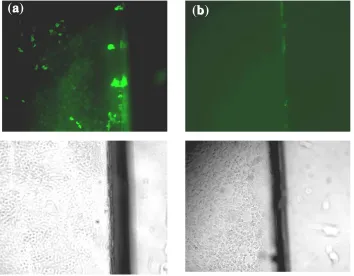
Fig 2. Transport of GFP-VP22 into the nontransfected cells. The translocation of GFP-VP22 from pGFP-VP22 transfected cells cultured on half of a coverslip into the non-transfected BHK-T7 cells plated on the other half of the coverslip (a) comparing to the pEGFP transfected control cells that show no GFP in the cells (b). The pictures show the non-transfected halves of coverslips. The images were taken from one microscopic field with fluorescent (above) and normal light microscopes (below).
Transport of GFP-VP22 into the non-transfected
cells. BHK-T7 cells were grown on the two halves
of each coverslip. After 24 h, the transfected half was completely closed to the non-transfected one and 24 h later cells were monitored for GFP translocation. Translocation of the GFP-VP22 into the non-transfected cells was detected in live and fixed cells. A gradient of GFP translocation from the edge of the coverslip to the center was seen. No GFP was detected in non-transfected cells in pEGFP control experiment (Fig. 2).
Distribution of GFP-VP22 between different cell
types. To confirm the translocation of VP22 between
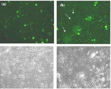
different cell types, BHK-T7 and Vero cells with completely distinguishable morphology were chosen. Non-transfected Vero cells in a ratio of 1:5 were added on the transfected BHK-T7 cells. The ratio of 1:5 was the best ratio of cells in which cells were morphologically distinguished from each other in a co-culture. Vero cells showed GFP when added on the pGFP-VP22 transfected cells while, in pEGFP transfected cells no translocation of GFP into the Vero cells was detected (Fig. 3).
DISCUSSION
Expression of VP22 in transfected or infected cells results in distribution of the protein through the neighboring cells where it accumulates in the nucleus. Although several studies have addressed the intracellular trafficking and nuclear localization of VP22 in non-expressing cells [4-6, 12], some reports have claimed that an artifact during methanol fixation results in the misinterpretation of the experiment [7, 13]. To be able to judge about the confused results surrounding the property of VP22, the gene for VP22 protein was fused to GFP gene and used to address the behavior of the VP22. The intrinsic GFP fluorescence property has been successfully used as a marker for detection of proteins fused to GFP [14]. In the first study, we detected the GFP-VP22 protein in the nucleus of the cells. Because of having a huge nucleus, BHK-T7 cell line was chosen for transfection and nuclear detection of the expressed protein. The localization of the GFP-VP22 in the nucleus and also in the cytoplasm of the cells was detected only in the pGFP-VP22 transfected cells but not in the pEGFP transfected control cells. It is note worthy that the
(a)
(b)
Fig. 3. Translocation of VP22-GFP between different cell types. The distribution of GFP-VP22 into the non-transfected Vero cells from BHK-T7 transfected cells (b). No GFP was detected in Vero cells in pEGFP transfected control test (a). Vero cells have been indicated by arrows. The images were taken from one microscopic field with fluorescent (above) and normal light (below) microscopes.
percentage of the cells with nuclear localization of GFP was not as high as the percentage of the non-expressing cells showing GFP-VP22. In the previous reports supporting intracellular trafficking of VP22, it has been shown that the majority of VP22 is localized to the nuclei of non-transfected cells [13], the phenomenon that has been rejected by some other research groups [7]. Our data suggest that the protein translocates between cells but not accumulates mainly in the nucleus. On the other hand, although GFP-VP22 spreads into the many cells of the monolayer but nuclear localization is not the fate of the translocated proteins in all of the non-expressing cells. It may depend on the stages of cell cycle. The previous data have suggested that the localization of the VP22 is regulated by the process of mitosis. During interphase, VP22 localizes predominantly to the cytoplasm. At the early stage of mitosis, VP22 translocates to the nucleus and binds to the chromatin [15]. Regarding this phenomenon, it can be explained that why some but not all of the non-synthesizing cells showed nuclear localization of the VP22 in a non-synchronized
confluent cell population. Anyhow, although rare but GFP-VP22 was detected in the nucleus comparing to the pEGFP transfected control cells that any GFP was detected in the nucleus. In live cells, nuclear localization of the protein was not detectable because of the reflection from very bright cells at the high magnifications.
In the second and the third experiments, we showed the transport of the GFP-VP22 into the non-transfected cells in live and fixed cells. The novel methods for detection of VP22 in non-transfected cells applied in this study strongly support the translocation of VP22. In the previous reports [7, 10], rejecting this property of VP22, ethanol fixation may facilitate spread of the protein between cells, but in our experiments especially the second one during methanol fixation, there was no trace of expressed VP22-GFP to be able to penetrate into the non-transfected cells. It confirms that nuclear localization is not an artifact raised during the process of methanol fixation
In conclusion, the present study consistent with previous results show that VP22 can easily spread
(a)
(b)
between cells, but in a new approach suggests that the protein dose not always localize in the nucleus of non-expressing cells. Nucleus localization of VP22 is completely a mitosis-dependent phenomenon but not a methanol fixation artifact.
ACKNOWLEDMENTS
We thank Dr. Stojdl, D.F. for his helpful advice and Pateson J. and Marius R. for their experimental assistance.
REFRENCES
1. Roizman, B. and Knipe, D.M. (2001) Herpes simplex
virus and their replication. In: Fields Virology.
(Knipe, D.M. and Howley, P.M., eds.), Lippicott
Williams & Wilkins, Philadelphia, Vol. 2, 4th ed. pp:
2391-2510.
2. Elliott, G. and O’Hare, P. (1997) Intercellular
Trafficking and Protein Delivery by a Herpesvirus
Structural Protein. Cell 88: 223-233.
3. Aints, A., Dilber, M.S., and Smith, E. (1999)
Intercellular Spread of GFP-VP22. J. Gene Med. 1:
275-279.
4. Cashman, M.S., Sadowski, L.S., Morris, J.D.,
Frederick, J. and Kumar-Singh, R. (2002) Intercellular trafficking of adenovirus-delivered HSV VP22 from the retinal pigment epithelium to the
Photoreceptors-implications for gene therapy. Mol.
Ther. 6: 813-823.
5. Greco, O., Joiner, M.C., Doleh, A. and Scott, S.D.
(2005) VP22-mediated intercellular transport for suicide gene therapy under oxic and hypoxic
conditions. Gene Ther. 12 (12): 974-979.
6. Phelan, A., Elliott, G. and O'Hare, P. (1998)
Intercellular delivery of functional p53 by the herpes
virus protein VP22. Natl. Biotechnol. 16 (5):
440-443.
7. Lundberg, M. and Johansson, M. (2001) Is VP22
nuclear homing an artifact? Nature Biotech. 19: 713.
8. Hakkarainen, T., Wahlfors, T., Merilainen, O.,
Loimas, S., Hemminki, A. and Wahlfors, J. (2005) VP22 does not significantly enhance enzyme prodrug cancer gene therapy as a part of a VP22-HSVTk-GFP
triple fusion construct. J. Gene Med. 7 (7): 898-907.
9. Zavaglia, D., Lin, E.H., Guidetti, M., Pluquet, O.,
Hainaut, P., Favrot, M.C. and Coll, J.L. (2005) Poor intercellular transport and absence of enhanced antiproliferative activity after non-viral gene transfer of VP22-P53 or P53-VP22 fusions into p53 null cell
lines in vitro or in vivo. J. Gene Med. 7 (7):
936-944.
10. Perkins, S.D., Hartley, M.G., Lukaszewski, R.A.,
Phillpotts, R.J., Stevenson, F.K. and Bennett, A.M. (2005) VP22 enhances antibody responses from DNA vaccines but not by intercellular spread.
Vaccine 23 (16): 1931-1940.
11. Sambrok, J. and Russell, D.W. (2001) Molecular
cloning- a laboratory manual, third ed., CSHL press.
12. Zheng, C.F., Brownline, R., Huang, D.Y,. Babiuk,
K.A. and Van, S. (2006) Intercellular trafficking of the tegument protein VP22 of bovine herpesvirus-1
and its application to improve a DNA vaccine. Arch.
Virol. 151 (5): 985-993.
13. Fang, B., Xu, B., Koch, P. and Roth, J.A. (1998)
Intercellular trafficking of VP22-GFP fusion proteins
is not observed in cultured mammalian cells. Gene
Ther. 5 (10): 1420-1424.
14. Elliott, G. and O’Hare, P. (1999) Intercellular
trafficking of VP22-GFP fusion proteins. Gene Ther.
6 (1): 149-151.
15. Elliott, G., and O’Hare, P. (2000)
Cytoplasm-to-nucleus translocation of a herpes virus tegument
protein during cell division. J. Virology 74 (5):
2131-2141.