*Corresponding author: Masoumeh Bahreini, PhD, De-partment of Biology, Faculty of Sciences, Ferdowsi Univer-sity of Mashhad, Mashhad, Iran.
Tel: +98 9153152856 Fax: +985138796416 Email: [email protected]
A rapid method for separating and concentration of food-borne pathogens
using elution from ready-to-eat vegetables
Safieh Rajabzadeh¹, Masoumeh Bahreini2*, Mohammad Reza Sharifmoghadam2
¹Department of Food Sciences and Technology, Resarch Institute of Food Sciences and Technology, Mashhad, Iran
2Department of Biology, Faculty of Sciences, Ferdowsi University of Mashhad, Mashhad, Iran
Received: April 2018, Accepted: November 2018 ABSTRACT
Background and Objectives: Traditional culture methods for detection of food-borne pathogens, a major public health problem, are simple, easily adaptable and very practical, but they can be laborious and time consuming. In this study, we eliminated culturing steps by developing a new separation method and therefore, decreased the detection time of food-borne pathogens (Salmonella enterica serovar Typhimurium, Escherichia coli O157:H7 and Listeria monocytogenes) to a few hours.
Materials and Methods: We used alkaline water and different alkaline buffers to elute bacteria from the lettuce surface as a model for ready-to-eat vegetables. Buffers used were as follows: 1) 0.05 M glycine; 2) 0.05 M glycine -100 mM Tris base -1% (w/v) beef extract; 3) buffer peptone water; 4) buffer phosphate saline. Buffers were adjusted to pH of 9, 9.5 and 10. In order to elute the bacteria, the lettuce pieces were suspended into buffers and shacked for 30, 45 and 60 min. Moreover, a multiplex PCR method for the simultaneous detection of food-borne pathogens was performed.
Results: The results showed that buffer peptone water at pH 9.5 for 45 min have high ability to elute bacteria from the lettuce surface and the bacteria can be detected using multiplex PCR.
Conclusion: We developed a new rapid and efficient method for simultaneous separation of food-borne pathogens. This method eliminates culturing stages and permits the detection and identification of target pathogens in a few hours.
Keywords: Rapid detection, Elution, Multiplex polymerase chain reaction, Food-borne pathogens, Ready-to-eat vegetables
ORIGINAL
AR
TICLE
INTRODUCTION
of vegetables (1). Raw and minimally processed ready to eat vegetables could be hazardous for the safety and health of the consumers. According to Korean FDA, the number of food-borne diseases in 2008 increased 3.8-fold compared to 2003 (2). In Eu-rope, for instance, human food-borne cases and out-breaks were reported yearly (3-5). The global
inci-dence of foodborne outbreaks is difficult to estimate,
although authors generally agree with the estimate that the percentage of the population affecting from food-borne disease each year could be up to 30% in industrialized countries and it could be worse in de-veloping countries (6). Therefore, there is an urgent and serious need for a rapid, sensitive and reliable method to detect food-borne pathogens, especially for high-risk organisms such as Salmonella, Listeria monocytogenes and Escherichia coli O157:H7 (7). It is also necessary to develop a new method, which could considerably decreases the spending time and the cost.
Detection of foodborne pathogens based on con-ventional culture methods, culture media and bio-chemical kits, are simple, easily adaptable, very practical, and generally inexpensive, however, they can be laborious and time-consuming (7-9).
Advances in technology have led to the use of mo-lecular methods and new nanomaterials to detect
pathogens. Molecular techniques would significant -ly decrease the resources required in routine labo-ratory operations, and would enhance the overall
efficiency of detection in food supervision and in -spection (2, 10-17). In these methods, however, due to the importance of presence of even 1 cfu/25 g of some pathogens in foodstuffs, especially Salmonella spp., Listeria monocytogenes and E. coli O157:H7, the pre-enrichment and enrichment stages are used (10). These methods are laborious and increase the detection time to 2-3 days (7, 2, 18). The incu-bation time of 6 hours is the minimum incuincu-bation period that has been used by Thapa and co-workers (2).
Recently, food-borne viruses have become an im-portant food safety concern and various studies have dealt with the development of standardized methods for detection of enteric viruses in foods. Viruses are able to attach to food surfaces through their nega-tively charged surface proteins. Changing the pH from neutral to alkaline alters the electrical charge that separates the viral particles from the food sur-face (19, 20). Many papers have been published using
alkaline pH for elution of food-borne viruses from the vegetable and food surfaces (21-23).
Bacteria, like viruses, also attach to the different surfaces such as vegetables and food by their ad-hesions (24). Using alkaline pH and consequently, changes in conformation of bacterial cell surface proteins, bacteria can be eluted from the vegetable surfaces.
In this study, the elution method of viruses from the food surfaces was used to develop a new method for separating food-borne bacteria including
Salmo-nella enterica serovar Typhimurium, Escherichia
coli O157:H7, Listeria monocytogenes from the sur-faces of lettuce (as a model for ready-to-eat vegeta-bles), so culturing stages were eliminated. Then, the food-borne pathogens were detected by multiplex PCR in a few hours. The proposed method is unique in that it eliminates culturing stages using a new sep-aration method, which permits the rapid detection
and identification of target pathogens by multiplex
PCR.
MATERIALS AND METHODS
Bacterial strains and culture conditions. Bacte-rial strains Salmonella enterica serovar Typhimuri-um (PTCC 1709) and Listeria monocytogenes (PTCC 1298) were purchased from Persian Type Culture Collection, Iran, and E. coli O157:H7 (NCTC 12900) was purchased from National Collection of Type Cultures, UK. The bacteria were grown in Trypti-case Soy Broth (Oxoid, UK) at 37°C and then, se-rial dilutions of strains from 100 to 106 cfu/ml (1 to 1000000 cfu/ml) were prepared using normal saline (0.85 g/l).
Buffers preparation. In order to elute bacteria from the surface of lettuce, alkaline distilled water (ADW) and four elution buffers were chosen. The elution buffers were prepared as follows: 1) 0.05 M glycine; 2) 0.05 M glycine-100 mM Tris base-1% (w/v) beef extract (Gly-T-BE); 3) buffer peptone wa-ter (BPW); 4) phosphate buffer saline (PBS): 145 mM NaCl; 7.7 mM Na2HPO4; 2.3 mM NaH2PO4. The pH of buffers and alkaline distilled water was adjusted to 9, 9.5 and 10 using 5N NaOH (20-23, 25).
pieces overwhelm in 3% sodium hypochlorite solu-tion for 15 min and in order to eliminate extra chlo-ride ions, the samples were moved to sterile distilled water containing some drops of 1% sodium thiosul-fate for 2 min. Afterward, the lettuce pieces were washed three times with sterile distilled water and dried under the laminar hood and sterile condition. Then samples were tested for the possible presence of E. coli O157:H7, S. enterica serovar Typhimurium and L. monocytogenes according to ISO standard mi-crobiological methods (26-28).
The strains were inoculated on lettuce as pure and mixed cultures, separately. Lettuce contamination was performed inoculating 100 µl of each serial di-lution to the 25 grams sample of lettuce, dried
un-der a laminar flow hood for 60 min unun-der the sterile
condition and stored at 4°C overnight (29, 30). For the mixed cultures, 100 µl of each bacterial strain inoculated on lettuce (300 µl in total).
Effect of alkaline pH on the bacterial survival. In order to evaluate the effect of alkaline pH on the bacterial survival after elution, the pellets were sus-pended in 100 µl of sterile normal saline, plated on the general and selective media, incubated at 37°C
for 24-48 h and finally the colonies were counted.
The used Media were; nutrient agar [Himedia, India (NA), trypticase soy agar [Oxoid, UK (TSA)], sorbi-tol-MacConky agar [Merck, Germany (SMAK)] for E. coli O157:H7; XLD agar [xylose lysine deoxycho-late agar (Merck, Germany)] for S. enterica serovar Typhimurium; and PALCAM Listeria selective agar [Sigma-Aldrich, Germany (PALCAM)] for L. mono-cytogenes].
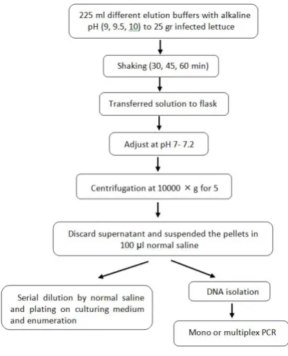
Elution method. To optimize the elution meth-od, the inoculated samples were transferred into the stomacher bag containing 225 ml of each buffer solu-tion with different pH (9, 9.5 and 10) and shaked for 30, 45 and 60 min at 150 rpm at room temperature.
Then, the elution buffers were transferred into flasks
and the pH of buffers was adjusted to pH 7 ± 0.2 with 1N HCl. Next, the samples were centrifuged at 10000 rpm for 5 min and supernatants were discarded. The pellets were suspended in 100 µl of sterile normal saline, plated on the nutrient agar, incubated at 37°C
for 24 h and finally, the colonies were counted (Fig.
1). The best pH and time in which the most bacteria were eluted from lettuce surfaces used as a standard elution method for the next steps.
DNA extraction. The eluted bacteria were re-sus-pended in 100 µl of sterile normal saline and used for DNA extraction. DNA extraction was performed on each bacterial strain before and after inoculation to lettuce using Bioneer genomic DNA isolation kit (Bioneer, Korea).
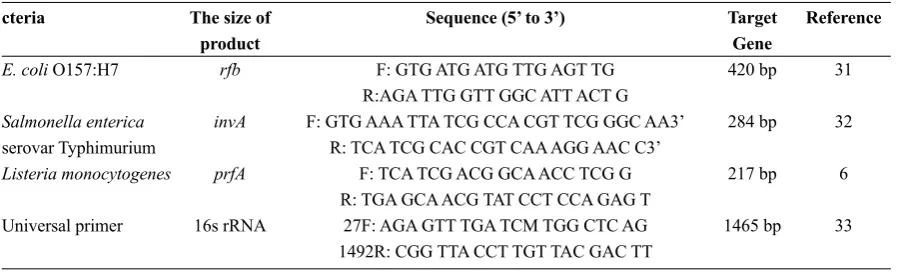
Mono and Multiplex PCR. The primer pairs used in this study were shown in Table 1. The target genes were the rfb gene (antigen O157 producer) for E. coli O157:H7 (31), the invA gene (invasion protein A) for S. enterica serovar Typhimurium (32), and the prfA gene (transcriptional activator of the virulence fac-tor) for L. monocytogenes (6). These genes described
here are known as the most specific and reliable ge -netic targets for the above microorganisms. As an internal control, the 16S rRNA gene was targeted in the presence of bacterial DNA (33). An uninoculated control was used in all steps as a negative control and all the experiments were performed three times.
Condition of monoplex PCR. All monoplex PCR reactions were conducted using GenetBio kit
(Genet-Bio, Korea) in a final volume of 25 µl. Master mix
composition was as follows: PCR buffer 10X, 2.5 µl; MgCl2 25 mM, 2.5 µl; Taq DNA Polymerase 5 U/µl,
Table 1. Primer pairs selected for the single and multiplex PCR cteria
E. coli O157:H7
Salmonella enterica serovar Typhimurium Listeria monocytogenes
Universal primer
The size of product
rfb
invA
prfA
16s rRNA
Sequence (5’ to 3’)
F: GTG ATG ATG TTG AGT TG R:AGA TTG GTT GGC ATT ACT G F: GTG AAA TTA TCG CCA CGT TCG GGC AA3’
R: TCA TCG CAC CGT CAA AGG AAC C3’ F: TCA TCG ACG GCA ACC TCG G R: TGA GCA ACG TAT CCT CCA GAG T
27F: AGA GTT TGA TCM TGG CTC AG 1492R: CGG TTA CCT TGT TAC GAC TT
Target Gene 420 bp
284 bp
217 bp
1465 bp
Reference 31
32
6
33
0.2 µl; dNTPs 10 mM, 0.4 µl; F/R primers 10 pmol, 1 µl; extracted DNA as template, 2 µl and distilled water, 15.4 µl.
Thermal cycler (Bio-Rad T100, thermal cycler, Germany) conditions were as follows: predenatur-ation at 94°C for 5 min; 35 cycles consisting of de-naturation at 94°C for 30 s, annealing at 55°C for 30
s, extension at 72°C for 60 s; final elongation at 72°C
for 7 min.
Condition of multiplex PCR. All multiplex PCR
reactions were performed in a final volume of 25 µl
using 4 µl of total extracted DNA from mixture of three pathogens as template. Master mix composi-tion was as follows: PCR buffer 10X, 2.5 µl; MgCl2 25 mM, 2.5 µl; Taq DNA Polymerase 5U/µl, 0.5 µl; dNTPs 10 mM, 1 µl; EC-F/R primer, 1 µl; SAL-F/R primer, 0.8 µl and LIS-F/R primer, 1 µl (10 pmol concentration of each primer), and distilled water, 8.9 µl.
Thermal cycler conditions were as follows: prede-naturation at 94°C for 3min; 35 cycles consisting of denaturation at 94°C for 30 s, annealing at 57°C for
30 s, extension at 72°C for 90 s; final elongation at
72°C for 10 min. PCR products were visualized via gel electrophoresis with 1% agarose gels.
RESULTS
The aim of this work was the development of an elution method to enhanced rapid detection of food-borne pathogens from ready-to-eat vegetables. For evaluation and development of the elution step, let-tuce was used as a model for ready-to-eat vegetables and inoculated with E. coli O157:H7, S. entrica
sero-var Typhimurium and L. monocytogenes. ADW and four different elution buffers (PBS, 0.05 M Glycine, Gly-T-BE and BPW) were tested for their ability to elute the food-borne bacteria from the surfaces of lettuce.
At first, the elution of bacteria using alkaline pH
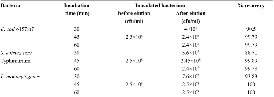
and their survival after elution were investigated. For recovery, the eluted bacteria were plated on differ-ent general and selective culture media. The results showed that alkaline pH has no bactericidal effects and the bacteria can be recovered by culturing on a general media such as nutrient agar (Table 2). Elution at different times, 30, 45 and 60 min were studied; the optimum time to elute the bacteria was 45 min (Table 3). The results shown in Tables 4-6 reveal that BPW and ADW at pH 9.5 for 45 min at 150 rpm and room temperature have high ability to elute the bac-teria from the lettuce surface. Both of them were able to recover 99.85-100% of inoculated bacteria at pH 9.5. However, BPW was chosen due to its constant pH during the experiments compare to ADW, which was very variable and the results were not reproduc-ible, therefore, it had not been used in the following experiments.
After separating and concentration of bacteria, we used mono and multiplex PCR for detection of
patho-genic bacteria. To verifying the method and confirm
Table 2. The effect of alkaline pH (9.5) and different culturing media on the bacterial survival after elution.
Buffers and alkaline water
PBS Gly Gly-T-BE BPW ADW
% recovery 0 100 0 99.63 77.50 100 number cfu/ml NG 1×10⁸ NG 9.3×10⁷ 1.6×10⁶ 1×10⁸ % recovery 63.85 100 0 100 78.19 100 number cfu/ml 1.3×10⁵ 1×10⁸ NG 1×10⁸ 1.8×10⁶ 1×10⁸ % recovery 0 67.89 0 80.88 71.50 82.88 number cfu/ml NG 2.7×10⁵ NG 3×10⁶ 5.3×10⁵ 4.3×10⁶ % recovery 0 68.5 0 75.00 74.63 69.25 number cfu/ml NG 3×10⁵ NG 1×10⁶ 9.4×10⁵ 3.5×10⁵ % recovery 80 80.13 79.25 86.75 75.00 81.00 number cfu/ml 8×10⁶ 2.6×10⁶ 2.2×10⁶ 8.9×10⁶ 1×10⁶ 3×10⁶ Culture medium SMAC NA XLD NA PALCAM NA Bateria Initial inoculation E. coli O157:H7 1×10⁸ cfu/ml S. entrica serv. Typhimurium 1×10⁸ cfu/ml L. monocytogenes
1×10⁸ cfu/ml
NG= not growth; cfu/ml= colony forming units per milliliter; % recovery= log final cfu/ml / log initial cfu/ml × 100; PBS=
phosphate buffer saline; Gly= 0.05 M glycin; Gly-T-BE= 0.05 M glysin-100 mM Triss base-1% (w/v) beaf extract; BPW=buf-fer peptone water; ADW= alkaline distilled water; SMAC= sorbitol-MacConky agar; NA= nutrient agar; XLD= xylose lysine deoxychlate agar; PALCAM= PALCAM Listeria selective agar.
Table 3. Percentage of bacterial recovery from lettuce surface using BPW at different incubation time. Inoculated bacterium After elution (cfu/ml) 4×10⁷ 2.4×10⁸ 2.4×10⁸ 5.6×10⁷ 2.45×10⁸ 2.4×10⁸ 7.6×10⁷ 2.5×10⁸ 2.5×10⁸ % recovery 90.5 99.79 99.79 88.71 99.89 99.78 93.83 100 100 Incubation time (min) 30 45 60 30 45 60 30 45 60 before elution (cfu/ml) 2.5×10⁸ 2.5×10⁸ 2.5×10⁸ Bacteria
E. coli o157:h7
S. entrica serv. Typhimurium
L. monocytogenes
cfu/ml= colony forming units per milliliter; % recovery= log final cfu/ml / log initial cfu/ml × 100.
DISCUSSION
The growing consumption of fresh and ready-to-eat vegetables has caused an increase in the number of food-borne disease outbreaks that could be haz-ardous for public health. Traditional culture methods for detection of food-borne pathogens can be labo-rious and time consuming. Hence, it is necessary to develop a new and rapid method, which could con-siderably decreases the time consumed.
Table 6. The numbers of eluted Listeria monocytogenes from surface of lettuce by alkaline water and different alkaline buffers.
% recovery 77.80 77.80 0 0 90.70 cfu/ml
2.3 ×10⁶ 2.3 ×10⁶
NG NG 2.6 ×10⁷ % recovery
79.70 57.50 73.90 99.90 100 cfu/ml
3.2 ×10⁶ 5 ×10⁴ 1 ×10⁶ 1.45 ×10⁸
1.5 ×10⁸ % recovery
0 88.10
0 0 91.80 cfu/ml
NG 1.6 ×10⁷
NG NG 3.2 ×10⁷
pH=9 pH=9.5 pH=10
Listeria monocytogenes
initial inoculation: 1.5×10⁸ cfu/ml
PBS Gly 0.05 M Gly-T-BE BPW ADW Buffers pH
NG= not growth; cfu/ml= colony forming units per milliliter; % recovery= log final cfu/ml ∕ log initial cfu/ml×100; PBS=
phosphate buffer saline; Gly= 0.05 M glycine; Gly-T-BE= 0.05 M glycine-100 mM Tris base-%1 (w/v) beef extract; BP-W=buffer peptone water; ADW= alkaline distilled water.
Table 4. The numbers of eluted E. coli O157:H7 from surface of lettuce by alkaline water and different alkaline buffers.
% recovery 79.7
0 0 0 77 cfu/ml
3.3×10⁶ NG NG NG 2×10⁶ % recovery
78.89 66.3 65.68
100 100 cfu/ml
2.8×10⁶ 2.2×10⁵ 2.35×10⁵
1.5×10⁸ 1.5×10⁸ % recovery
0 73.34
0 98.81 90.37 cfu/ml
NG 1×10⁶
NG 1.2×10⁸ 2.45×10⁷
pH=9 pH=9.5 pH=10
E. coli O157:H7 initial inoculation: 1.5×10⁸ cfu/ml
PBS Gly Gly-T-BE BPW ADW Buffers pH
NG= not growth; cfu/ml= colony forming units per milliliter; % recovery = log final cfu/ml ∕ log initial cfu/ml×100; PBS=
phosphate buffer saline; Gly= 0.05 M glycine; Gly-T-BE= 0.05 M glycine-100 mM Tris base-%1 (w/v) beef extract; BP-W=buffer peptone water; ADW= alkaline distilled water.
Table 5. The numbers of eluted Salmonella entrica serovar Typhimurium from surface of lettuce by alkaline water and differ-ent alkaline buffers.
% recovery 63.30
0 0 98.81 82.44 cfu/ml
1.5×10⁵ NG NG 1.2×10⁸ 5.5×10⁶ % recovery
85.62 74.35 79.73 99.85 99.70 cfu/ml
1×10⁷ 1.2×10⁶ 3.3×10⁶ 1.46×10⁸ 1.42×10⁸ % recovery
0 73.38
0 95.15
96 cfu/ml
NG 1×10⁶
NG 6×10⁷ 7×10⁷
pH= 9 pH= 9.5 pH= 10
Salmonella entrica serovar Typhimurium initial inoculation: 1.5×10⁸ cfu/ml
PBS Gly Gly-T-BE BPW ADW Buffers pH
NG= not growth; cfu/ml= colony forming units per milliliter; % recovery= log final cfu/ml ∕ log initial cfu/ml×100; PBS=
bacteria from the food surfaces (20). The results also show that altering in electrical charge at pH 9.5 is higher than pH 9 resulting in better elution of bac-teria and less bactericidal effect compared with pH 10 ending in higher recovery of bacteria. PWB and ADW were able to recover the most inoculated bac-teria. Out of four buffers, however, 0.05 M glycine and Gly-T-BE (0.05 M glycine-100 mM Tris base-1% (w/v) beef extract) buffers had the lowest bacterial recovery showing that buffer composition plays a key role in the bacterial elution and BPW was able to recover of inoculated bacteria. By this new method, we are able to separate and concentrate the patho-genic bacteria from food matrix and eliminate the homogenization of food stuff and the culturing steps. Immunomagnetic separation (IMS) technology is a promising candidate for food pretreatment sys-tems (10, 12, 15, 18, 34). Antibody-functionalized magnetic beads enable selective separation and con-centration of target bacteria from a range of sample matrices. Although commercialized IMS platforms can automatically separate and concentrate immu-nomagnetic beads with target bacteria, these meth-ods have limitations, such as longer per-enrichment step, small sample volume capacity (18), inability to serotype between 5 to 8% of isolates, incorrect typing due to the loss of bacterial cell surface an-tigens and being expensive (35). In additional, the capturing ability of the immunomagnetic beads is
affected by the presence of inhibitors to specific im -munoreactions. Food components such as carbohy-drates, proteins and fats normally inhibit the specif-ic binding of the antibody to target molecules (36). Using IMS-mPCR technique, Yang et al. were able to detect S. Typhimourium, L. monocytogenes and E. coli with the detection limit of 103 CFU/g in the artificially contaminated lettuce, tomato and ground
beef without any pre-enrichment (12). Ma et al. used IMS-RT-PCR for detection of Salmonella spp., Shi-gella spp., and S. aureus with the detection limit of 2-9.6 CFU/g, using pre-enrichment for 6 hours (37).
These findings showed that the IMS technology re -quired pre-enrichment to have lower limit of detec-tion and good results. Our new eludetec-tion method can be a promising candidate to replace food per-enrich-ment and immunomagnetic beads for separating and concentration of pathogenic bacteria.
Many molecular-based methods have been devel-oped for rapid detection of pathogenic bacteria, (12, 13, 30, 31, 36, 37) and, considering the possibility of
the coexistence of different pathogens in one sample,
multiplex detection and rapid identification of the
pathogens in a single analysis is very important and desirable (11).
Polymerase chain reaction (PCR) as a nucleic ac-id-based method widely used in food-borne patho-gens detection. The reported detection limits using mPCR after pre-enrichment have been 1-10 cfu/ml (or per 25 gr) of foodstuffs. Lee et al. (38) and Thapa et al. (2) reported the lowest detection limit of 1 and 10 cfu/ml in food stuff after 12 and 6 hours pre-en-richments, respectively; that is similar to our results, which are without pre-enrichment step. Guan et al.
(39) could detect five food-borne pathogens on in -fected pork with a detection limit of 103 cfu/mL for the simultaneous detection of the five target patho -gens and less than 10 cfu/mL for detection of a single pathogen. Such varied results could be due to vari-ous PCR inhibitors that can be found in foodstuffs and culture media. Some inhibitors that may affect different steps of the PCR method include: phenolic compounds, fats and glycogen (40). Different meth-ods were used for elimination of the inhibitors such
as microfiltration membrane and immunomagnetic
separation to improve the detection limits of the PCR assay (10, 14). However, there are also many draw-backs of using this method.
In new elution method, since homogenization of foodstuffs and culturing steps are eliminated, PCR inhibitory factors are in minimal and thus increasing sensitivity. In addition, our new method in combina-tion with the molecular method can be used for rapid detection of the food-borne bacteria and at the same time, for the detection of the food-borne viruses us-ing the same sample.
Other advantages of using our new method are as follows: the minimum equipment requirements, in-expensive, reliable and the most importantly decreas-ing the time required for the test (preparation time decreases to about 1 hour; overall time decreases to 4 hours). It can be expanded and employed in real sam-ples for the detection of multiple viable pathogens in food products.
CONCLUSION
lettuce surface using alkaline buffers. In addition, a multiplex PCR method for the simultaneous detec-tion of E. coli O157:H7, S. enterica serovar Typh-imurium and L. monocytogenes has been described. The elution of bacteria by alkaline buffers and then performing multiplex PCR was allowed us to set a robust method with high performances to de-crease detection time to less than 4 hours compare with other methods, when tested on a complex food system. The sensitivity and robustness of the meth-od proposed together with its ability to perform on a complex food matrix make it a suitable method to be implemented in quality control laboratories for the detection of the target pathogens in food samples.
ACKNOWLEDGEMENTS
This work was supported by the Ferdowsi Univer-sity of Mashhad, Mashhad, Iran (Grant number of 3/31036).
REfERENCES
1. Elizaquıvel P, Aznar R. A multiplex RTi-PCR reac -tion for simultaneous detec-tion of Escherichia coli O157:H7, Salmonella spp. and Staphylococcus aureus on fresh, minimally processed vegetables. Food Micro-biol 2008; 25: 705-713.
2. Thapa SP, Han R, Cho JM, Hur JH. Multiplex PCR and DNA array for the detection of Bacillus cereus, Staph-ylococcus aureus, Listeria monocytogenes, Escherich-ia coli O157:H7 and Salmonella spp. Targeting viru-lence-related genes. Ann Microbiol 2013; 63: 725-731. 3. CDC (Center for disease Control), Investigation Update:
Multistate Outbreak of E. coli O157:H7 infections as-sociated with In-shell Hazelnuts. 2011. available from: http://www.cdc.gov/ecoli/2011/hazelnuts-4-7-11.html 4. CDC (Center for disease Control), Cholera and
Oth-erVibrioIllness Surveillance System. 2017. avaiable at: https://www.cdc.gov/nationalsurveillance/pdfs/sur-veillance-system-overview_covis.pdf
5. CDC (Center for disease Control), Multistate Outbreak of Human Salmonella Montevideo Infections Linked to Live Poultry. 2012b. available at: https://www.cdc.gov/ salmonella/montevideo-06-12/signs-symptoms.html 6. Germini A, Masola A, Carnevali P, Marhelli R.
Simul-taneous detection of Escherichia coli O157:H7, Salmo-nella spp. and Listeria monocytogenes by multiplex PCR. Food Control 2009; 20: 733-738.
7. Yu Q, Zhai L, Bie X, Lu Z, Zhang C, Tao T, et al.
Sur-vey of five food-borne pathogens in commercial cold
food dishes and their detection by multiplex PCR. Food Control 2016, 59: 862-869.
8. Ryu J, Park SH, Yeom YS, Shrivastav A, Lee SH, Kim YR, et al. Simultaneous detection of Listeria species isolated from meat processed foods using multiplex PCR. Food Control 2013; 32: 659-664.
9. Jasson V, Jacxsens L, Luning P, Rajkovic A, Uytten-daele M. Alternative microbial methods: An overview and selection criteria. Food Microbiol 2010; 27: 710-730.
10. Zhang QY, Zhou WW, Zhou Y, Wang XF, Xu JF. Re-sponse surface methodology to design a selective co-enrichment broth of Escherichia coli, Salmonella spp. and Staphylococcus aureus for simultaneous de-tection by multiplex PCR. Microbiol Res 2012; 167: 405-412.
11. Ohk SH, Bhunia AK. Multiplex fiber optic biosensor
for detection of Listeria monocytogenes, Escherichia coli O157:H7 and Salmonella entrica from ready-to-eat meat samples. Food Microbiol 2013; 33: 166-171. 12. Yang Y, Xu F, Xu H, Aguilar ZP, Niu R, Yuan Y, et al.
Magnetic nano-beads based separation combined with propidium monoazide treatment and multiplex PCR assay for simultaneous detection of viable Salmonella Typhimurium, Escherichia coli O157:H7 and Listeria monocytogenes in food products. Food Microbiol 2013; 34: 418-424.
13. Donhauser SC, Niessner R, Seidel M. Sensitive
quanti-fication of Escherichia coli O157:H7, Salmonella enter-ica, and Campylobacter jejuni by combining stopped polymerase chain reaction with chemiluminescence
flow-through DNA microarray analysis. Anal Chem 2011; 83: 3153-3160.
14. Vasanth D, Pugazhenthi G, Uppaluri R. Fabrication and
properties of low cost ceramic microfiltration mem -branes for separation of oil and bacteria from its solu-tion. J Membr Sci 2011; 379: 154-163.
15. Coklin T, Farber JM, Parrington LJ, Bin Kingombe CI, Ross WH, Dixon BR. Immunomagnetic separation
sig-nificantly improves the sensitivity of polymerase chain
reaction in detecting Giardia duodenalis and Crypto-sporidium spp. in dairy cattle. J Vet Diagn Invest 2011; 23: 260-267.
16. Hagren V, Von Lode P, Syrjala A, Korpimaki T, Tuo-mola M, Kauko O, et al. An 8-hour system for Salmo-nella detection with immunomagnetic separation and
homogeneous time-resolved fluorescence PCR. Int J Food Microbiol 2008; 125: 158-161.
17. Brandao D, Liebana S, Pividori MI. Multiplexed detec-tion of food-borne pathogens based on magnetic par-ticles. N Biotechnol 2015; 32: 511-520.
automated system for separation and concentration of food-borne pathogens using immunomagnetic separa-tion. Food Control 2017; 73: 1541-1547.
19. Croci L, Dubois E, Cook N, Medici D de, Schultz AC, China B, et al. Current methods for extraction and con-centration of enteric viruses from fresh fruit and veg-etables: Towards international standards. Food Anal Methods 2008; 1: 73-84.
20. Butot S, Putallaz T, Sanchez G. Procedure for rapid concentration and detection of enteric viruses from berries and vegetables. Appl Environ Microbiol 2007; 73: 186-192.
21. Kim H, Kwak I, Hwang I, Ko G. Optimization of meth-ods for detecting norovirus on various fruit. J Virol Methods 2008; 153: 104-110.
22. Dubois E, Hennechart C, Deboosere N, Merle G, Lege-ay O, Burger C, et al. Intra-laboratory validation of a concentration method adapted for the enumeration of
infectious F-specific RNA coliphage, entrovirus and
hepatitis A virus from inoculated leaves of salad vege-tables. Int J Food Microbiol 2006; 108: 164-171.
23. Bahreini M, Habibi Najafi MB, Bassami MR, Yavar -manesh M. Optimization of extraction and concen-tration methods of enteric viruses from the surface of ready to eat vegetables using MS2 coliphage as a mod-el. J Food Sci Tech 2014; 11: 1-10.
24. Sapers GM. Efficacy of washing and sanitizing meth -ods for disinfection of fresh fruit and vegetable prod-ucts. Food Technol Biotechnol 2001; 39: 305-311. 25. Love DC, Casteel MJ, Meschke JS, Sobsey MD.
Meth-ods for recovery of hepatitis A virus (HAV) and other viruses from processed foods and detection of HAV by nested RT-PCR and TaqMan RT-PCR. Int J Food Mi-crobiol 2008; 126: 221-226.
26. ISO, 11290-2: Microbiology of food and animal feeding stuffs – Horizontal method for the detection and enu-meration of Listeria monocytogenes– Part 2: Enumer-ation method. 1998.
27. ISO. 16654: Microbiology of food and animal feeding stuffs – Horizontal method for the detection and enu-meration of Escherichia coli O157:H7. 2002.
28. ISO. 6579: Microbiology of food and animal feeding stuffs – Horizontal method for the detection of Salmo-nella spp. 2002.
29. Coudray-Meunier C, Fraisse A, Martin-Latil S, Guilli-er L, Delannoy S, Fach P, et al. A comparative study
of digital RT-PCR and RT-qPCR for quantification of
Hepatitis A virus and Norovirus in lettuce and water samples. Int J Food Microbiol 2015; 201: 17-26. 30. Casteel MG, Schmidt CE, Sobsey MD. Chlorine
disin-fection of produce to inactivate hepatitis A virus and coliphage MS2. Int J Food Microbiol 2008; 125: 267-273.
31. Maurer JJ, Schmidt D, Petrosko P, Sanchez S, Bolton L, Lee MD. Development of primers to O-antigen
bio-synthesis genes for specific detection of Escherichia coli O157:H7 by PCR. Appl Environ Microbiol 1999; 65: 2954-2960.
32. Rahn K, De Grandis SA, Clarke RC, McEwen SA,
Galán JE, Ginocchio C, et al. Amplification of an invA gene sequence of Salmonella typhimurium by
poly-merase chain reaction as a specific method of detection
of Salmonella. Mol Cell Probes 1992; 6: 271-279. 33. Chiang YC, Yang CY, Li C, Ho YC, Lin CK, Tsen HY.
Identification of Bacillus spp., Escherichia coli, Salmo-nella spp., Staphylococcus spp. and Vibrio spp. with 16S ribosomal DNA-based oligonucleotide array hy-bridization. Int J Food Microbiol 2006; 107: 131-137. 34. Wang L, Li Y, Mustaphai A. Rapid and simultaneous
quantitation of Escherichia coli 0157:H7, Salmonella, and Shigella in ground beef by multiplex real-time PCR and immunomagnetic separation. J Food Prot 2007; 70: 1366-1372.
35. Schrader KN, Fernandez-Castro A, Cheung WKW, Crandall CM, Abbott SL. Evaluation of commercial antisera for Salmonella serotyping. J Clin Microbiol 2008; 46: 685-688.
36. Kim TH, Park J, Kim CJ, ChoYK. Fully integrated lab-on-a-disk for nucleic acid analysis of food-borne patho-genes. Anal Chem 2014; 86: 3841-3848.
37. Ma K, Deng Y, Bai Y, Xu D, Chen E, Wu H, et al. Rap-id and simultaneous detection of Salmonella, Shigella, and Staphylococcus aureus in fresh pork using a mul-tiplex real-time PCR assay based on immunomagnetic seperation. Food Control 2014; 42: 87-93.
38. Lee N, Kwon KY, Oh SK, Chang HJ, Chun SH, Choi SW. A multiplex PCR assay for simultaneous detection of Escherichia coli O157:H7, Bacillus cereus, Vibrio parahaemolyticus, Salmonella spp., Listeria monocy-togenes, and Staphylococcus aureus in Korean ready-to-eat food. Foodborne Pathog Dis 2014; 11: 574-580. 39. Guan ZP, Jiang Y, Gao F, Zhang L, Zhou GH, Guan
ZJ. Rapid and simultaneous analysis of five food-borne
pathogenic bacteria using multiplex PCR. Eur Food Res Technol 2013; 237: 627-637.