P R I M A R Y R E S E A R C H
Open Access
Si-RNA mediated knockdown of CELF1 gene
suppressed the proliferation of human lung
cancer cells
Li-Na Wu
1†, Yi-Jun Xue
2†, Li-Jian Zhang
3, Xue-Mei Ma
2and Jin-Feng Chen
3*Abstract
Background:Lung cancer is the leading cause of cancer-related death in the world, with metastasis as the main reason for the mortality. CELF1 is an RNA-binding protein controlling the post-transcriptional regulation of genes related to cell survival. As yet, there is little knowledge of CELF1 expression and biological function in lung cancer. This study investigated the expression levels of CELF1 in lung cancer tissues and the biological function of CELF1 in lung cancer cells.
Methods:CELF1 mRNA expression was determined in lung cancer and normal tissues, and the relationship
between the expression level of CELF1 and clinicopathological parameters was evaluated. The biological function of CELF1 in A549 and H1299 lung cancer cell lines growth was examined.
Results:The expression of CELF1 was higher in human lung cancer tissues compared with the normal lung tissue. Lentiviral-mediated transfection of CELF1 siRNA effectively silenced the expression of CELF1 in both A549 and H1299 cells. Moreover, CELF1 knockdown markedly reduced the survival rate of lung cancer cells. Colony formation assays revealed a reduction in the number and size of lung cancer cell colonies from CELF1 knockdown.
Conclusion:These results indicated that CELF1 may have significant roles in the progression of lung cancer, and suggested that siRNA mediated silencing of CELF1 could be an effective tool in lung cancer treatment.
Keywords:Lung cancer, CELF1 gene, Proliferation
Introduction
Lung cancer is one of the leading causes of cancer-related deaths worldwide [1]. Studies have shown that the genes and target proteins involved in lung cancer function in cell proliferation [2], apoptosis [3], and angiogenesis [4] pathways. Identifying a mechanism that inhibits the growth of lung cancer metastasis would be useful in developing potential treatments for lung can-cer. Veale D et al. first reported the epidermal growth factor receptor (EGFR) was associated with spread of human non-small cell lung cancer and might be a poten-tial therapeutic target in many carcinomas [5]. Now, the EGFR superfamily is well known to promote cancer cell
growth, and has become a therapeutic target for lung cancer and changed the lung cancer treatment model. By exploring new cancer-related genes and clearly iden-tifying the roles of these genes in tumor development and progression, not only can we obtain a deeper under-standing of the nature of tumors, but we can also dis-cover new tumor therapeutic targets.
The CELF (CUGBP and Etr-like factors) family pro-teins are major sequence-specific RNA binding propro-teins that control alternative splicing and mRNA translation and stability [6,7]. Some reports have demonstrated that CELF1 protein regulates pre-mRNA alternative splicing and is involved in mRNA editing and translation [8-10]. Whether the expression of the CELF1 gene is related to the proliferation of human lung cancer has not been investigated.
Here we investigated the relationship between CELF1 expression and lung cancer clinicopathological factors at * Correspondence:[email protected]
†Equal contributors
3Key laboratory of Carcinogenesis and Translational Research (Ministry of Education), Department of Thoracic Surgery II, Peking University Cancer Hospital & Institute, Beijing 100142, China
Full list of author information is available at the end of the article
the RNA level and clarified the physiological impact of CELF1 on lung cancer cell growth at the cellular level.
Results
Expression of CELF1 in lung cancer tissues
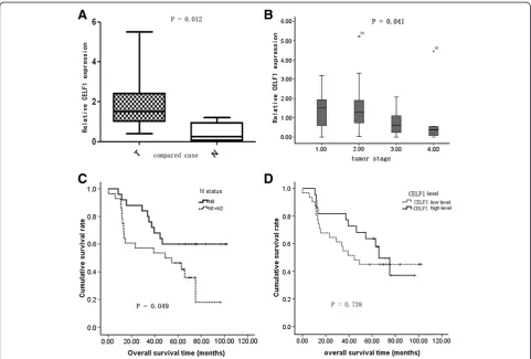
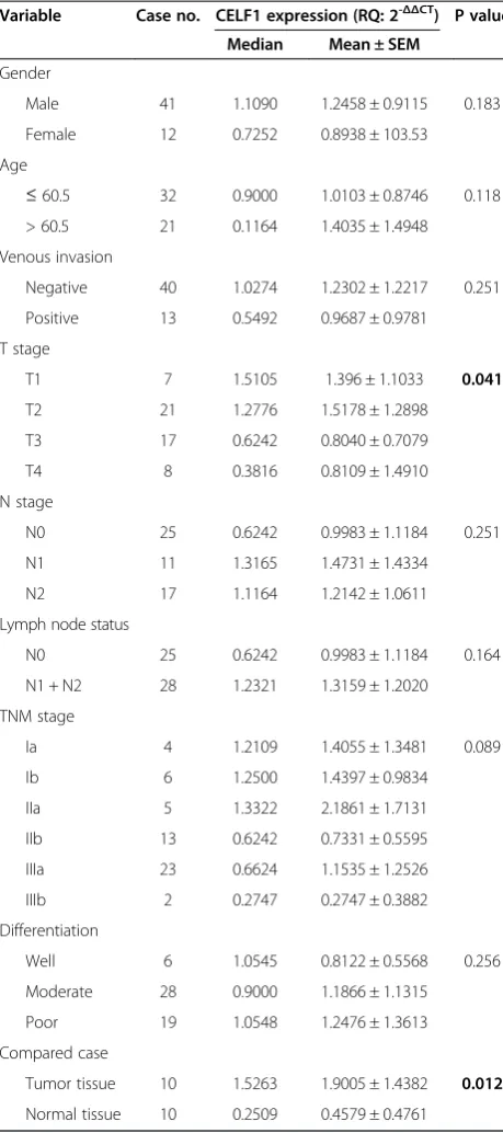
To evaluate the levels of CELF1 expression in lung can-cer tissues and normal tissues, real time PCR was per-formed in 10 lung cancer tissues and 10 normal tissues. Results showed that the relative expression levels of CELF1 were higher in lung cancer tissues compared with the normal tissues (Figure 1A and Table 1). Moreover, the expression levels of CELF1 in lung cancer tumors varied depending on tumor grade. CELF1 expression levels were higher in low-grade cancers compared with high-grade cancers (Figure 1B). A higher expression level of CELF1 was observed in patients with the in-crease of T stage, indicating that when tumors begin to grow larger, gradually some other mechanism(s) may play a more important role to promote cancer cell growth, so the CELF1 expression level had some de-crease. Furthermore, comparison of the survival rate of
patients with N1 and N2 lymph node metastasis and without lymph node metastasis showed that the survival rates were significantly higher in the absence of lymph node metastasis (Figure 1C). In comparing the survival rate with CELF1 expression levels, no significant dif-ference in survival rate was observed between patients with higher CELF1 expression and lower expression (Figure 1D). Together these data indicate that CELF1 ex-pression is not related to postoperative survival in lung cancer patients.
Effect of CELF1 siRNA on the expression levels of CELF1 in lung cancer cells
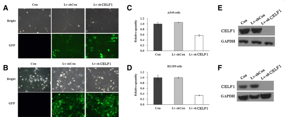
Next, CELF1 mRNA and protein levels in A549 and H1299 lung cancer cells were evaluated by real time PCR and western blot, respectively. As shown in Figure 2, both gene and protein expression of CELF1 were detected in A549 and H1299 cells. The CELF1expressing lung can-cer cells were then infected with lentivirus containing CELF1 shRNA or non-silencing control shRNA, and suc-cessful infection was confirmed by green fluorescence of
infected cells (Figure 3A and B). Fluorescence analysis showed that the lentiviral infection rate was higher in H1299 cells than in A549 cells. Infection of cells with lentivirus containing CELF1 shRNA significantly reduced CELF1 gene and protein expression levels in both A549 and H1299 cells (Figure 3C-F). In contrast, the non-silencing siRNA infection had no effect on CELF1 levels,
confirming that CELF1 expression levels were reduced specifically from CELF1 siRNA.
Effect of CELF1 knockdown on the survival of lung cancer cells
The effect of CELF1 knockdown on lung cancer cell sur-vival was analyzed using a MTT assay performed over a five-day time course. In both cell lines, significant differ-ences in cell survival were not observed until three days after infection. From the 4th day of infection onwards, significant differences in cell survival were observed in both cell lines. The survival rate of CELF1 knockdown cells was markedly (P< 0.0001) reduced at the 5thday of infection compared with the non-infected and control siRNA infected A549 and H1299 cells (Figure 4). The re-duction in cell survival due to CELF1 knockdown was higher in A549 cells than in H1299 cells.
Effect of CELF1 knockdown on the colony forming ability of lung cancer cells
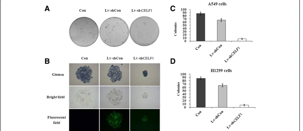
Lung cancer cells tend to form large cell colonies while in culture; therefore, we next evaluated the effect of CELF1 silencing on the colony forming ability of lung cancer cells. As shown in Figure 5A and B, the size of colonies in CELF1-silenced A549 and H1299 cells was markedly reduced compared with the control groups. Similarly, the number of colonies was also significantly (P< 0.001) reduced with the CELF1 gene knockdown. Together these results indicate that CELF1 plays an important role in the growth progression of lung can-cer cells.
Discussion
With increasing mortality rates, lung carcinoma has already become the leading cause of cancer mortality in the world [11]. Many genes are subjected to post-transcriptional regulation via control of the rate of mRNA turnover for transcripts bearing destabilizingcis -elements [12]. Among the very few regulatory factors identified thus far, CELF1 regulates post-transcriptional gene expression by facilitating alternative splicing, trans-lation [13], and mRNA degradation, and it functions by binding directly to RNA [14]. Rattenbacher et al. identi-fied the CELF1 gene and its target proteins as a critical posttranscriptional regulatory network that may play a role in the development of cancer [8]. In addition to re-ports of involvement in breast cancer and leukemia de-velopment, the CELF1 gene may also play a significant role in tumorigenesis and the deterioration of certain tu-mors [15], which is also confirmed by the results in our present study.
Timchenko et al. first identified CELF1 function in the regulation of translation of C/EBP beta isoforms [6]. Subsequent research demonstrated that members of this
Table 1 Expression level of CELF1 in lung carcinoma and relationship to clinicopathological parameters
Variable Case no. CELF1 expression (RQ: 2-ΔΔCT) P value
Median Mean ± SEM
Gender
Male 41 1.1090 1.2458 ± 0.9115 0.183
Female 12 0.7252 0.8938 ± 103.53
Age
≤60.5 32 0.9000 1.0103 ± 0.8746 0.118
> 60.5 21 0.1164 1.4035 ± 1.4948
Venous invasion
Negative 40 1.0274 1.2302 ± 1.2217 0.251
Positive 13 0.5492 0.9687 ± 0.9781
T stage
T1 7 1.5105 1.396 ± 1.1033 0.041
T2 21 1.2776 1.5178 ± 1.2898
T3 17 0.6242 0.8040 ± 0.7079
T4 8 0.3816 0.8109 ± 1.4910
N stage
N0 25 0.6242 0.9983 ± 1.1184 0.251
N1 11 1.3165 1.4731 ± 1.4334
N2 17 1.1164 1.2142 ± 1.0611
Lymph node status
N0 25 0.6242 0.9983 ± 1.1184 0.164
N1 + N2 28 1.2321 1.3159 ± 1.2020
TNM stage
Ia 4 1.2109 1.4055 ± 1.3481 0.089
Ib 6 1.2500 1.4397 ± 0.9834
IIa 5 1.3322 2.1861 ± 1.7131
IIb 13 0.6242 0.7331 ± 0.5595
IIIa 23 0.6624 1.1535 ± 1.2526
IIIb 2 0.2747 0.2747 ± 0.3882
Differentiation
Well 6 1.0545 0.8122 ± 0.5568 0.256
Moderate 28 0.9000 1.1866 ± 1.1315
Poor 19 1.0548 1.2476 ± 1.3613
Compared case
Tumor tissue 10 1.5263 1.9005 ± 1.4382 0.012
protein family regulate pre-mRNA alternative splicing and may also be involved in mRNA editing and transla-tion [16]. The CELF1 gene may play a role in myotonic dystrophy type 1 (DM1) via interactions with the dys-trophia myotonica-protein kinase (DMPK) gene [10]. A previous report identified a correlation between the ex-pression of CELF1 and human lung cancer [17]. How-ever, the cellular mechanism underlying how the CELF1 gene causes this phenomenon has not been clarified. To-gether these findings implicate possible involvement of the CELF1 gene in cell growth. So far there is no litera-ture reporting the biological function of CELF1 gene in lung cancer cell. We speculate that CELF1 may also play an important role in lung cancer proliferation.
Our research primarily focused on the effect of CELF1 knockdown on the viability of lung cancer cells. As shown in the results, the expression of CELF1 was higher
in human lung cancer tissues compared with normal tis-sues. Moreover, A549 and H1299 lung cancer cells also exhibited CELF1 expression in mRNA and protein level. Lentiviral-mediated delivery of CELF1 silencing siRNA significantly inhibited these upregulated levels of CELF1 expression, demonstrating that the CELF1 knockdown method was successful. Furtherin vivostudies should be performed to confirm the use of this siRNA method as a potential therapeutic tool.
Interestingly, upon knockdown of CELF1, the survival rates and colony forming ability of lung cancer cells were markedly reduced, indicating pivotal roles of CELF1 in the survival of lung cancer cells. Reports in the literature have suggested that upregulation of CELF1 increased the turnover of oncogenes related to the proliferation of lung cancer cells [7,9,18]. Hence, in the absence of CELF1, the turnover of possible oncogenes could presumably Figure 2Expression levels of CELF1 in lung cancer cells. (A). Expression levels of CELF1 in A549 and H1299 cells analyzed by real time PCR.
(B). Protein expression levels of CELF1 in A549 and H1299 cells analyzed by western blotting. The values represents the mean from three independent experiments; bars represent SD. *P< 0.05, **P< 0.01 compared with controls.
decrease, consistent with the cancer cells showing de-creased capacity of proliferation and colony formation. Our study showed that CELF1 is overexpressed in lung cancer tissue on RNA level compared with the normal lung tissue and tumor grades had relationship with CELF1 expression level, which is line with the hypothesis men-tioned above.
From these results, we can conclude that CELF1 can affect the growth of lung cancer cells and plays an im-portant role in the tumor development process. Further research on the molecular mechanisms of the CELF1 gene is required, particularly in identifying CELF1-interacting proteins, elucidating the molecular mecha-nisms underlying its biological effects, and determining
whether it plays a guiding role in the treatment of lung cancer.
Conclusion
In summary, CELF1 may have significant roles in the progression of lung carcinoma. The CELF1 siRNA me-thod has emerged as a potentially powerful tool for can-cer therapeutics in silencing genes responsible for cancan-cer progression and tumorigenesis.
Materials and methods Human specimens and reagents
Fifty-three pulmonary cancer samples of fresh frozen tis-sue were acquired from the Department of Thoracic Figure 4Effect of CELF1 gene knockdown on the proliferation of lung cancer cells.Proliferation of A549(A)and A1299(B)cells were treated as indicated and measured by MTT assay. The values represents the mean from three independent experiments; bars represent SD. ***P< 0.001 compared with controls.
Surgery, Beijing cancer hospital, under approval from the Ethical Committee. Written consent statements were obtained from all patients before operation. None of the patients received any neoadjuvant therapy prior to surgery. The tissues were collected immediately after surgical resection at the Beijing Cancer Hospital and stored at the Tissue Bank of Peking University Oncology School. Clinicopathological characteristics of the tumors were defined according to the TNM staging system criteria of UICC. Clinicopathological factors are shown in Table 1.
AgeI, EcoRI, and SYBRGreen Master Mix Kits were purchased from TaKaRa (Dalian, China). pHelper1.0, pHelper 2.0, and pGCSIL-GFP plasmids were purchased from Genechem Co. Ltd (Shanghai, China). The RNeasy Midi Kit was from Qiagen (Valencia, CA, USA). Dulbec-co’s modified Eagle’s medium (DMEM), Roswell Park Memorial Institute 1640 (RPMI 1640) and fetal bovine serum (FBS) were obtained from Hyclone (Logan, UT, USA). Lipofectamine2000, TRIzol and SuperScriptII re-verse transcriptase were purchased from Invitrogen (Carlsbad, CA, USA). All other chemicals were obtained from Sigma (St. Louis, MO, USA). The following anti-bodies were obtained from Santa Cruz: anti-CELF1 (1:1000 dilution), anti-GAPDH (glyceraldehyde-3-phosphate dehydrogenase, 1:3000 dilution) and anti-mouse HRP (1:5000 dilution).
Cell culture
Human lung cancer cells (A549 and H1299) and human embryonic kidney (HEK) 293 T cell lines were obtained from the cell bank of Shanghai Institute of Cell Biology. A549 and 293 T cells were maintained in DMEM sup-plemented with 10% heat-inactivated FBS and penicillin/ streptomycin at 37°C in a humidified atmosphere of 5% CO2. H1299 cells were maintained in RPMI 1640 me-dium supplemented with 10% heat-inactivated FBS and penicillin/streptomycin at 37°C in a humidified atmos-phere of 5% CO2.
Construction of CELF1 shRNA-containing lentivirus and infection
The sequences of CELF1 siRNA and non-silencing
con-trol siRNA were 5′-CTAGCCGGGATTGAAGAATGCC
GGATATTCAAGAGATATCCGGCATTCTTCAATCT
TTTTAAT-3′ and 5′-CTAGCCCGGTTCTCCGAAC
GTGTCACGTATCTCGAGATACGTGACACGTTCGG
AGAATTTTTTTAAT-3′, respectively. The nucleotide
sequences were inserted into the plasmid through the pFH-L vector (Shanghai Hollybio, China) and the gen-erated lentiviral-based shRNA-expressing vectors were confirmed by DNA sequencing. Lentiviruses were genera-ted by transfection of 293 T cells at 80% confluence with generated plasmids. The cells were starved for 2 h before
transfection, and pFH-L-shCELF1 or -shCTRL and the
packaging vector carriers pVSVG-I and PCMVΔR8.92
were transfected into cells using Lipofectamine 2000. The supernatant was collected 48 h after transfection and len-tiviral particles were harvested by ultracentrifugation (4000g) at 4°C for 10 min. The collected virus particles were filtered through a 45 μM filter and the filtrate was centrifuged (4000g at 4°C) for 15 min to collect the viral concentrate.
A549 and H1299 cancer cells were then infected with CELF1 shRNA- or control shRNA-expressing lentivi-ruses at a MOI of 30 for A549 cells or MOI of 15 for H1299 cells. The cells were seeded (5 × 10⁴ cells per well) in six-well plates, and after 24 h of incubation, the culture medium was replaced with Opti-MEM medium containing the appropriate amount of the virus. The cells were then incubated with the virus for another 48 h. Successful transfection was confirmed by observa-tion through a fluorescence microscope (Leica, Germany) for expression of green fluorescence protein.
Real-time PCR analysis
RNA was obtained using the Total RNA Isolation Rea-gent (ABgene™) according to the manufacturer’s instruc-tions (Abgene, Surrey, UK). Total RNA was converted to cDNA using DuraScript™, a commercial reverse tran-scription kit from Sigma Aldrich. Real-time quantitative PCR analysis for the RNA extracted from lung cancer tissues and cultured lung cancer cells was performed using the SYBR Green Master Mix Kit (Applied Biosys-tems, Foster City, CA). In brief, each PCR reaction mix-ture contained 10 μl of 2 × SYBR Green Master Mix, 0.4μl of sense and antisense primers (2.5μM) and 10 ng of cDNA. Reactions were run for 40 cycles, including de-naturation at 95°C for 10 min and annealing at 60°C for 1 min in a total volume of 20μl using an ABI 7500 Real Time PCR platform. The primer sequences for PCR
amplification of the CELF1 gene were 5′-ACCTGTTC
ATCTACCACCTG-3′ and 5′-GGCTTGCTGTCATTC
TTCG-3′. Primer sequences for the internal control
GAPDH were 5′-GACCCCTTCATTGACCTCAAC-3′
and 5′-CTTCTCCATGGTGGTGAAGA-3′. Relative gene
expression levels were calculated using 2-ΔΔCT analysis.
Western blot analysis
collected after centrifuging the cell lysate (12,000 ×gfor 15 min) and the protein content was measured by the Lowry method. The protein concentration of each sam-ple was adjusted to 2 μg/μl and a 20 μl volume was mixed with 2 × SDS sample buffer (100 mM Tris–HCl, pH 6.8, 10 mM EDTA, 4% SDS, 10% glycine) and se-parated by electrophoresis on a 10% SDS-PAGE gel at 50 V for 3 h. The gel was transferred to a PVDF membrane at 300 mA for 1.5 h, and proteins were detected after primary antibody treatment overnight at 4°C and secondary antibody treatment for 2 h at room temperature using an Amersham ECL kit (GE Healthcare, UK) and exposure to X-ray film. The bands obtained were quantified with an Image Quant densitometric scanner (Molecular Dynamics, Amersham Biosciences).
MTT analysis
For the cell viability analysis, A549 and H1299 cells were first seeded (2 × 10³ cells/well) into a 96-well plate and infected with CELF1 silencing or non-silencing siRNA-containing lentivirus for 72 h. Following infection, 20μl of MTT solution (5 mg/ml) was added to each well and cells were incubated at 37°C for 4 h. The medium and
MTT from the wells was removed and 200μl of DMSO
was added to each well. The optical density was mea-sured using a microplate reader at 490 nm. Experiments were performed in triplicate.
Colony formation assay
Lung cancer cells seeded in six-well plates (2 × 10²cells/ well) were infected with CELF1 silencing and non-silencing siRNA-containing lentivirus for 72 h. The cells were continuously incubated, and medium was replaced with new medium every three days until 8 days of cul-ture. The cells were then washed with PBS and fixed with 4% paraformaldehyde. The fixed cells were stained with freshly prepared diluted Giemsa stain for 20 min. The cells were washed with double distilled water and colonies were counted using a fluorescence microscope.
Consent
Written informed consent was obtained from the patients before operation.
Competing interests
The authors declare that they have no competing interests.
Authors’contributions
LNW and YJX contributed equally to the study design, experimental work, data analysis and preparation of the manuscript. JFC, LJZ and XMM participated in the design of the study and performed the statistical analysis. JFC conceived of the study, and participated in its design and coordination and helped to draft the manuscript. All authors read and approved the final manuscript.
Acknowledgements
The authors wish to thank Yinan Liu for clinical data collection support; Xinyuan Lao for checking grammar error. This study was supported by Beijing Science New Star Plan (Z11111005450000).
Author details
1Key laboratory of Carcinogenesis and Translational Research (Ministry of Education), Central Laboratory, Peking University Cancer Hospital & Institute, Beijing 100142, China.2The College of Life Science and Bio-engineering, Beijing University of Technology, Beijing 100022, People’s Republic of China. 3Key laboratory of Carcinogenesis and Translational Research (Ministry of Education), Department of Thoracic Surgery II, Peking University Cancer Hospital & Institute, Beijing 100142, China.
Received: 17 June 2013 Accepted: 14 November 2013 Published: 15 November 2013
References
1. Parkin DM, Pisani P, Ferlay J:Global cancer statistics.CA: Cancer J Clin1999,
49(1):33–64. 31.
2. Caldon CE, Lee CS, Sutherland RL, Musgrove EA:Wilms’tumor protein 1: an early target of progestin regulation in T-47D breast cancer cells that modulates proliferation and differentiation.Oncogene2008,
27(1):126–138.
3. Yang M, Yuan F, Li P, Chen Z, Chen A, Li S, Hu C:Interferon regulatory factor 4 binding protein is a novel p53 target gene and suppresses cisplatin-induced apoptosis of breast cancer cells.
Mol Cancer2012,11:54.
4. Klos KS, Wyszomierski SL, Sun M, Tan M, Zhou X, Li P, Yang W, Yin G, Hittelman WN, Yu D:ErbB2 increases vascular endothelial growth factor protein synthesis via activation of mammalian target of rapamycin/ p70S6K leading to increased angiogenesis and spontaneous metastasis of human breast cancer cells.Cancer Res2006,66(4):2028–2037. 5. Veale D, Ashcroft T, Marsh C, Gibson GJ, Harris AL:Epidermal growth
factor receptors in non-small cell lung cancer.British J Cancer1987,
55(5):513–516.
6. Timchenko NA, Welm AL, Lu X, Timchenko LT:CUG repeat binding protein (CUGBP1) interacts with the 5’region of C/EBPbeta mRNA and regulates translation of C/EBPbeta isoforms.Nucleic Acids Res1999,
27(22):4517–4525.
7. Chang ET, Donahue JM, Xiao L, Cui Y, Rao JN, Turner DJ, Twaddell WS, Wang JY, Battafarano RJ:The RNA-binding protein CUG-BP1 increases survivin expression in oesophageal cancer cells through enhanced mRNA stability.Biochem J2012,446(1):113–123.
8. Rattenbacher B, Beisang D, Wiesner DL, Jeschke JC, Von Hohenberg M, St Louis-Vlasova IA, Bohjanen PR:Analysis of CUGBP1 targets identifies GU-repeat sequences that mediate rapid mRNA decay.Mol Cell Biol2010,
30(16):3970–3980.
9. Zheng Y, Miskimins WK:CUG-binding protein represses translation of p27Kip1 mRNA through its internal ribosomal entry site.RNA Biol2011,
8(3):365–371.
10. Ward AJ, Rimer M, Killian JM, Dowling JJ, Cooper TA:CUGBP1 overexpression in mouse skeletal muscle reproduces features of myotonic dystrophy type 1.Hum Mol Gen2010,19(18):3614–3622. 11. Kang S, Koh ES, Vinod SK, Jalaludin B:Cost analysis of lung cancer
management in South Western Sydney.J Med Imaging Radiat Oncol2012,
56(2):235–241.
12. Benjamin D, Moroni C:mRNA stability and cancer: an emerging link?
Expert Opinion Biol Ther2007,7(10):1515–1529.
13. Cui YH, Xiao L, Rao JN, Zou T, Liu L, Chen Y, Turner DJ, Gorospe M, Wang JY:miR-503 represses CUG-binding protein 1 translation by recruiting CUGBP1 mRNA to processing bodies.Mol Biol Cell2012,
23(1):151–162.
14. Vlasova-St Louis I, Bohjanen PR:Coordinate regulation of mRNA decay networks by GU-rich elements and CELF1.Curr Opin Gen Dev2011,
21(4):444–451.
15. Jones K, Timchenko L, Timchenko NA:The role of CUGBP1 in age-dependent changes of liver functions.Ageing Res Rev2012,11(4):442–449. 16. Devi GR:siRNA-based approaches in cancer therapy.Cancer Gene Ther
17. Jiao W, Zhao J, Wang M, Wang Y, Luo Y, Zhao Y, Tang D, Shen Y:
CUG-binding protein 1 (CUGBP1) expression and prognosis of non-small cell lung cancer.Clin Transl Oncol2013,15(10):789–795.
18. Talwar S, Balasubramanian S, Sundaramurthy S, House R, Wilusz CJ, Kuppuswamy D, D’Silva N, Gillespie MB, Hill EG, Palanisamy V:
Overexpression of RNA-binding protein CELF1 prevents apoptosis and destabilizes pro-apoptotic mRNAs in oral cancer cells.RNA Biol 2013,10(2):277–286.
doi:10.1186/1475-2867-13-115
Cite this article as:Wuet al.:Si-RNA mediated knockdown of CELF1
gene suppressed the proliferation of human lung cancer cells.Cancer
Cell International201313:115.
Submit your next manuscript to BioMed Central and take full advantage of:
• Convenient online submission
• Thorough peer review
• No space constraints or color figure charges
• Immediate publication on acceptance
• Inclusion in PubMed, CAS, Scopus and Google Scholar
• Research which is freely available for redistribution