Lesław W. Zub
1, B, C, F, Małgorzata Szymczyk
2, B–D, Anna Pokryszko-Dragan
2, B, C,
Małgorzata Bilińska
2, A, E, FEvaluation of Pain in Patients with Lumbar Disc Surgery
Using VAS Scale and Quantitative Sensory Testing
Ocena bólu u pacjentów operowanych z powodu dyskopatii lędźwiowej
za pomocą skali VAS i ilościowego pomiaru czucia
1 Department of Neurosurgery, Wroclaw Medical University, Poland 2 Department of Neurology, Wroclaw Medical University, Poland
A – research concept and design; B – collection and/or assembly of data; C – data analysis and interpretation;
D – writing the article; E – critical revision of the article; F – final approval of article; G – other
Abstract
Background. Pain is interpreted at the cortex level, however, pain signaling stimuli arise at the periphery and are conveyed by nociceptive A delta and C fibers.
Objectives. Evaluation of pain using the VAS scale in pre- and postoperative S1 sciatica patients with regard to thermal thresholds in the corresponding dermatome.
Material and Methods. Twenty-six S1 sciatica patients with herniated disc on an MRI, non-responsive to
con-servative care, were involved in the study. Pain in the affected leg was measured using the VAS scale and thermal thresholds in S1 symptomatic dermatome using Quantitative Sensory Testing (QST).
Results. Pain intensity as well as thermal thresholds were increased in sciatica patients compared to controls. Disc surgery resulted in a pronounced lowering of pain in each of the operated patients. From the whole group, 21 subjects were examined postoperatively six months later. In the group with complete clinical recovery, thermal thresholds were within normal limits. In those patients with residual pain disability, normalization of thresholds has not been achieved
Conclusions. Pain in sciatica patients may be objectively measured by QST (Adv Clin Exp Med 2013, 22, 3, 411–419).
Key words: monoradicular sciatica, VAS scale, QST, disc surgery.
Streszczenie
Wprowadzenie. Ból jest zjawiskiem złożonym i jego interpretacja następuje na poziomie kory mózgowej, do której informacja o szkodliwych bodźcach jest przewodzona z obwodu nocyceptywnymi włóknami A delta i C.
Cel pracy. Ocena bólu z wykorzystaniem skali VAS w odniesieniu do funkcji włókien A delta i C w uszkodzonym der-matomie u chorych z jednopoziomowym bólowym zespołem korzeniowym przed i po zabiegu dekompresji korzenia.
Materiał i metody. 26 chorych na ischialgię S1, nieodpowiadających na leczenie zachowawcze, z potwierdzonym
uciskiem korzenia przez dysk poddano zabiegom discektomii. Ból w zajętej kończynie oceniano w skali VAS, a progi czucia metodą ilościowego pomiaru (QST) w dermatomie S1 bezpośrednio przed i 6 miesięcy po zabiegu.
Wyniki. U wszystkich chorych przed zabiegiem wykazano podwyższenie progów czucia. Po zabiegu u wszystkich
badanych intensywność bólu znacznie zmalała. 21 chorych poddano powtórnym badaniom 6 miesięcy po zabiegu. U pacjentów z całkowitym ustąpieniem bólów progi wróciły do wartości prawidłowej. U chorych z niepełną popra-wą progi czucia pozostały nieprawidłowe.
Wnioski. Badanie progów czucia metodą QST może służyć obiektywizacji doznań bólowych (Adv Clin Exp Med
2013, 22, 3, 411–419).
Słowa kluczowe: jednopoziomowe uszkodzenie korzeniowe, skala VAS, ilościowy pomiar czucia (QST),
dekom-presja korzenia.
Adv Clin Exp Med 2013, 22, 3, 411–419 ISSN 1899–5276
ORIgINAL PAPERS
Sciatica is characterized by radiating leg pain and related disabilities in dermatomal distribution. The disease results mainly from nerve root com-pression by herniated disc [1]. The conservative management of pain should be introduced first. However, with persistent, intractable pain non-re-sponsive to 6 to 8-week pharmacological treatment with rehabilitation, disc surgery should be taken into consideration [2]. Pain reported by patients differs in intensity which depends on many factors such as self-experience, personality, cultural and en-vironmental conditions. From a neuropsychologi-cal point of view, pain is a complex phenomenon and its interpretation takes place at the cortex level. However, pain information is transmitted from the periphery by afferent nociceptive pathways into the central nervous system (CNS). At the periphery, small myelinated A delta fibers and unmyelinated C ones are involved in the process of pain signal-ing, which has been proven on animal models and confirmed among people by selective blocking of the nerve’s fibers in sensory cutaneous nerves [3]. Approaches towards pain assessment have result-ed in the introduction of different types of scales, among them the numerical Visual Analogue Scale (VAS) and scales which determine the intensity of pain based on the degree of restrictions in everyday activities [4, 5]. Recently, a new neurophysiologi-cal method, Quantitative Sensory Testing (QST), has been applied to study the functional changes in the sensory fibers of peripheral nerves [6, 7]. QST determines skin thermal thresholds in the area supplied by nerve. Each modality of thermal sensation is conveyed by selective types of sensory fibers. The threshold for cold sensations (CS) is tied to the A delta fibers’ function, and for warm sensations (WS) the function of C fibers specific to temperature in the innocuous range, respectively, while thresholds for cold pain (CP) and heat pain (HP) determine the function of polymodal C fibers responsive to noxious cold and heat, respective-ly [8–11]. Bearing in mind that pain information is transmitted from the periphery by A delta and C fibers, the authors have decided to compare the pain intensity estimated by the VAS scale in the af-fected leg with the function of A delta and C fibers in the affected dermatome in S1 sciatica patients just prior and 6 months after disc surgery.
Material and Methods
Twenty-six patients (14 males and 12 females, aged 22–59 years, mean 36.8 ± 8.7 years) with monoradicular sciatica corresponding to S1 root distribution were included in the study. All of them complained of severe, intractable pain which
cycle was repeated. The mean from 4 cycles stands for the threshold for CS. The same procedure was performed in order to obtained WS threshold but, instead of lowering, an increase of temperature was applied [14]. The thresholds for CP and HP (first, unpleasant feeling of temperature with changes of 2°C per sec) were obtained in the same way as the three cycles for each modality. The electrophysi-ological evaluation was performed in a silent room at a stable temperature of 25°C. The above listed exams were performed 6 months later. The con-trol group consisted of 30 age- and sex-matched healthy controls (aged from 21 to 50 years, mean 36.6 ± 7.6 years, 13 women and 17 men). Neuro-graphic studies were performed on the left side in the control group. All subjects signed informed consent. The study was approved by the Local Eth-ics Committee at the Wroclaw Medical University, no 530/2009.
The statistical analyses were performed using STATISTICA 10.0 PL. The t-Student test for inde-pendents samples was used to compare the means in the two groups. The t-Student test for dependent samples was used to compare the means before and after operations. The results are presented as means ± SD. The association between two variables was examined by Pearson’s correlation coefficient. Statistical significance was accepted at p < 0.05.
Results
All patients complained of radiating pain along the affected leg in the distribution of S1
der-matome. Upon neurological examination, mild hypoaesthesia localized to S1 was presented in 18 cases. All patients experienced pain at straight leg raising. The Lasegue’s sign was positive at a mean value of 30.2 ± 11.7 degrees. There was also a dis-crete lowering of dorsiflexion (4 or 4/5 point) on the 6-point Lovett’s scale in 3 and diminished an-kle reflex in 11 patients. The mean score of pain intensity on the VAS scale in the affected limb was 8.04 ± 2.13. The mean value of VAS on the first visit when the neurosurgeon classified patients for disc surgery was 9.25 ± 1.01. Conventionally-per-formed motor nerve conduction studies on both sides in the S1 sciatica group did not reveal any abnormalities (results not included in paper). The preoperative parameters of the sural n. study and CS, WS, CP, and HP thresholds on the affected side in patients with S1 sciatica and in controls are presented in Tab. 1 and Tab. 2, respectively. The parameters of sural n. did not differ between the examined groups, while there was significant increase in each examined threshold in patients compared with the controls (for WS p = 0.0001, for CS, CP, and HP p < 0.0001, respectively).
The correlations between thermal thresholds in symptomatic S1 dermatome and the value of VAS and Lasegue’s sign are listed in Tab. 3. CS did not correlate with either VAS or the Lasegue’s sign. WS correlated with VAS (p = 0.007) while not with Lasegue. Both CP and HP were highly correlated with VAS (p < 0.0001) and Lasegue (p < 0.0001, and p = 0.0006, respectively).
Twenty-one patients who underwent S1 de-compression were evaluated 6 months later. The
Table 1. Sural nerve conduction studies on the symptomatic side in preoperative S1 sciatica patients and healthy controls
Tabela 1. Parametry przewodnictwa czuciowego w n. łydkowym po stronie korzeniowego zespołu bólowego S1 przed zabie-giem i w grupie kontrolnej
Sural nerve (Nerw łydkowy)
Distal latency (Latencja dystalna) (msec)
SNAP Amplitude (Amplituda) (µV)
Sensory velocity
(Szybkość przewodzenia czuciowego) (m/sec)
S1 sciatica group (grupa chorych) (n = 26)
2.86 ± 0.62 15.92 ± 5.15 50.82 ± 6.97
Control group (grupa kontrolna) (n = 30)
2.65 ± 0.47 16.17 ± 5.63 51.37 ± 5.20
p ns ns ns
n – number of patients/healthy controls. ns – not significant.
Table 2. Thermal thresholds in the S1 dermatome on the symptomatic side in preoperative S1 sciatica patients and healthy controls
Tabela 2. Progi czucia w dermatomie S1 po stronie korzeniowego zespołu bólowego S1 przed zabiegiem i w grupie kontrolnej
Thermal tresholds (Progi czucia)
Cold Sensation (CS) (Próg czucia zimna) [oC]
Warm Sensation (WS) (Próg czucia ciepła) [oC]
Cold Pain (CP) (Próg czucia bólowego zimna)
[oC]
Heat Pain (HP) (Próg czucia bólowego ciepła)
[oC]
S1 sciatica group (grupa chorych) (n = 26)
24.80 ± 5.36 38.77 ± 3.95 11.90 ± 9.59 45.24 ± 3.71
Control group (grupa kontrolna) (n = 30)
29.36 ± 0.90 35.67 ± 0.94 21.65 ± 3.54 41.33 ± 1.79
p < 0.0001 < 0.0001 < 0.0001 < 0.0001
n – number of patients/healthy controls. ns – not significant.
n – liczba pacjentów/osób z grupy kontrolnej. ns – bez istotności statystycznej.
Table 3. Correlations between thermal thresholds in S1 dermatome and pain intensity on VAS scale, and Lasegue’s sign on the symptomatic leg in S1 sciatica patients
Tabela 3. Korelacje między progami czucia w dermatomie S1, natężeniem bólu w skali VAS i objawem Lasegue’a u chorych z jednopoziomowym zespołem bólowym korzeniowym S1 po stronie uszkodzenia
Thermal tresholds
(Progi czucia) Visual Analogue Scale (VAS)(Skala VAS) Lasegue’s sign(Objaw Lasegue’a) Cold Sensation (CS)
(Próg czucia zimna) r = 0.35p = 0.076 ns
Warm Senation (WS)
(Próg czucia ciepła) r = 0.52p = 0.007 ns
Cold Pain (CP)
(Próg czucia bólowego zimna) r = –0.75p < 0.0001 r = 0.52P = 0.0006 Heat Pain (HP)
(Próg czucia bólowego ciepła) r = 0.69P < 0.0001 r = –0.52P = 0.006
mean VAS value in this group in the affected leg was 0.38 ± 1.02 points, which was significantly lower than before the operation (7.81 ± 2.23, p < 0.0001). The residual features of sensation disability (pain) rec-ognizes a division of patients into 2 groups: 1) with complete recovery (A1) which means with VAS = 0,
and 2) with incomplete recovery (B1). The mean
VAS value in the A1 group before disc surgery (A0
group) was 7.21 ± 2.22. In the B1 group before the
operation (B0 group), the mean VAS value was 9.0.
The preoperative values of VAS in A0 and B0 groups
did not differ significantly. Upon neurological ex-amination after disc surgery no motor deficit was found. Repeated conventionally-performed MNCS and SNCS in the A1 group did not differ from the
results obtained in the same group just prior to the operation (A0 group). There were also no
differenc-es in the MNCS and SNCS parameters between B1
and B0 groups (data not included in this paper).
The postoperative QST data in the A1 group
with preoperative QST findings (A0) and the
post-operative QST data in the B1 group with
preopera-tive QST findings (B0) are listed in Tab. 4. There
was significant improvement in all four thermal thresholds in the A1 group compared to the results
obtained before disc surgery (A0) (for CS p = 0.02,
for WS p = 0.01, for CP p = 0.01, for HP p = 0.04). The thresholds in the A1 group did not differ from
signifi-cantly from the results obtained in the B0 group.
These postoperative thresholds were significantly increased when compared to the controls (for all modalities p < 0.0001). The graphically presented preoperative and postoperative thermal thresholds in patient with complete recovery are presented in Fig. 1 and Fig. 2, respectively. The graphically presented preoperative and postoperative thermal thresholds in patients with incomplete recovery are presented in Fig. 3 and Fig. 4, respectively.
Next, the comparison of preoperative thresh-olds between the group with complete postopera-tive recovery (A0) and the group with incomplete
recovery (B0) was performed. The preoperative
thermal thresholds for CS, WS, CP in the B0 group
were significantly increased compared to the cor-responding thresholds in the A0 group (p = 0.01,
p = 0.0006 and p = 0.02, respectively). There was no difference in HP threshold between the A0 and
B0 groups.
Discussion
Disabling pain with highly restricted self-service together with one-level evident root com-pression by a herniated disc on the MRI were es-sentials for the decision to perform surgical root decompression. In each case, root compression
was confirmed intraoperatively. The approach to the treatment of acute radicular pain depends on clinical findings [2]. In cases of pain which ac-company cauda equina syndrome or in cases with severe muscle paresis, surgical treatment should be introduced. Management of cases with mono-radicular pain caused by a herniated disc, which means conservative vs. surgical decompression, is mainly based on an individual assessment of each case by a neurosurgeon and on the results of the remote effects of each type of treatment [15]. The efficacy of surgical decompression was demon-strated by Weber [16]. The cited author in the pro-spective study has estimated the remote results of conservative vs. surgical management in patients with lumbo-sacral radiculopathies. After one year of observation, 66% of operated patients were free of symptoms while in the conservative treatment group, full recovery was achieved only in 33%. Af-ter a four-year observation, there was still a preva-lence of good results of surgical management vs. conservative (70% vs. 51%). Atlas et al. [17, 18] compared the long-term outcomes (five-year and ten-year observation) of surgical and non-surgical management of sciatica secondary to a lumbar disc herniation. Similarly to Weber [16], after both pe-riods of time they found better functional status in operated patients compared with those treated with non-surgical management. A routine examination
Table 4. Preoperative (A0) and postoperative (A1) thermal thresholds in S1 dermatome in the group of patients with complete
recovery and preoperative (B0) and postoperative (B1) thermal thresholds in the group of patients with incomplete recovery
Tabela 4. Przedoperacyjne (A0) i pooperacyjne (A1) wartości progów czucia w dermatomie S1 u chorych z pełną poprawą po
zabiegu oraz przedoperacyjne (B0) i pooperacyjne (B1) wartości progów czucia w dermatomie S1 u chorych z niepełną poprawą
Cold sensation (CS) [oC]
(Próg zimna)
Warm sensation (WS) [oC]
(Próg ciepła)
Cold Pain (CP) [oC]
(Próg bólowego zimna)
Heat Pain (HP) [oC]
(Próg bólowego ciepła)
A0 (n = 14) 26.79 ± 2.73 36.94 ± 2.42 15.11 ± 8.74 44.06 ± 3.26
A1 (n = 14) 28.63 ± 1.68 35.98 ± 1.74 21.49 ± 3.88 41.92 ± 3.03
B0 (n = 7) 24.59 ± 3.37 40.36 ± 3.88 8.14 ± 10.11 45.74 ± 4.65
B1 (n = 7) 25.3 ± 4.96 41.74 ± 3.21 9.56 ± 10.0 46.91 ± 2.85
p A0 vs A1 :0.02
B0 vs B1: ns
A0 vs B0: 0.01
A0 vs A1 : 0.01
B0 vs B1: ns
A0 vs B0: 0.0006
A0 vs A1 : 0.01
B0 vs B1: ns
A0 vs B0: 0.02
A0 vs A1 : 0.04
B0 vs B1: ns
A0 vs B0: ns
A0 – preoperative thermal tresholds in a group of S1 sciatica with complete recovery.
A1 – postoperative thermal tresholds in a group of S1 sciatica with complete recovery.
B0 – preoperative thermal tresholds in a group of S1 sciatica with incomplete recovery.
B1 – postoperative thermal tresholds in a group of S1 sciatica with incomplete recovery.
n – number of patients.
A0 – przedoperacyjne wartości progów czucia w dermatomie S1 u chorych z pełną poprawą po zabiegu.
A1 – pooperacyjne wartości progów czucia w dermatomie S1 u chorych z pełną poprawą po zabiegu.
B0 – przedoperacyjne wartości progów czucia w dermatomie S1 u chorych z niepełną poprawą po zabiegu.
B1 – pooperacyjne wartości progów czucia w dermatomie S1 u chorych z niepełną poprawą po zabiegu.
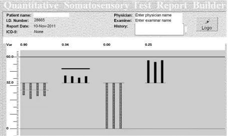
Fig. 2. graphical presentation of postoperative thermal thresholds in the same patient as in Fig. 1 (examination no. 29055). Mean values of examined thresholds: CS – 28.6oC, WS – 34.5oC, CP – 20.7oC, HP – 43.5oC. CS – cold
sen-sation, WS – warm sensen-sation, CP – pain induced by cold, HP – pain induced by hot
Ryc. 2. graficzne przedstawienie pooperacyjnych wartości termicznych progów u chorego z ryc. 1 (badanie nr 29055).
Średnie wartości badanych progów: CS – 28.6oC, WS – 34.5oC, CP – 20.7oC, HP – 43.5oC. CS – zimno, WS – ciepło,
CP – ból wywołany zimnem, HP – ból wywołany wysoką temperaturą
Fig. 1. graphical presentation of preoperative thermal thresholds in S1 sciatica patient with complete recovery after disc surgery (examination no. 28665). Mean values of examined thresholds: CS – 23.4oC, WS – 37.2oC, CP – 0oC, HP
– 47.7oC. CS – cold sensation, WS – warm sensation, CP – pain induced by cold, HP – pain induced by hot
Ryc. 1. graficzne przedstawienie przedoperacyjnych wartości termicznych progów u chorego z jednopoziomowym zespołem bólowym S1 z całkowitą poprawą po zabiegu (badanie nr 28665). Średnie wartości badanych progów: CS – 23.4oC, WS – 37.2oC,
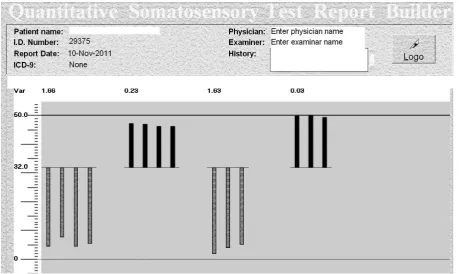
Fig. 3. graphical presentation of preoperative thermal thresholds in S1 sciatica patient with incomplete recovery after disc surgery (examination no. 28982). Mean values of examined thresholds: CS – 2.9oC, WS – 44.1oC, CP – 0oC,
HP – 50.0oC. CS – cold sensation, WS – warm sensation, CP – pain induced by cold, HP – pain induced by hot
Ryc. 3. graficzne przedstawienie przedoperacyjnych wartości termicznych progów u chorego z jednopoziomowym zespo-łem bólowym S1 z niecałkowitą poprawą po zabiegu (badanie nr 28982). Średnie wartości badanych progów: CS – 2.9oC, WS
– 44.1oC, CP – 0oC, HP –50.0oC. CS – zimno, WS – ciepło, CP – ból wywołany zimnem, HP – ból wywołany wysoką
temperatu-rą
Fig. 4. graphical presentation of postoperative thermal thresholds in the same patient as in Fig. 3 (examination no. 29375). Mean values of examined thresholds: CS – 5.6oC, WS – 46.8oC, CP – 3.7oC, HP – 49.9oC. CS – cold
sensa-tion, WS – warm sensasensa-tion, CP – pain induced by cold, HP – pain induced by hot
Ryc. 4. graficzne przedstawienie pooperacyjnych wartości termicznych progów u chorego z ryc. 3 (badanie nr 29375).
Średnie wartości badanych progów: CS – 5.6oC, WS – 46.8oC, CP –3.7oC, HP – 49.9oC. CS – zimno, WS – ciepło,
of sural n. conduction studies has not revealed any abnormalities on the affected side, which is in ac-cordance with data from literature [19, 20]. The sparing of SNAP probably resulted from a lack of damage of the dorsal root ganglia (DRg) [21, 22]. DRgs are usually unaffected by disc herniation due to their location in the intravertebral foram-ina. However, Moon et al. [23] found the intra-vertebral location of DRg at S1 level in 81% of the examined patients.
Preoperative QST studies have revealed an increase in thermal thresholds of all modalities in present S1 sciatica group, pointing to the state-ment that all types of small nociceptive fibers are affected. The results concerning the involvement of different populations of nociceptive fibers in ra-dicular pain differ. Nygaard et al. [24] confirmed the increase in CS and WS thresholds in the af-fected dermatome while HP thresholds were the same in sciatica patients and controls. Mosek et al. [25] found selective damage to A delta fibers (increased CS threshold) while not to C ones in root compression. Zwart et al. [26] revealed in the subgroup of sciatica patients with operative-ly confirmed disc herniation much more severe damage to A delta than C fibers. This is explained by a higher susceptibility of myelinated fibers to compression [22]. In present patients, increased thermal thresholds correlated with pain intensity in the affected leg measured using VAS. Similarly, Quarishi et al. [27] found a correlation between CS and WS thresholds, and VAS intensity. In pres-ent group, the Lasegue’s sign only correlated with C polymodal fibers conveying heat and cold pain.
Five patients were lost for subsequent evalua-tion, which took place 6 months after disc surgery. Similarly to the results of Peul et al. [15], the pain levels reported by the remaining 21 patients were much lower compared to the level in the same group in the preoperative period. On the basis of
VAS evaluation, the S1 sciatica patients were sub-divided into 2 groups. Patients who did not suffer pain (VAS = 0) made a group with complete recov-ery while those with residual pain sensation in the affected dermatome comprised a group with in-complete recovery. There was a normalization of all thermal thresholds postoperatively in the group of full recovery. On the other hand, after disc surgery, patients who reported even discrete levels of pain presented on a postoperative QST exam sustained abnormal thresholds indicating a prolonged dys-function of both A delta and C fibers in the affected dermatome. The normalization of C fiber function in the affected dermatome in fully recovery sciatica patients shortly after decompression was reported by Nygaard et al. [28]. Normalization was obtained within the first six weeks after the operation. In opposition to the improvement of C fiber func-tion, A delta did not reach normalization for up to 12 months. In present patients with incomplete recovery at six months after disc surgery, they did not reach normalization of all thermal thresholds, which is in agreement with data from the litera-ture [25, 28]. The discrepancy between full clini-cal recovery and QST thresholds was reported by Quraishi et al. [27]. The authors have claimed that, despite the relief of pain, normalization of thermal thresholds could be achieved not earlier than in 12 months after decompression.
The authors have also found that preoperative thermal thresholds have been less abnormal in pa-tients with complete recovery at six months after disc surgery than the preoperative thresholds in sciatica patients who have not fully improved after the same period of time.
The results of this study point to the statement that pain in sciatica syndrome is accompanied by changes in the function of nociceptive fibers in the affected dermatome, which may be recorded by QST.
Acknowledgments
The authors thank Mr. Leszek Noga for his assistance in the statistical analysis in this study.
References
Konstantinou K, Dunn KM:
[1] Sciatica: review of epidemiological studies and prevalence estimates. Spine 2008, 33,
2464–2472.
Koes BW, van Tulder MW, Peul WC:
[2] Diagnosis and treatment of sciatica. BMJ 2007, 334, 1313–1317.
Mackenzie RA, Burke D, Skuse NF, Lethlean K:
[3] Fibre function and perception during cutaneous nerve block.
J Neurol Neurosurg Psychiatry 1975, 38, 865–873.
Farrar JT, Young JP Jr, LaMoreaux L, Werth JL, Poole RM:
[4] Clinical importance in chronic pain intensity
mea-sured on an 11-point numerical pain rating scale. Pain 2001, 94, 149–158.
Little DG, MacDonald D:
[5] The use of the percentage change in Oswestry Disability Index score as an outcome
measure in lumbar spinal surgery. Spine 1994, 19, 2139–2143.
Rolke R, Magerl W, Campbell KA, Schalber C, Caspari S, Birklein F, Treede RD:
[6] Quantitative sensory testing:
Yarnitsky D, Sprecher E:
[7] Thermal testing: normative data and repeatability for various test algorithms. J Neurol
Sci 1994, 125, 39–45.
Campero M, Serra J, Ochoa JL:
[8] C-polymodal nociceptors activated by noxious low temperature in human skin.
J Physiol 1996, 497, 565–572.
Chen CC, Rainville P, Bushnell MC:
[9] Noxious and innocuous cold discrimination in humans: Evidence of
sepa-rate afferent channels. Pain 1996, 68, 33–43.
Hallin RG, Torebjork HE, Wiesenfeld Z:
[10] Nociceptors and warm receptors innervated by C fibres in human skin.
J Neurol Neurosurg Psychiatry 1981, 44, 313–319.
Ochoa J, Torebjork E:
[11] Sensations evoked by intraneural microstimulation of C nociceptor fibres in human skin
nerves. J Physiol 1989, 415, 583–599.
Shin J:
[12] Clinical electromyography. Nerve conduction studies. Lippincott Williams and Wilkins. A Wolter Kluwer
Company, Philadelphia, Baltimore, New York, London, Buenos Aires, Hong Kong, Sydney, Tokyo, 2003, 3rd ed.,
37–45.
Yarnitsky D:
[13] Quantitative sensory testing. Muscle Nerve 1997, 20, 198–204.
Levy D, Abraham R, Reid G:
[14] A comparison of two methods for measuring thermal thresholds in diabetic
neu-ropathy. J Neurol Neurosurg Psychiatry 1989, 52, 1072–1077.
Peul WC
[15] , van Houwelingen HC, van den Hout WB, Brand R, Eekhof JA, Tans JT, Thomeer RT, Koes BW:
Leiden-The Hague Spine Intervention Prognostic Study group: Surgery versus prolonged conservative treatment for sciatica. N Engl J Med 2007, 356, 2245–2256.
Weber H:
[16] Lumbar disc herniation: a controlled, prospective study with ten years of observation. Spine 1983, 8,
131–140.
Atlas SJ, Keller RB, Chang Y, Deyo RA, Singer DE:
[17] Surgical and nonsurgical management of sciatica secondary to
a lumbar disc herniation: five-year outcomes from the Maine Lumbar Spine Study. Spine 2001, 26, 1179–1187.
Atlas SJ, Keller RB, Wu YA, Deyo RA, Singer DE:
[18] Long-term outcomes of surgical and nonsurgical management
of sciatica secondary to a lumbar disc herniation: 10 year results from the Maine Lumbar Spine Study. Spine 2005, 30, 927–935.
Albeck MJ, Taher G, Lauritzen M, Trojaborg W:
[19] Diagnostic value of electrophysiological tests in patients with
sciatica. Acta Neurol Scand 2000, 101, 249–254.
Plastaras CT:
[20] The electrodiagnostic evaluation of radiculopathy. Phys Med Rehabil Clin N AM 2011, 22, 59–74.
Fisher MA:
[21] Electrophysiology of radiculopathies. Clin Neurophysiol 2002, 113, 317–335.
Yoshizawa H, Kobayashi S, Morita T:
[22] Chronic nerve root compression pathophysiologic mechanism of nerve
root dysfunction. Spine 1995, 20, 397–407.
Moon HS, Kim YD, Song BH, Cha YD, Song JH, Lee MH:
[23] Position of dorsal root ganglia in the lumbosacral
region in patients with radiculopathy. Korean J Anaesthesiol 2010, 59, 398–402.
Nygaard QP, Mellgren SI:
[24] The function of sensory nerve fibres in lumbar radiculopathy: use of quantitative
sen-sory testing in exploration of different populations of nerve fibres and dermatomes. Spine 1998, 23, 348–352.
Mosek A, Yarnitsky D, Korczyn AD, Niv D:
[25] The assessment of radiating low back pain by thermal sensory
test-ing. Eur J Pain 2001, 5, 347–351.
Zwart JA, Sand T, Unsgaard G:
[26] Warm and cold sensory thresholds in patients with unilateral sciatica: C fibers
are more severely affected than A-delta fibers. Acta Neurol Scand 1998, 97, 41–45.
Quraishi NA, Taherzadeh O, McGregor AH, Hughes SP, Anand P:
[27] Correlation of nerve root pain with
derma-tomal sensory threshold and back pain with spinal movement in single level lumbar spondylosis. J Bone Joint Surg Br. 2004, 86, 74–80.
Nygaard QP, Kloster R, Mellgren SI:
[28] Recovery of sensory nerve fibres after surgical decompression in
lum-bar radiculopathy: use of quantitative sensory testing in the exploration of different populations of nerve fibers. J Neurol Neurosurg Psychiatry 1998, 64, 120–123.
Address for correspondence:
Małgorzata Bilińska Bacciarellego 35B/3 51-649 Wrocław Poland
Tel.: +48 71 734 31 00 E-mail: [email protected]
Conflict of interest: None declared Received: 21.05.2012