TIIE STRUCTURE AND FUNCTION OF
■PIIOTOSYSTEM ■ I
^ V O c i v € . M . « C c \ ( C o f
Û p\cuJt \
A thesis submitted by Matthew C. Berry for the degree of Doctor of Philosophy
ProQuest Number: 10017786
All rights reserved
INFORMATION TO ALL U SE R S
The quality of this reproduction is d ep en d en t upon the quality of the copy subm itted.
In the unlikely even t that the author did not sen d a com plete manuscript
and there are m issing p a g e s, th e se will be noted. Also, if material had to be rem oved, a note will indicate the deletion.
uest.
ProQ uest 10017786
Published by ProQ uest LLC(2016). Copyright of the Dissertation is held by the Author.
All rights reserved.
This work is protected against unauthorized copying under Title 17, United S ta tes C ode. Microform Edition © ProQ uest LLC.
ProQ uest LLC
789 East E isenhow er Parkway P.O. Box 1346
ABSTRACT
This thesis reports structural and functional studies of the photosystem I reaction
centre of higher plants. The main techniques used were reaction centre preparation by
detergent fractionation and modification by chaotrope treatment, optical and pulsed electron spin polarised kinetic spectroscopies at room and at cryogenic temperatures; continuous wave and time - resolved (ns time scale) pulsed electron spin resonance spectroscopies, and electron
spin envelope echo modulation spectroscopy.
The kinetic behaviour of spinach PSI particles was examined where the iron - sulphur
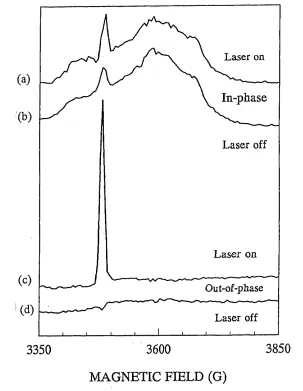
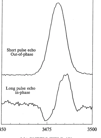
clusters were chemically prereduced at room temperature and at 4 K. A flash - induced electron spin polarised signal was observed in the out - of - phase detection channel using a short microwave pulse sequence, which decayed with a rate constant of 23 ps. Using microwave pulses of longer duration a spin polarised signal was observed in the in - phase
channel decaying with the same rate constant as the former and spectrally identical to the spin polarised signal attributed to the P700^A/' radical pair. It is concluded that at 4 K forward
electron transfer from is inhibited and that the observed spin polarised signal decays by a charge recombination and that the phasing of this signal depends on the characteristics of the pulse sequence used in detection.
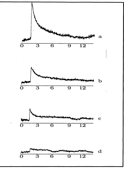
At room temperature the flash - induced out - of - phase spin polarised signal was
found to decay with a rate constant of 130 ns +/_ 50 ns and its decay was accompanied by the rise of a second spin polarised signal attributed to the radical pair, P700TeS" which in turn decays with a rate constant of 2 ps. When the iron - sulphur clusters, Fe-S^s, were extracted the room temperature kinetic behaviour of the PSI core particles was largely
well, the decay of the out - of - phase signal was slowed down to give a rate constant of 1.3 ps and the in - phase signal was abolished. This demonstrates that in intact PSI particles at room temperature electron transfer takes place from Apo the next acceptor and establishes unambiguously that this acceptor is the iron - sulphur cluster, Fe-S^ and means that the "200 ns" kinetic widely reported in the literature can be attributed to the reaction A /' FeS%— >AiFeSx". The same measurements were also carried out on PSI reaction centres prepared from the cyanobacterium, Synechocystis, and very similar kinetic behaviour was observed, implying that there is structural and mechanistic conservation between the two species.
Acknowledgements
I would like to thank Professor M.C.W. Evans for his patient supervision and encouragement over the three years. The spectroscopic work presented in this thesis is
collaborative and in this regard I am greatly indebted to the practical and theoretical expertise of Dr, P. Bratt, Dr. P. Moenne - Loccos, Dr.P. Heathco te. I would also like to express my gratitude to Dr. J. Nugent and Dr. S. Rigby for their invaluable advice.
CONTENTS Page Number
Title page 1
Abstract 2
Acknowledgements 4
Contents 5
Index of table and figures 10
1.0 INTRODUCTION 13
1.1 General Introduction 13
1.1.1 Historical Background 14
1.1.2 The Photosynthetic Unit 16
1.1.4 Prokaryotic Photosynthetic Organisms: 23
(a) Rhodospirallales 23
(b) Cyanobacteriales 24
(c) Prochlorophyta 25
1.1.5 The Evolution of Photosynthesis 25
1.1.6 The Chloroplast 31
1.2 The Redox Components of the Reaction Centres of Higher Plants 35
1.2.1 Photosystem 2 36
1.2.2 Photosystem 1 41
(a) P700 special pair, primary donor 46
(b) Aq - chlorin primary acceptor 47
(c) Aj- quinone secondary acceptor 49
(d) Iron - sulphur cluster, Fe-Sx 53
(e) Intrinsic Peptides of PSI 57
(f) Psa C - iron - sulphur cluster binding 59
(g) Psa D 61
(h) Psa E 63
(i) Psa H 65
2.0 A KINETIC STUDY OF SPINACH PHOTOSYSTEM I USING PUT.SRD ESR SPECTROSCOPY
2.1 SUB - INTRODUCTION 68
(a) Spin Polarised Resonance Signals of PSI 70
(b) The Kinetics of Electron Transfer in PSI 70
2.2 MATERIALS AND METHODS 76
(a) Preparation of BBYs and Triton - X- 100 - Fractionated PSI Particles 76
(b) Preparation of Digitonin Fractionated PSI Particles 77
(c) Chlorophyll assay 78
(d) P700 Assay 79
(e) SDS Polyacrylamide Gel Electrophoresis (PAGE) 79
(f) Removal of Psa C, the Preparation of P700FeSx Core Particles 81 (g) Removal of FeSx, the Preparation of P700A, Core Particles 81
(h) Room Temperature Optical Kinetics Spectroscopy 82
(i) Preparation of Samples for ESR Spectroscopy 83
(j) Induction, Detection and Measurement of the Decay of PSI Electron Spin
Polarised (ESP) Signals 83
(k) Fitting Procedure - Determination of Rate Constants 86
(1) NADP^ Reduction Assay 87
2.3 RESULTS
2.3.1 Polypeptide Composition of PSI Preparations 87
2.3.2 NADP^ Reduction Assay 88
2.3.3 Removal of Psa C 91
(a) Room Temperature Optical Kinetic Spectroscopy 91
(b) Ultrascan Densitometry of Protein Gels 91
(c) CW ESR Spectroscopy 95
2.3.4 Oxidative Dénaturation of Iron Sulphur Cluster, FeSx 98
2.3.5 Pulsed ESR Kinetic Measurements
(a) Low Temperature 100
(b) Room Temperature 107
2.4 DISCUSSION
(a) Low Temperature Kinetics 114
(b) Room Temperature 116
3.0 Sub - Introduction II 123
Distance Determination in PSI Using Pulsed ESR Spectroscopy
3.1 Use of Tj Saturation Recovery Measurements to Study the Structure of Photosystem I
3.1.1 The Principle of Saturation Recovery 124
3.1.2 Relaxation Enhancement Determination as a Method of Distance Determination
in PSI 128
3.2 MATERIALS AND METHODS
(a)Preparation of PSI Particles Containing Radicals Under Different Redox Conditions 129
(c)Saturation Recovery 132
(d)Cw Microwave Power Saturation Recovery Measurements 133
3.3 Theory 133
3.4 RESULTS
3.4.1 Saturation Recovery Measurements on FeS% 136
3.4.2 Cw ESR Spectroscopy of the Chlorin and Quinone Acceptors
in Intact PSI Particles 142
3.4.3 ESEEM Spectroscopy of Aq and A, 143
3.4.4 ESEEM Spectroscopy of the P700 Cation 147
3.4.5 Saturation Recovery Measurements of g = 2.00 Radicals 154 3.4.6 Power Saturation Measurements on the FeS% at 8 K 164
3.5 DISCUSSION 166
List of Figures and Tables Chapter 1
Figures:
1.1 The Z - Scheme 18
1.2 The Structural Formula of Bacteriochlorophyll a 21
1.3 Schematic of Section through a Chloroplast 33
1.4 Model of Photosystem II 38
1.5 Model of Photosystem I 43
Chapter 2 Figures:
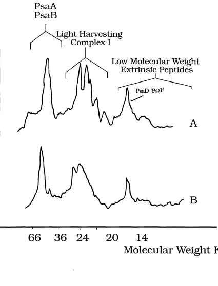
2.1 Densitometric Scans Showing Peptide Composition of Triton X 100 and
Digitonin PSI Particles 89
2.2 NADP'^Reduction Rates of Different PSI Preparations 90
2.3 Room Temperature Optical Kinetic Spectra - Chaotropic Removal of
Iron - Sulphur Clusters, FeS^B 93
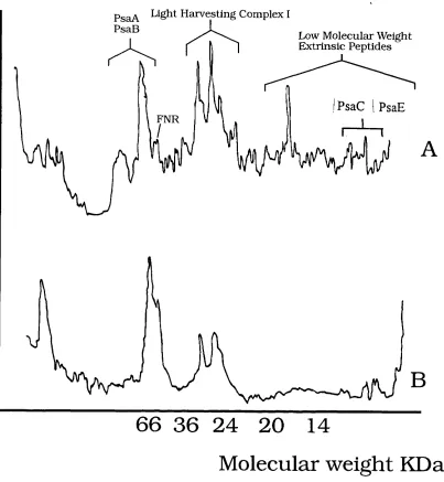
2.4 Densitometric Scans Showing Peptide Composition of Intact and
Urea Extracted PSI 94
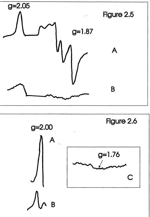
2.5 CW ESR Spectra of Intact and Urea Extracted PSI Particles 97 2.6 CW ESR Spectrum of PSI Core Particles in the g = 2.00 Region 97 2.7 Room Temperature Optical Kinetic Spectra - Chaotropic Removal of
Iron - Sulphur Cluster, FeSx 99
2.8 4 K Field Swept Spin Echo ESR Spectrum of Intact Spinach PSI Prereduced
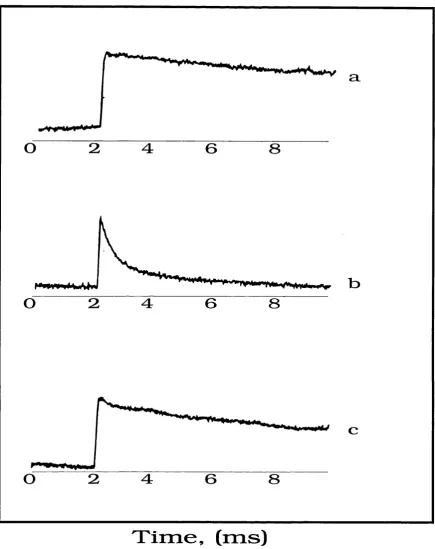
2.9 In - Phase and Out - of - Phase Kinetic Traces of the Laser Induced Signal in
Prereduced Intact Spinach Digitonin PSI Particles at 4K 103
2.10 Field Swept Spin Echo ESR Spectra of Laser Induced Signals Observed
Using Different Microwave Pulse Patterns 104
2.11 4 K Kinetic Trace of Out - of - Phase Laser Induced Signal in Spinach Digitonin
PSI Depleted of its Iron - Sulphur Clusters, FeS^B 105
2.12 4 K Out - of Phase Field Swept Spin Echo ESR Spectrum and Kinetic Trace of Laser Induced Signal in Spinach Digitonin PSI Depleted of
its Iron - Sulphur Clusters, F e S ^ 106
2.13 Room Temperature Field Swept Spin Echo ESR Spectra of Laser
Induced Signals in Intact Spinach Digitonin PSI 109
2.14 Room Temperature In - Phase / Out - of - Phase Kinetic Traces of
Laser Induced Signals in Intact and Urea Extracted Spinach Digitonin PSI 110 2.15 Room Temperature Out - of - Phase Kinetic Trace of Laser Induced
Signal in Intact Spinach Digitonin PSI, Prereduced with Sodium Dithionite 112 2.16 Room Temperature In - Phase Kinetic Trace of Laser Induced Signal
in Intact Synechocystis PSI 113
Chapter 3 Figures :
3.1 Schematic of 6 Â X- Ray Crystal Structure of Synechococcus PSI 126 3.2 Schematic of Microwave Pulse Sequence Used in Spin - Lattice
Saturation Recovery Measurements 127
3.3 Field Swept Spin Echo ESR Spectrum of prereduced Spinach 138 PSI, Frozen Under Illumination
3.4 Fitted Spin - Lattice Saturation Recovery Curves of Iron - Sulphur Cluster, FeS% at 3.7 and 5.5 K
3.5 CW ESR Spectra of the Aq' and A f Radicals in Intact Spinach PSI
3.6 1 Dimensional ESEEM Spectra of Aq' and A {
3.7 Field Swept Spin Echo ESR Spectrum of Prereduced Spinach PSI, Illuminated so as to Form A {
3.8 Field Swept Spin Echo ESR Spectrum of Prereduced Spinach Digitonin PSI, Illuminated in Instrument Cavity to Form P700+ 3.9 Field Swept Spin Echo ESR Spectmm of Ascorbate Dark Reduced Spinach PSI, Illuminated at 77 K to Form P700^FeS/
3.10 1 Dimensional ESEEM Spectrum of P700+ Radical 3.11 Fitted Saturation Recovery Curves for Enhanced and Unenhanced Radicals, P700^, A {, Aq' at 8 K
3.12 CW ESR Spectra of Aq' and A { formed in Spinach PSI Depleted of all its Iron - Sulphur Clusters
3.13 Microwave Power Saturation Plot Tables
3.1 Spin Lattice Relaxation Rates of FeSx 3.2 Spin Lattice Relaxation Rates of P700+ and FeS^ , 4 - 14 K
3.3 Enhanced and Intrinsic Spin Lattice Relaxation Rates of g = 2.00 Radicals at 8 K
139 144 145 149 150 151 152
156 - 161
162 165
140
153
1.0 INTRODUCTION
1.1 General Introduction
Photosynthesis is the process whereby light energy is utilised for chemical means. The process is light dependent and involves the oxidation of an inorganic or organic molecule, acting as an electron donor. The overall reaction may be represented by the simple equation given below:
2 H2A + LIGHT --- > A2 + 4e + 4H+
In oxygenic photosynthesis the electron donor is water which is oxidised to oxygen. In anoxygenic photosynthesis the donor is an inorganic molecule other than water, eg H2S
which is oxidised without the production of oxygen. Photosynthetic organisms use the electrons to reduce fixed carbon dioxide for the generation of amino acids and sugars. Such organisms may be prokaryotes or eukaryotes, marine or terrestrial, they can all grow autotrophically though some are capable of feeding heterotrophically and occur in a wide range of environments.
The reactions of photosynthesis occur in pigment protein complexes which have a broadly similar design and bind electron carriers which are chemically similar in both eukaryotes and prokaryotes. For convenience sake photosynthesis can be broken down into
the daik reaction and the light reaction. The first of these refers to the processes that fix carbon dioxide and produce organic molecules required by the organism for its normal functioning and growth. The energy needed for this is supplied by the light reaction, an electron transport reaction generating reduced cofactors and a proton gradient.
1.1.1 Historical Background
In the early eighteenth century it was proved that green plant photosynthesis involved carbon dioxide fixation into carbohydrates with the evolution of oxygen. Ingenhousz (1779) showed that photosynthesis needed the illumination of the chlorophyll - containing parts of plants. The work of de Saussure (1804) dealt with the stoichiometry of the overall process, resulting in the well-known photosynthetic equation:
6 CO2 + 6 H2O --- > CgHj20g + 6 O2
shows that the light reactions are insensitive to temperature changes, a feature typical of photochemical processes.
Emerson and Arnold (1932) demonstrated using the alga Chlorella that the dark and light reactions can be separated in time. The cells were exposed to a 3ms pulse of light, followed by periods of darkness of varying lengths. During the dark period CO^ was fixed, using energy generated by photochemical processes occurring during the light pulse. For the efficient utilisation of light energy it was found that a dark period considerably longer than the light pulse was needed. From these results it was deduced that the system carrying out the photochemistry was regenerated during this "longer" dark period. It is now known the light and dark reactions are not only separated in time but also in space in the chloroplast of higher plants.
Localisation of photosynthetic activity to the chloroplast was first achieved by Engelmann (1894). His work also proved the chloroplast to be the site of oxygen evolution. This was extended by Hill (1937). His experiments used isolated chloroplasts which produced oxygen under illumination only if a suitable electron acceptor was added to the system. The acceptor was a ferric salt which was reduced to the ferrous form in the course of the reaction. The donor oxidised to produce oxygen gas was identified as water by Ruben et ol (1941) using '^O-labelling experiments. The reaction by which water oxidation is coupled to the reduction of a ferric salt acceptor resulting in the production of oxygen gas, when isolated chloroplasts are illuminated became known as the Hill reaction. Hill could not show that CO2
can serve as the terminal electron acceptor. It was Amon et al (1954) who demonstrated chloroplasts are also the site of CO2 fixation, using ^"*C0 2 .
It was Hill along with Bendall(1960) who put forward the so-called Z-scheme which
was the first time a thermodynamic framework was placed around the electron transfer
reactions of photosynthesis and incorporated new information concerning the cytochrome b^f complex and ferredoxin as electron carriers (figure 1.1).
1.1.2 The Photosynthetic Unit
This is a term applied to the complex of pigments and other molecules involved in the
Figure 1.1 The Z - scheme showing the electron pathway from the primary electron donor, water, to NADP^ in photosynthesis. The process is light dependent. The schematic also indicates the relationship between the two photosystems, PSI and PSII. Plastoquinol (QH2)
formed by PSII, serves as electron donor to the cytochrome b^f complex, which reduces plastocyanin (PC), which acts as the electron donor to PSI. PSI reduces the iron sulphur protein ferredoxin (Fd), which in turn reduces NADP^, forming NADPH. The proton gradient across the thylakoid membrane is formed when electrons pass through bgf complex and is further enhanced by the fact that water oxidation and NADP^reduction occur on opposite sides of the thylakoid membrane. P680 - primary donor of PSII, a chlorophyll dimer. P700 -
primary donor of PSI . Z - tyrosine residue mediating electron flow between the manganese cluster (Mn), and P680. Ph - phaeophytin, the primary electron acceptor of PSII. and Qg are plastoquinone binding proteins. Aq and Aj are electron acceptors in PSI. Fp is the flavoprotein, ferredoxin - dependent - NADP^ reductase.
- 1.6 -1 .4
- 1.2
- 1.0
- 0.8
-0 .6 S -0 .4
g
-h-> c£ g ’V & - 0.2 0 0.2 0 .4 0.60.8
H,P hotosystem II
P680' /iv, Mn cen te P 680 Ph
i
9al
9
.\
9 H :
Cytochrom e
b j
Com plex '
Proton
P hotosystem I
P 7 0 0 V ^ A.
\
hv.
PC P 7 0 0
A,
\
Fe-S \ Fd\
.■ NADP"
+
proton gradient
A quantum of light absorbed by any one of them will be transferred to the reaction centre, where it will result in a charge separation. The existence of a photosynthetic unit is supported by the experiments of various workers :
Emerson and Arnold, using Chlorella suspensions showed a maximum of IO2 molecule
is produced per light flash per 2500 chlorophylls, which means one quantum of light is absorbed by one chlorophyll molecule out of about 300. This inference follows on from an earlier observation indicating that about 8 quanta of light must be absorbed by chlorophylls
in order to reduce 1 CO2 molecule and release 1 O2 molecule.
Gaffron and Wohl showed a large difference to exist between the theoretical rates of CO2 reduction and O2 evolution (calculated assuming the presence of a single chlorophyll
molecule) and the actual rates measured in plant material. This led them to infer the presence of many other chlorophyll molecules whose function is to channel light energy to a single reaction centre.
The technique of difference spectroscopy has led to the discovery of the P700 special pair (a chlorophyll a dimer) of photosystem I (Kok et al) and also implicated cytochrome (Duysens et al) in the photochemistry. In algae and higher plants there is one light reacting cytochrome and one P700 dimer per 600 chlorophyll molecules.
The idea of a photosynthetic unit is rather outdated and the experimental work mentioned above which gave rise to it is at least forty years old, however it can still be crudely defined as a protein pigment complex consisting of about 600 chlorophyll molecules, and the redox components required to use harvested light energy to evolve oxygen and reduce
NADP.
There are two types of photosystem. The first incorporates chlorophylls, phaeophytins (both kinds of tetrapyrrole pigment) and quinones as acceptors. It includes the photosystem
2 (PS II) of higher plants and the reaction centre found in purple bacteria. The other incorporates chlorophylls (or bacteriochlorophylls), quinones and iron-sulphur centres as acceptors. It includes the photosystem 1 (PSI) of higher plants and the reaction centre of green sulphur bacteria. The first type is functionally associated with water oxidation/oxygen evolution, the second type with ferredoxin and NADP reduction.
1.1.3 Pigments utilised in photosynthesis
Twsett (1906) was one of the first to extract and chromatographically separate leaf pigments involved in photosynthesis. There are three main groups of photosynthetic pigments : chlorophylls, carotenoids and phycobil ins. The first two are soluble only in organic solvents, the third group are water soluble. Carotenoids and phycobil ins are termed accessory pigments because they transfer light energy to chlorophylls.
In higher plants there are two kinds of chlorophylls a and b, which differ chemically. The structure of chlorophyll was worked out by Fischer (1940) and first synthesised by Woodward and co-workers (1960). All chlorophylls consist of a tetrapyrrole ring surrounding a magnesium atom (the polar porphyrin nucleus) and a lipophilic phytol tail. Phaeophytins, a third variety of pigment found in reaction centres, lack a magnesium atom but are otherwise chemically identical to chlorophylls. The molecular formula for chlorophyll a is C55H72N405Mg, for chlorophyll b, Cg^H^oN^O^Mg. Chlorophyll a has a methyl group at position 3 on the tetrapyrrole ring, where b has a carbonyl group. Chlorophyll a and b also display characteristic absorption spectra. The absorption maxima observed for the pigments are different for different solvents. In ether the absorption maximum for chlorophyll a is 660nm and for chlorophyll b, 643nm; in acetone 663nm for chlorophyll a, 645nm for
CH
CHoCH
Mg
CH
c=o
CH
'igure 1 .2 T h e s tr u c tu r a l fo r m u la o f b a cterio ch lo ro p h y U a. C h lo ro p h y ll a la c k s th e a rb o n y l g r o u p s h o w n in th e to p 'left h a n d co rn er o f t h is str u c tu r e , h a v in g
L 0 = 0 b o n d in s te a d . R is a O2 0h y d ro c a rb o n c h a in .
Carotenoids are pigments found in plants and purple bacteria. When dissolved in organic solvents they are yellow or orange in colour. Carotenoids are unsaturated organic compounds, containing alternating double and single C to C bonds. Broadly speaking there are two kinds - those which are hydrocarbons (the carotenes) and oxygenated hydrocarbons (the carotenols or xanthophylls) typically of 40 carbon, atoms in length and composed of isoprene subunits. Their absorption spectra display three peaks, lying between 400 and 520nm.
They are found in the lamellae of chloroplasts where they are closely associated with chlorophylls. Their functions are believed to include energy transfer to other pigments as
already mentioned and also to protect against the photoinhibitory effects of high light
The most important function cmried out by carotenoids is the quenching o f chlorophyll triplet intensity, and therefore prevention of singlet oxygen formation. Singlet oxygen is a highly reactive
species which can cause damage to proteins.
Cyanobacteria and red marine algae aaaiiionally contain a class of pigments called
phycobil ins. These are linear tetrapyrroles resembling chlorophyll a, which contains a cyclic tetrapyrrole ring, but lacking a phytol ring and central magnesium atom. They may be covalently bonded to polypeptides to form phycobil iproteins. There are three kinds of phycobil in - phycoerythrins, phycocyanins and allophycocyanins. Phycoerythrins give red algae their distinctive colour and absorb in the middle of visible range. It is the presence of these pigments at high levels that allows red algae to photosynthesise deep beneath the ocean surface. The greater the depth at which they occur, the higher their phycoerythrin:chlorophyll ratio. The blue phycocyanins and allophycocyanins are found in cyanobacteria (blue green algae) which occur terrestrially and close to the surface of lakes. As with carotenoids they are
accessory pigments involved in energy transfer. Studies on Porphyridium species (red algae) using picosecond spectroscopy have established the following sequence of energy transfer:
phycoerythrin >phycocyanin >allophycocyanin >chlorophyll a.
Chlorobiineae (Green bacteria) contain bacteriochlorophylls and bacteriophaeophytins as well as chlorophylls), which are very much like their eukaryotic equivalents with only slight
structural modifications.
1.1.4 Prokaiyotic Photosynthetic Oiganisms
There are three groups of prokaryotic organism that can carry out phtosynthesis:
{2i)Rhodospirallales.
{b)Cyanobacteriales. {c)Prochlorophyta
The organisms are grouped in this way according to pigment content and whether or not they evolve oxygen when they photosynthesise. They are all gram-negative bacteria apart from
Heliobacterium chlorum.
( 1 )Rhodospiralles..
They are split into the purple bacteria {Rhodospirillineae) and the green bacteria
{Chlorobiineae). Purple bacteria are sub-divided into the purple sulphur bacteria
{Chromatiaceae) and purple non-sulphur bacteria {Rhodospirillaceae). Purple sulphur bacteria only grow under anaerobic conditions as autotrophs using hydrogen sulphide as an inorganic electron donor to reduce carbon dioxide in the Calvin cycle. Hydrogen sulphide is oxidised
in the process without oxygen formation, i.e. photosynthesis in these organisms is anoxygenic. The bulk of purple non-sulphur bacteria are aerobic heterotrophs requiring an organic compound such as acetate for a carbon source in their medium which they metabolise in the
dark. In the light they can function as photoautotrophs using carbon dioxide as a carbon source. The purple non-sulphur bacteria have been more intensively studied than any other photosynthetic organism, this is partly due to their nutritional flexibility meaning they can be easily cultured in the laboratory and are amenable to genetic manipulation techniques., such as the production of mutants.
Green bacteria are sub-divided into the green sulphur bacteria (Chlorobiaceae) and the
green non-sulphur bacteria (Chloroflexaceae). The green sulphur are obligate anaerobic photoautotrophs using hydrogen sulphide etc. as electron donors and possess a PS I-type reaction centre. The green non-sulphur bacteria are facultative aerobes which only photosynthesise under anaerobic conditions. They possess PS II- type reaction centres. The only representative of this group to be studied is Chloroflexus auranticus.
(2)CyanobacteriaIes
They are predominantly aerobic photoautotrophs as is the case for higher plants and algae and like all photosynthetic eukaryotes they possess both types of reaction centre,
(3)Prochlojpphy^ta
The photosynthesis of organisms in this group is oxygenic. A representative species is Prochloron didemni, an endosymbiont associated with certain snail species. It has thylakoid membranes organised into a structure closely resembling the chloroplasts of eukaryotes,
binding both PSI- and PSII-type reaction centres incorporating chlorophylls a and b, lacking the phycobilins encountered in cyanobacteria. Proclorothrix is a genus of free-living organisms belonging to this phylum.
1.1.5 Evolution of Photosynthesis.
There are a number of important similarities between the reaction centres of prokaryotic and eukaryotic photosynthetic organisms. Naturally this has led to the search for evolutionary relationships between the reaction centres through the comparison of sequence data. This has identified conserved residues and motifs involved in the binding of redox components. Detailed high resolution crystal structures are available for the reaction centres of the purple non-sulphur bacteria, Rhodopseudomonas viridis (Michel, 1982, Deisenhofer
et al, 1984) and Rhodobacter sphaeroides R26 (Chang et al, 1986/91, Allen et al 1986). Because of the similarity of these systems to the PSU of eukaryotic algae and higher plants, the X-ray crystal structures have been used as the basis for homology modelling of the latter, e.g. Ruffle et al (1992), S tyring et al (1990), excluding the donor sides which are very different in the two systems with the purple bacteria lacking a manganese cluster and oxygen evolving complex(OEC). Such work as this suggests future experiments, eg by identifying informative targets for mutagenesis and generally aids functional and structural studies of
PSII. X-ray crystallography studies are being carried out on PSI (Witt et oT) and PSII (Feher
et al). There follows a brief survey of the similarities and differences between the different reaction centres and their possible evolutionary importance.
Photosynthetic bacteria, algae and plants all have light harvesting apparatus. In all cases the amounts of light harvesting material is regulated in response to changing levels of light intensity. In most cases (the exception being chlorosomes) the light harvesting pigments are associated with polypeptides. The differences between the groups arise in the organisation and pigment composition of the light harvesting apparatus. In green sulphur bacteria for example these pigments are localised to p r i d e s called chlorosomes, which are in turn associated with the cell membrane. In the purple bacteria and photosynthetic eukaryotes the light harvesting and reaction centre complexes are intimately associated integral thylakoid membrane proteins. In cyanobacteria and red algae, phycobilisomes found in the outer surface of the photosynthetic membranes contain the water-soluble phycobilins. These structures contain in the region of 300-800 chromophores absorbing visible light over a range of wavelengths. The light quanta absorbed by the phycobilisomes are transferred to the reaction centre pigments with high efficiency.
The reaction centres of plants and bacteria show a similar pattern of organisation. In all reaction centres the first photochemical event of photosynthesis proper (i.e. excluding light
harvesting processes) is the transfer of an electron from a donor molecule to an acceptor molecule. In purple bacteria and PSII of eukaryotes the primary donor is a dimer of
plants these quinone acceptors are closely associated with non-haem iron atom. The relatively low energies of the reaction centre chlorophylls a and bacteriochlorophylls means that the loss of excitation energy through fluorescence is reduced. Quantum yields of electron transfer through the carriers of the reaction centre are high and the likelihood of back reactions is reduced by having a series of carriers arranged across the complex, of increasingly positive redox potentials and at increasing distances from the primary donor, leading to a stabilisation of charge separation.
There are significant amounts of amino acid sequence homology and similarity between the L and M subunits of Rhodopseudomonas species and the 32kDa D2 subunit (herbicide binding protein) of PSII. From such homology a shared ancestor has been inferred. There are other experimental observations which support this relationship - both reaction centres consist of polypeptides able to bind herbicides such as azidoatrazine (Pfister et al
1991); they both have similar quinone-iron cw EPR signals, with metal and radical magnetically coupled in both cases. There are also important differences between them however- as already mentioned the donor side of PSII ( the manganese cluster, tyrosine radicals ie the components involved in water oxidation/oxygen evolution) is peculiar to it; the redox potential of P680 (a chlorophyll a dimer, the primary donor of PSII) is about 600- 800mV more positive than the purple bacteria equivalent (P870). The histidine ligands believed to bind the chlorophyll molecules of the primary donors in each case are conserved in the L and M subunits of purple bacteria and the D1 and D2 subunits of PSII (Trebst et al
1986). This has led to the postulation that the PSII primary donor is a chlorophyll dimer orientated perpendicularly to the plane of the membrane as in the bacterial reaction centre,
this has some spectroscopic support (van Kan et al, 1990). However there is also spectroscopic evidence which indicates the P680 has properties different to the purple
bacterial reaction centre and that it exhibits considerable monomeric character (iLosche ei al^ 1988, van Mieghem et cd, 1991). ENDOR(electron nuclear double resonance) studies indicate that the oxidised P680 is a dimer but that the bulk of the electron spin is localised on one of the chlorophyll molecules (5:1 ratio), (Rigby etol, 1994). Mutational studies in cyanobacterial PSII have also shown a difference in the primary donor ligand usage between the two system types. When one of the histidines in D1 protein of the Synechocystis 6803 reaction centre thought to ligand P680 is changed to a residue that does not ligate magnesium no change in the chlorophyll content is observed. When the equivalent residue is mutated in the purple bacterium reaction centre the magnesium is not inserted, so the chlorophyll is pheophytinised. This suggests strongly the analogy between the purple bacteria reaction centre and PSII does not extend to the primary donor site. It does seem to hold for the primary acceptor site, with the equivalent histidine residues ligating bacteriophaeophytin in purple bacteria and phaeophytin in PSII. The work done so far on P680 infers a distorted dimer. Such distortion would help to explain P680’s high redox potential, a property necessary for its water oxidising function.
Inspite of these and other observations indicating sequence/structural differences between PSII and purple bacterial reaction centres it is generally believed the former has evolved from the latter. There are also significant similarities between these reaction centres and that of the green non-sulphur bacterium Chloroflexus ouranticus (Trebst et ai, 1986, Barber et al, 1988,1 Michel and Deisenhofer, 1988, Rutherford). There are also differences
' \et al, 198»
between the latter and the purple bacteria. In Chloroflexus one of the accessory bacteriochlorophylls is replaced by a bacteriophaeophytin and the non-haem iron found
between the primary and secondary quinones is replaced by a manganese (Blankenship et al,
It has been assumed that PSI and the reaction centre of the green sulphur bacteria are related based on similarities of redox component composition and organisation. Two structural genes from the green sulphur bacteria, Chlorobium limicola, have been identified j one which at the nucleotide level and amino acid level correspondsto the psaA, psaB (encoding the major
polypeptides of PSI) and one to the psaC (encoding the peptidej which binds the extrinsic iron sulphur centres, FeS^g) genes (Buttner et al 1992a). However there is only about 15% identity
between the psaA/B and the corresponding Chlorobium protein when whole sequences are compared, with the most striking conservation of the region including the two cysteines thought to bind the intrinsic iron sulphur centre, FeSx- Spectroscopic analysis of Chlorobium
reaction centres prepared anaerobically has revealed the presence of two 4Fe4S-type iron- sulphur clusters with spectra resembling those produced by Fe-S^g clusters of PSI (Kjaer et al, 1994). From the fact only one psaA-Xypt gene has been identified in Chlorobium it was inferred the reaction centre of this species is a homodimer and not a heterodimer as in higher plants (Buttner et al, 1992b). The homodimeric structure of the Chlorobium reaction centre is supported further by ENDOR studies on its primary donor, a chlorophyll dimer P840 (Rigby et al, 1994), which indicates the electron spin is equally distributed over the two halves of the dimer. The reaction centre of heliobacteria, on the basis of sequence homology, is also thought to be related to green-sulphur bacterial reaction centre and PSI. There is now optical spectroscopic evidence for an Fe-Sx-type iron-sulphur cluster obtained from heliobacterial reaction centres depleted of their Fe-S^g-type clusters (Kleinherenbrink et al,
1994), which further support this postulated relationship.
If one ignores the iron sulphur centres F eS ^ there are obvious similarities between PSI and the purple bacterial reaction centre. In both there is a dimer of tetrapyrrole chromophores acting as the primary donor, with a tetrapyrrole monomer and quinones acting
as acceptors. The non-haem iron of purple bacteria can be seen as being analogous to the intrinsically bound iron sulphur centre, FeSx (though the former is not redox active). Indeed
from the low resolution X-ray crystal structure of PSI currently available Fe-Sx can be seen to occupy an equivalent position in the reaction centre core as the non-haem iron in the purple bacterial reaction centre and it has been proposed the latter is derived from the former. It is well known from the X-ray crystal structure that the purple bacterial reaction centre has two
paths for electron transfer from the primary donor to a quinone acceptor but that in vivo only one of them is used. A similar C-2 symmetry exists in PSI (Heathcote et al 1993), there being two molecules of each electron carrier per reaction centre and with only one of the paths being active under physiological conditions.
Other efforts have been made to construct a phylogenetic tree depicting the relatedness of the three PSI-type reaction centres : of cyanobacteria and higher plants, green sulphur bacteria and heliobacteria through careful sequence alignment. Taking the complete amino acid sequences there is only about 20%homology between the three types. Limiting the comparisons to known conserved regions gave more accurate and informative alignments (Liebl et ol, 1993). The heliobacterial sequence shows 40%and 48%identity with PsaA and PsaB, respectively, compared to 32%and 39%between the Chlorobium sequences and PsaA/PsaB sequences. From this it is inferred the green sulphur bacteria diverged from the ancestral reaction centre, followed by the heliobacteria which is thus more closely related to the PSI subunits. This study also showed both green sulphur and heliobacterial sequences
share greater homology with PsaB than with PsaA. This can be interpreted to mean that PsaB derives directly from a homodimeric ancestral reaction centre which later underwent a
Chloroplasts are the specialised organelles found in eukaryotic photosynthetic algae and higher plants which house the thylakoid membranes that are the sites of the photochemistry of photosynthesis. They have their own genome encoding many of the protein subunits required for photosynthetic function and also proteins responsible for the regulation
of their expression and assembly. As has already been mentioned neither the purple and green bacteria or the cyanobacteria have their photosynthetic membranes organised as chloroplasts. In the former, the pigment-containing bodies can be isolated from disrupted bacterial cells by differential centrifugation. Under the electron microscope these bodies known as
chromatophores, appear spherical and are 30-100nm across. Each one contains a number of photosynthetic units, e.g. a chromatophore from the purple non-sulphur bacterium,
Rhodobacter sphaeroides contains about 40 reaction centre complexes, 500 light harvesting complexes, 1000 carotenoid molecules and 1000 ubiquinone molecules. Chromatophores are believed to originate from elaborate infolding of the cytoplasmic membrane. In cyanobacteria, which are also prokaryotes, the accessory pigments are located in phycobilisomes.
In eukaryotes that photosynthesise, the pigments required for the photochemistry are found in chloroplasts (figure 1.3) which are organelles in their own right. In the unicellular green alga Chlamydomonas f einhordtii the single chloroplast is seen under the microscope in cross section as a cup-shaped structure enclosing the nucleus. In higher plants chloroplasts occur in large numbers in all the green parts of the plant, though their abundance varies seasonally and according to species. They can be isolated from the leaves of higher plants in large amounts using standard methods. Under the electron microscope the higher plant
Granum
Stroma
Inner
membrane
Outer
embrane
Intermembrane
" Spaee
Thylakoid
membrane
Thylakoid
spaee
chloroplast is a saucer-shaped structure of 4-lOpm across and about 1pm in thickness and has an outer double unit envelope separating its contents from that of the cytoplasm. In all plant species the chloroplast can undergo simple division.
Inside the chloroplast the photosynthetic membranes or thylakoids are arranged in stacks called grana which at lower resolutions appear as dense green areas within the chloroplast. The grana are embedded in a colourless matrix called the stroma{\hQ chloroplast "cytoplasm"). The grana are interconnected by a system of loosely arranged membranes known as the stromal lamellae. Using electron microscopy it can be seen the thylakoids have embedded in them the protein-pigment complexes. There is a difference in the complex organisation between the stacked granal lamellae and the loosely packed stromal lamellae. This is because the granal membranes contain mainly PSII, and the stromal membranes contain mainly PSI.
It is possible to separate the pigment-containing thylakoid membranes from the stromal matrix and to analyse its protein/pigment content. The thylakoids are about half lipid and half protein. The proteins bind the redox components needed for electron transfer and also strengthen the membranes. Some of these membrane proteins bind the light harvesting chlorophylls a and b. The stroma contains the soluble enzymes involved in the Calvin cycle which reductively fixes CO2 to produce sugars. Because the thylakoid lumen is separated from
the rest of the chloroplast by a double unit membrane, its internal environment can be regulated independently to some extent and optimised for the photosynthetic reactions. As a result the stroma of the chloroplast has a different chemical composition and pH to that of the surrounding cytoplasm.
similar dimensions to a photosynthetic bacterium or cyanobacterium and has its own nuclear material. The DNA of chloroplasts is a circular double stranded molecule containing enough genetic information for approximately 125 polypeptides, however it contains quite a lot of non - coding DNA and the characterisation of chloroplast genes and their products is by no means
complete. Chloroplasts have all the machinery required for protein synthesis, including ribosomes in both the thylakoids and the stroma. These ribosomes resemble those of prokaryotes rather than those of eukaryotes, providing further support for the endosymbiotic
hypothesis for the origin of chloroplasts. The primary structure of chloroplast DNA is significantly different to that of nuclear DNA.To begin with it contains a long inverted repeat
sequence. Additionally chloroplast DNA is not complexed to histones as is nuclear DNA, another feature more characteristic of prokaryotes than of eukaryotes.
1.2 Redox Components of the Reaction Centres of Higher Plants.
General Features of Photosynthetic Reaction Centres
All reaction centres have a similar organisation and chemical composition because they carry out the same basic function i.e. the promotion of a stable charge separation with a cationic primary donor and negatively charged terminal acceptor spatially separated on
opposing sides of a membrane. They are all integral membrane protein - pigment complexes. The most significant differences between reaction centres arise in relation to the additional functions that they carry out, such as water oxidation in the case of PS II.
There are two types of reaction centre : (l)Low potential - in which the electron passes from a chlorophyll dimer (or special pair) to a chlorophyll monomer, a quinone
acceptor and then onto three iron - sulphur clusters. The acceptors are bound by dimeric core comprising polypeptides, with a molecular weight of 70 - 80 kDa. PSI, the reaction centres of green sulphur bacteria and heliobacteria are of this type. (2)High potential - here the sequence of acceptors is as follows : a chlorophyll dimer, a phaeophytin, and a quinone terminal acceptor. In this type of reaction centre the acceptors are bound by dimeric core consisting of monomeric polypeptides with a molecular weight of about 30 kDa. There
follows brief accounts of the redox components and function of the reaction centres found in photosynthetic eukaryotes, PSI and PSII.
L2.1 Photosystem II ( PS II )
A schematic showing the proposed arrangement of the major components of PSII is shown in figure 1.4. PSII can be treated as an enzyme catalysing the photoinduced oxidation of water coupled to the reduction of the terminal acceptor, plastoquinone. Plastoquinone goes on to reduce plastocyanin through the b^f complex, reduced plastocyanin being the electron donor to PSI. The passage of electrons through asymmetrically arranged photosystems I and II generates a proton gradient across the thylakoid membrane. This constitutes part of the proton - motive force (which has a pH gradient and a membrane potential component) and this drives ATP formation by the membrane - bound ATP synthase.
Our knowledge of PSII has been greatly increased by a number of developments in the last decade : the availability of improved preparations of purified PSII particles; the application of molecular genetics and above all the determination of a high resolution X - ray crystal structure of the reaction centre of purplej non - sulphur which is closely related to
I
bacteriapsn.
Figure 1.4 Schematic of photosystem II. This is a model based roughly on the solved X-ray crystal structure of the purple bacterial reaction centre, showing the major polypeptides and the various redox components thought to be involved in PSII photochemistry. D1 and D2 are the core polypeptides that provide ligands for the chlorin and quinone electron acceptors. OL
and (3 are the subunits of the cytochrome.
Photosystem II Protein Complex
Stroma
cyt b-559
Lumen
of light energy and its transfer to the reaction centre core; the reaction centre which contains the redox components involved in the photochemistry of charge separation and including the oxygen - evolving complex (OEC) which is concerned with water oxidation; the regulatory cap which is composed of polypeptides bound lumenally to the thylakoid membrane.The antenna is made up of proximal and distal antennae. The former encloses the reaction centre
core and consists of two pigment - protein complexes, CP47 and CP43, which bind Chi a and P - carotene molecules, but not Chi b. The distal antenna consists of the light - harvesting complexes (LHCII) and binds Chi a, Chi b and xanthophylls. A 6 Â electron diffraction
structure of LHCII (Kuhlbrandt 1991) is available and from this it is clear that the
i and Wang,
complex is a trimer with each monomeric subunit binding 15 chlorophylls and some carotenoids. The LHCII can dissociate itself from PSII and associate itself more closely with PSI, so it plays a part in energy distribution by mediating between the two photosystems. The process is regulated by the phosphorylation and dephosphorylation of certain amino acid residues.
The regulatory cap is made up of three hydrophilic extrinsically bound proteins, BP33, BP24 and BP17. BP33 separates the OBC from the contents of the lumen. Cross - linking studies have shown that it is in contact with the reaction centre heterodimer. It is thought to have a role in water oxidation as its removal by Tris - washing results in reduced oxygen - evolving activity. It is sometimes referred to as the manganese - stabilising polypeptide because it is thought to be involved in the stable assembly of the manganese cluster and might even provide ligands to the manganese atoms.
The PSU core is the minimal functional unit. It is capable of carrying out the photoinducible charge separation reactions but is unable to evolve oxygen. The PSII core can be routinely isolated and has the following composition : 4 to 6 Chi a molecules, 2
phaeophytins, 2
(3
- carotenes, 1 heterodimer, 1 CP47, 1 CP43, 1 or 2 cytochrome bggg and 1 110 kDa I polypeptide per P680. The core preparation contains no quinone (Q^) and no OEC. PSII preparations that evolve oxygen also contain and a closely associated non -haem iron atom.
Kinetic studies have been carried out on the PSII reaction centre preparations using optical absorption techniques. The primary charge separation reaction which produces the radical pair P680^Phe" occurs in 3 ps. Charge recombination is prevented by the rapid transfer of the electron from Pheo" to in 200 to 500 ps and the rapid rereduction of P680^by the
redox active tyrosine residue Y^. Qa" is oxidised in 1 0 0 to 2 0 0 ps by a second quinone, Qg
to give the semiquinone radical. This semiquinone is reduced by a second Qa to give Qg^ . By analogy with the purple sulphur bacteria it is assumed that Qg^" is doubly protonated to plastoquinol which is released from its binding and migrates to the bgf complex. The vacant Qg niche in the Dj polypeptide is filled by another Qg drawn from the cell’s plastoquinone pool.
PSII contains a second redox active tyrosine residue, Y^, in the D2 polypeptide present.
This tyrosine residue is known to interact with the the oxygen evolving complex in various S states. For example, in the dark the OEC adopts the state from Sq by donating an electron to Y^^, or from 8 3/ S3 by accepting electrons from Yg. Y^ has also been implicated
in the light - activated assembly of the manganese cluster of the OEC.
Cytochrome b^^g is a heterodimer consisting of a 9 kDa CLsub-unit and a 4 kDa p- subunit which coordinate a haem prosthetic group. This cytochrome may play a role in reducing the likelihood of photoinhibition and / or proton pumping during cyclic electron transfer in PSII.
for water oxidation is thought to be a tetrad of manganese atoms. This manganese cluster is thought to provide the means of storing the oxidising equivalents needed to oxidise water, as well as containing the substrate binding site and site of oxygen evolution. As the reaction centre is turned over by the absorption of successive light quanta, electrons are stripped from
the OEC, leading to the accumulation of oxidising equivalents by a process known as the Kok cycle. According to this cycle the OEC goes through 5 distinct redox states, designated Sq to S4. It is known that oxygen gas is liberated at the S3 to Sq transition. The actual mechanism
of water oxidation, the identity of the S - state that binds water, which S - state transitions involve manganese oxidation are the subjects of ongoing investigation. The structure of the manganese cluster has been studied using X-ray absorption fine structure (XAFS) spectroscopy and should soon be known through X - ray diffraction studies of PSII crystals. The chemistry of the Kok cycle is being studied using ESR, XAFS and X - ray absorption near edge structure (XANES) spectroscopy.
1.2.2 Photosystem I (PSI)
Thanks to improvements in preparations/biochemical manipulation, the continuing application of spectroscopic techniques and more recently site directed mutagenesis, the structural and functional elucidation of PSI continues apace though several difficult questions remain to be answered. A schematic diagram of higher plant PSI is shown in figure 1.5.
The PSI reaction centre can be viewed as a complex integral membrane enzyme system, a light dependent plastocyanin : ferredoxin oxidoreductase to be exact. This reaction centre is found embedded in the thylakoid membranes of higher plants, green algae and the
prokaryotic cyanobacteria. Essentially an electron donated by lumenally bound plastocyanin,
NADPH
NADP
Fd
Stroma
sl-E
Psl-D
A/B
P700
Lumen
PC
a soluble copper-containing protein is transferred across the thylakoid membrane via a series
of redox components, whose chemical identity will be discussed below, to reduce ferredoxin, a stromally located soluble iron-sulphur protein. It is assumed the design of PSI, as with other reaction centres, has evolved to permit efficient electron transfer and high quantum yields i.e. charge separations per light quantum absorbed.
Although no PSI or analogous bacterial reaction centre crystal structure is available as yet there is a considerable body of experimental evidence to support the view that the
major redox components involved in charge separation are bound by the polypeptides encoded by the following chloroplast genes : psaA, psaB and psaC . The psaA and psaB polypeptides form the heterodimeric core of the reaction centre. These polypeptides have molecular masses of about 82-83kDa and bind, apart from the various redox components connecting plastocyanin and ferredoxin, about 100 accessory chlorophyll molecules. This is different from PSII, in which the core polypeptides bind hardly any accessory chlorophylls, which are bound mainly in the light harvesting complexes. The terminal membrane - bound electron acceptors are two 4Fe-4S iron- sulphur centres designated Fe-S^B which are bound by the 9kDa peptide, psaC which is in turn bound to the core polypeptides. Other extrinsic peptides are thought to stabilise the binding of psaC to the core. The PSI holocomplex also includes at least 10 other polypeptides, named PsaD to PsaO, some of which are nuclear and some of which are chloroplast encoded. In addition there are several light-harvesting chlorophyll proteins (LHCI) encoded by the cab-6AJ6B, cab-1, cab-S, and cab-11/12 genes. The cyanobacterial PSI holocomplex is very similar to that of algae and higher plants in terms of polypeptide composition, with the important difference that the former completely lacks any LHCI proteins.
(which search for stretches of hydrophobic amino acid residues) predict up to 1 1
membrane-spanning a-helical regions for each subunit (Fish et al, 1986, Fish et al, 1985). The two subunits are about 45% identical and about 55% similar, i.e. taking into account the conservative amino acid substitutions. Alignment of the available higher plant sequences shows there is 95% identity between them. The sequences of the cyanobacterium,
Synechococcus spp.PCC 7002, and the unicellular green algae, Euglena gracilis and
Chlamydomonas rheinhardtii PsaA and PsaB proteins indicate a 95% similarity to the corresponding sequences of higher plants (Andersen et al, 1990). The most highly conserved region of the PsaB protein contains the proposed helices VIII - IX, two of the four cysteine residues thought to ligate the internally bound iron-sulphur centre acceptor, Fe-Sx(Golbeck and Cornelius,
1986) and the three leucines separated by a heptad of other residues thought to constitute a leucine zipper (Kossel et al, 1990, Webber >, 1990). This last feature,
and Malkin
commonly associated with DNA-binding proteins, may here have a role in dimérisation and help explain the relative stability of PSI compared to PSII. From inspection of the sequences a case can be made for other putative leucine zipper motifs. However the discovery that the amino acid sequences of heliobacterium and Chlorobium (Liebl et al, 1993, Buttner et al,
1992, respectively) do not contain the zipper motif has cast doubt on the authenticity of the PSI leucine zippers. Modelling of this region (Nugent , unpublished) shows that there are
land Berry
structural arguments against the existence of leucine zippers. Mutational studies of the conserved leucine residues in Synechocystis PCC 6903 suggest strongly that the proposed leucine zippers are spurious (Smart et al, 11993). Helix IX contains three highly conserved histidine residues which have been put forward as candidate P700 / Aq ligands.
The Redox Components
1.2.2 (a) E700- the Primaiy Donor
The structure of P700 has not been worked out beyond all doubt. It is possible to
obtain ether - extracted PSI particles which have a chlorophyll:P700 ratio of 8 (Ikegami|and Katoh
cd, 1989). Resonance Raman spectroscopy has been carried out on the P700 signal of such preparations (Moenne-Loccoz, P. et cd, 1990). The spectrum so obtained can only be interpreted if P700 consists of two distinct chlorophyll molecules. This work also provides further structural information. In CPI particles (i.e. PSI core containing P700 and functional Aq only) P700 gives Raman bands at 1555 and 1612 cm'^ indicating that the central magnesium ion of each chlorophyll interacts with one external ligand. These results argue against a model in which the two chlorophyll molecules are bridged by water and supports a model in which the chlorophylls are hydrogen bonded to the surrounding protein. Further strong evidence for P700 being a dimer comes from "photochemical hole burning spectroscopy" (Ratajczak etcd, 1988). P700, P870 (the purple bacterial special pair), and P960 have optical reorganisation energies an order of magnitude bigger than those observed for accessory (i.e. monomeric) chlorophyll. Optical absorption studies on P700 yield a spectrum
the unoxidised chlorophyll of P700^ and a second chlorophyll absorbing at 6 8 6 nm are
orientated at 90° to the membrane normal (Brettel et cd, 1987). The orientation of these redox active components obviously has important consequences for the rates of electron transfer. Orientation studies on the P700 triplet in magnetically ordered PSI membranes also supported a 90° orientation (Rutherford et cd, 1990). ENDOR on P700 radical also favours its being a dimer albeit with most of the electron spin density localised on one chlorophyll of the pair in a ratio of 3:1 (O’Malley et cd, 1984, Rigby et cd, 1994). Analysis of electron spin echo envelope modulation (ESEBM) data has been carried out PSI reaction centre preparations (Davis et cd, 1992) and supported a 3:1 electron spin distribution. This work is currently being extended in a way that will further clarify the electronic structure of P700 (Bratt et cd,
unpublished).
(b) The Primaiy Chlorin Acceptor (Ap)
A chlorin primary acceptor was predicted for PSI after it was discovered a phaeophytin served as the first acceptor to P680 in PSII and because it was thought such an arrangement would maximise electron transfer rates for the initial photochemical reactions. ESR spectroscopy provided good evidence that this was indeed the case. Signals accumulated in PSI samples either illuminated as they are frozen or illuminated at 200K in the presence of
sodium dithionite, a reductant (conditions suggested by analogy with purple bacteria) were attributed initially to a monomeric chlorophyll anion and subsequently to a combination of the anion and another species, A, (Bonnerjea and, 1982). Further support for the Aq spectrum
Evans
belonging to a chlorophyll anion has come from optical data. Picosecond optical kinetic measurements support the identification of Ag as a chlorophyll. However these measurements are complicated by the presence within the sample of some 50 accessory chlorophylls per
P700 which transfer excitons at rates in the same time domain and give absorption spectra which are virtually the same as for Aq. Measurements have been made using ether-extracted PSI preparations which lack all functional acceptors after Aq and have fewer accessory
chlorophylls present (Mathis ^ 1988, Kim et al, 1989). Using such preparations Aq was land Setif
found to have a rise time of 14ps and a duration of Ins. Adding vitamin K to the samples
(i.e.effectively restoring A, which is known to be a molecule of phylloquinone, also known as vitamin K) shortens the duration of the Aq signal to 150ps. From experiments using less
harshly treated PSI preparations, a duration of 40ps was obtained (Wasielewski et al, 1987). The same measurements done on the reaction centre of Heliobacteria, believed to be closely related to PSI, strongly suggest the primary acceptor is a chlorophyll and in this system there is no interference from accessory chlorophylls because they absorb at a different wavelength to Ag(Van den Meent et al, 1991). In Heliobacterial membrane fragments a rise time of <36 ps was estimated for the primary acceptor (Nuijs et al, 1985). In the absence of any acceptors after Aq the electron of Aq" back reacts with P700^ and some of this charge recombination
occupies the so-called triplet state. This P700 triplet decays to the ground state with a half-life
of 3-5ps.The triplet signal is also associated with the back reaction between A { ' and P700^. More recently femtosecond transient absorption spectroscopy used with high intensity excitation under both oxidising and reducing conditions has been used to obtain a A q /A q ' difference spectrum (Hastings et al, 1994). From these observations it was estimated that Ag was reduced in 28ps and reoxidised in 21ps. A photovoltage technique has been used to obtain a rate for electron transfer from Aq to Aj in a PSII minus Synechocystis mutant. Using
this approach two phases were observed. A fast phase that gave a fluoresence decay time constant of 2 2 ps which is thought to reflect the rate of primary charge pair formation; and a
Aq"—>Ai(Hecks et al, 1994). The midpoint potential for the Aq/Aq' pair has been indirectly estimated as — I.OIV Vos et al, 1988). It must be emphasised the unambiguous identification of Aq has not been achieved as yet.
Inconsistencies between the evidence supplied by different experimental approaches have hampered the positive identification of Aj. Most of the spectroscopic data is best interpreted as arising from phylloquinone. The redox potential of A, has been estimated at around - 800 to - 810mV (Breton , 1989, Vos et al, .)• The EPR signal of reduced
I and Ikegami and Van Gorkum
Aj' can be photoaccumulated by illuminating PSI preparations at pH 8.0 in the presence of sodium dithionite at 205K. In experiments done at more alkaline pHs and at 230 K the signal accumulated is a mixture of Aj' and Aq'. The A { EPR signal occurs at g=2.0058 with a peak to peak line width of 0.95mT and is typical of the spectrum belonging to a semiquinone anion (Mansfield ,1988). There are two molecules of phylloquinone per reaction centre one of
and Evans
which can be doubly reduced by prolonged illumination and the other being more resistant to double reduction (Bottin , 1991, Heathcote et al, 1993). Such double reduction traps
and Setif
electrons in a high energy well with no forward or backward transfer possible. This phenomenon could provide clues to the photoinhibition seen in PSI under conditions of high light intensity and a highly reducing environment (Inoue et al, 1989). In the green algae
Euglena gracilis and the cyanobacterium, A nacystis nidulans, phylloquinone is replaced by 5’-monohydroxyphylloquinone (Ziegler et al, 1989). An optical flash induced signal assigned to Al is also thought to arise from phylloquinone (Anderson , 1991), though as with some
and McIntosh