Address for correspondence Dr. Praneet Awake,
Department of Dermatology, Government Medical College, Chandrapur, Maharashtra, India Email: [email protected]
Original Article
Clinical, Investigative and Therapeutic assessment of
patients with positive versus negative Autologous
Serum Skin Test (ASST) in chronic urticaria at a
tertiary care hospital
Introduction
Urticaria is defined as the repeated daily or
almost daily occurrence of cutaneous wheals which typically lasts for less than 24 hours and is accompanied by redness and itching.1Urticaria may be acute or chronic. Episodes of urticaria lasting for more than 6 weeks are termed as chronic urticaria (CU) (Figure 1).2 30-50% of patients with CSU have an autoimmune etiology which is referred to as chronic autoimmune urticaria (CAU).1,3,4
Shruti Dewang, Praneet Awake*, P.L. Chandravathi, Mir Mubbashir Ali
Department of Dermatology, Care Institute of Medical Sciences, Hyderabad, Telangana, India * Department of Dermatology, Government Medical College, Chandrapur, Maharashtra, India Abstract Background Chronic urticaria (CU) has prevalence of 0.1%-3%. About 30-50% of patients with
chronic spontaneous urticaria (CSU) have an autoimmune etiology, called as chronic autoimmune urticaria (CAU). These patients usually demonstrate wheal and flare response to intradermal injection of autologous serum.
Objective To compare clinical, investigative manifestations and therapeutic response in patients with positive versus negative ASST in patients of CSU.
Materials and Methods This was prospective comparative study comprising 45 CSU patients of age group 18-65 years, attending outpatient department of dermatology during a period of 2 years. Patients were subjected to ASST, complete blood count, urine routine examination, thyroid stimulating hormone, serum vitamin B12 levels and random blood sugar. All the patients were treated according to EAACI/GA2LEN/EDF/WAO 2013 guidelines and were followed up for a period of 90 days.
Results ASST was positive in 57.77% and negative in 42.22% of the patients. Significant association of ASST positivity was seen with longer duration, generalized distribution, angioedema and partial or no response to treatment. Whereas, shorter duration, localised disease and association with dermographism was significantly higher in ASST negative patients.
Conclusion ASST is a simple, cost effective screening test for detecting CAU patients. This test has good sensitivity and so can be used as a predictive clinical test to diagnose auto immune urticaria which is more likely to have longer, more widespread and severe disease and more likely to require higher doses of antihistamines and longer duration of treatment.
Key words
The lifetime prevalence of CU is approximately 0.1%-3% of the adult population.1 Mast cell degranulation is of central importance in pathogenesis of CIU.5 CAU is associated with the IgG antibodies against IgE receptor alpha subunit (FcεR1α) in 35-40% of the patients, and IgG antibodies against IgE in 5-10%.6
In about 60% of patients with chronic urticaria, injection of patient’s own sera intradermally has been shown to cause pink wheal which is called as positive autologous serum skin test (ASST).7
The sensitivity and specificity of the test proved to be 70% and 80% respectively under ideal conditions.8
Subjects and Methods
Study design and setting
This was prospective comparative study which included 45 patients (35 females and 11 males) of chronic urticaria, of the age group 18-65 years attending our dermatology OPD for the period of 2 years from July 2015 to June 2017.
Inclusion criteria
• Wheals appearing daily or almost daily for more than 6 weeks spontaneously.
• Patients with chronic urticaria consenting for the study and follow- up and ASST.
• Patients aged between 18-65 years.
• Old cases of chronic urticaria with exacerbations and newly diagnosed cases.
Exclusion criteria
• Subjects with history of physical urticaria other than simple dermographism-due to heat, cold, sun-induced, pressure induced or dermographism.
• Any other known etiology or urticaria due to medications, insect bites, food allergy.
• Pregnant and lactating women.
• Patients on corticosteroids (>20mg) and other immunosuppressive medications in the past 6 weeks.
Prerequisites
• Antihistamines and corticosteroids (<20mg) to be withdrawn 2 days (48 hours) prior to test.
• Doxepin and astemizole to be withdrawn 2 weeks beforehand.
Methods
Cases were taken up for study after receiving appropriate informed consent. Medical history was noted on a predesigned proforma. Laboratory investigations including complete blood picture, thyroid stimulating hormone, vitamin B12, random blood sugar and complete urine examination were performed.
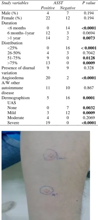
Figure 1 Patient with lesions of chronic urticaria
Figure 2 Patient showing positive ASST with a wheal formation of average diameter 34 mm at the end of 30 minutes
Figure 3 Patient showing ASST negativity with average diameter of serum induced wheal = diameter of normal saline induced wheal at the end of 30 minutes
wheals induced by serum and normal saline. The average diameter of serum induced wheal more than 1.5mm than that of saline induced was considered to be positive (Figure 2 & 3).
Based on this patients were divided into CAU (ASST positive) and CSU (negative groups).
Treatment to all the patients was given according to EAACI/GA2LEN/EDF/WAO 2013 guidelines. All patients were given single dose of second generation H1 antihistamines and were monitored for treatment response based on Urticaria Actirily Score (UAS) and clinical response at each visit. Patients were followed up on day 15, 30, 60 and 90.
Statistical analysis
The Statistical software namely SPSS version 23, and Microsoft word and Excel have been used. For inferential statistics, chi square test and test of proportions was used. In all instances, p< 0.05 was taken as statistically significant.
Results
The mean age of presentation in our study was 35.38 years. Maximum numbers of patients belonged to age group 18-29. Out of total patients included in the study, 11 (24.4%) were males and 34 (75.6%) were females.
The mean duration of the disease in our study was 8 months, with maximum number of patients (16) with the duration of disease >1 year
(Table 1).
Table 1 Disease duration ASST < 6
months
6 month to 1 year
>1 year
Total
Positive 0 12 14 26
Negative 14 3 2 19
Total 14 15 16 45
Table 2 Comparison of study variables between ASST positive and ASST negative
Study variables ASST P value
Positive Negative
Male (%) 4 7 0.194
Female (%) 22 12 0.194
Duration <6 months 6 months-1year >1 year
0 12 14
14 3 2
<0.0001 0.0694 0.0073 Distribution
<25% 26-50% 51-75% >75%
0 4 9 13
16 3 0 0
< 0.0001 0.7042 0.0128 0.0009 Presence of diurnal
variation
9 9 0.328
Angioedema 20 2 <0.0001
A/W other autoimmune disease
11 10 0.867
Dermographism 5 16 0.0001
UAS None Mild Moderate Severe
0 3 4 19
7 12
0 0
0.0032 0.0009 0.2069 <0.0001
Figure 4 A patients showing negative ASST with positive dermographism at the site of marking
>75% and all being ASST positive. 7 (4 positive, 3 negative) and 9 (all positive) patients had area of distribution 26-50% and 51-75% respectively. 57.8% (26) of patients did not have any diurnal variations. 4.4% (2) patients had exacerbations in the evening, 26.7% (12) in the night, 11.1% (5) patients had in the early morning. 22 (48.9%) patients had history of angioedema. Out of which 20 patients were ASST positive and 2 were negative (Table 2). Positive ASST was seen in 7 (26.9%) patients with hypothyroidism, 2 (7.7%) patients with hyperthyroidism, 0 patients with vitiligo, 1 (3.8%) patient with other autoimmune diseases, 1 (3.8%) patient with DM and 15 (57.7%) patients with no autoimmune disease. Negative ASST was seen in 7 (36.8%) patients with hypothyroidism, 1 (5.3%) patient with hyperthyroidism, 1 (5.3%) patient with vitiligo, 0 with other AI diseases, 1 (5.3%) patient with DM and 9 (47.4%).
21 (46.66%) patients had various medical conditions. Hypothyroidism was seen in 14 (31.3%), 7 were tested positive and 7 negative. Hyperthyroidism in 3 (6.7%) patients, 2 positive and 1 ASST negative. Vitiligo seen in 1 (2.2%) ASST negative patient. 1 patient had rheumatoid arthritis and 2 (4.4%) patients (1 ASST positive and 1 ASST negative) had diabetes mellitus.
Positive ASST was noted in 5 (19.2%) patients with dermographism and 21 (80.8%) patients with no dermographism. Negative ASST was noted in 16 (84.2%) patients with dermographism and 3 (15.8%) patients with no dermographism (Figure 4).
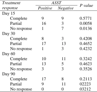
Table 3 Comparison of laboratory parameters and treatment between ASST positive and ASST negative
Study variables ASST P value
Positive Negative
Deranged TSH 6 1 0.226
S. B12 10 1 0.232
RBS 1 0 0.423
CUE 1 2 0.328
Treatment required
- Single dose antihistamines - Up dosing of antihistamines Immuno suppressants
Short term CS
9 14 3 0
9 7 2 1
0.775 0.408 0.707 0.867
Negative ASST was seen in 7 (36.8%) patients with 0 UAS score, 12 (63.2%) patients with mild UAS score, 0 patients with moderate UAS score and 0 patients with severe UAS score.
Positive ASST was seen in 20 (76.9%) patients with normal TSH, 4 (15.4%) patients with raised TSH and 2 (7.7%) patients with decreased TSH. Negative ASST was seen in 18 (94.7%) patients with normal TSH, 1 (5.3%) patients with raised TSH and 0 patients with decreased TSH. Positive ASST was seen in 16 (61.5%) patients with normal serum vitamin B12 level and 10 (38.5%) patients with low serum vitamin B12 level. Negative ASST was seen in 18 (94.7%) patients with normal serum vitamin B12 level and 1 (5.3%) patient with low serum vitamin B12 level. Positive ASST was seen in 25 (96.2%) patients with normal RBS and 1 (3.8%) patients with high RBS. Negative ASST was seen in 19 (100%) patients with normal RBS and 0 patients with high RBS. Positive ASST was seen in 25 (96.2%) patients with normal CUE report and 1 (3.8%) patients with abnormal CUE report. Negative ASST was seen in 17 (89.5%) patients with normal CUE report and 2 (10.5%) patients with abnormal CUE report (Table 3). Disease control was achieved in 18 (40%) patients with single dose of antihistamines out of which 9 (50%) patients were ASST positive and 9 (50%) were ASST negative. Up dosing of antihistamines was required in 21 (46.7%)
patients of which 14 (66.7%) patients were ASST positive and & 7 (33.3%) were ASST negative. 3 (60%) ASST positive and 2 (40%) were started on immune suppressive. One ASST negative patient required short term course of oral corticosteroids.
At D15, out of 26 (57.8%) ASST positive patients, 9 (34.6%) patients showed complete response, 16 (61.5%) patients showed partial response and 1 (3.8) patient was non responder. Out of 19 (42.2%) ASST negative patients, 9 (47.4%) patients responded completely, 3 (15.8%) patients responded partially and 7 (36.8%) patients were non responders.
At D30, out of 26 (57.8%) ASST positive patients, 8 (30.8%) patients showed complete response, 17 (65.4%) patients showed partial response and 1 (3.8%) patient was non responder. Out of 19 (42.2%) ASST negative patients, 3 (15.8%) patients responded completely, 13 (68.4%) patients responded partially and 3 (15.8%) patients were non responders.
Table 4 comparison of therapeutic response in ASST positive and negative patients
Treatment response
ASST
P value Positive Negative
Day 15 Complete Partial No response 9 16 1 9 3 7 0.5771 0.0058 0.0136 Day 30 Complete Partial No response 8 17 1 3 13 3 0.4208 0.4652 0.4232 Day 60 Complete Partial No response 10 13 3 11 5 3 0.3242 0.4623 0.3526 Day 90 Complete Partial No response 17 9 0 8 11 0 0.2113 02223 03212
partially and 3 (15.8%) patients were non responders.
At D90, out of 26 (57.8%) ASST positive patients, 17 (65.4%) patients showed complete response and 9 (34.6%) patients showed partial response. Out of 19 (42.2%) ASST negative patients, 8 (42.0%) patients responded completely and 11 (57.9%) patients responded partially (Table 4).
In our study, significantly higher number of patients with longer duration, generalized distribution, angioedema and partial or no response to treatment was observed. Whereas, shorter duration, localised disease and
association with dermographism was
significantly higher in ASST negative patients. Table 5 Comparison of our study with previous studies
Sr no.
Study Year Population ASST
positivity
Parameters studied Significant associations
1 Kumar, et al
[9]
2016 110,
37 males and 73 females.
43.62% Mean age, gender, frequency of attacks, distribution of wheals, associated diseases, angioedema, diurnal variation, family history and treatment outcome.
Angioedema was significantly higher in patients with CAU.(p=0.031)
2 Vikramkumar,
et al[3]
2014 48,
20 males and 28 females
41.6% Age,gender,duration,size of
wheals, atopy, associated AI conditions, angioedema and disease severity.
Statistically significant differences were not noted.
3 AL-Hamamy,
et al[8]
2013 54,
7 males, 15 females
40.7% Age, gender, UAS, duration,
distribution, frequency of attacks, angioedema, atopy and associated AI conditions
Distribution of wheals on the body showed significant association with positivity(p=0.004)
4 Krupashankar,
et al[10]
2012 80,
36 males and 44 females.
58.75% Age, gender, UAS, duration,
distribution, angioedema, atopy diurnal variation, associated AI conditions and thyroid abnormalities.
Angioedema was significantly higher in patients with CAU.(0.040)
5 Vohra, et al[7] 2011 100,
31 males and 69 females.
46% Age, gender, UAS, duration,
distribution, angioedema and associated conditions.
Widespread distribution ,especially on palms and soles, higher UAS and angioedema significantly higher in CAU patients.
6 Our study 45,
11 males and 34 females
57.77% Age, sex, duration, angioedema, distribution, dermographism, UAS, associated autoimmune diseases, diurnal variation, TSH, serum B12, RBS, CUE, treatment requirement and outcome.
Discussion
Chronic urticaria has a profound impact on the quality of life and causes immense distress to patients. CAU denotes a subset of population that has an increased potential to develop urticaria due to endogenous causes and fits many, but not all criteria for autoimmune disease and can be detected by ASST.
In our study, female patients were more than males and percentage of ASST positivity was also more in females. We found statistically significant association with longer duration, generalised distribution, higher UAS and angioedema in ASST positive patients.
On literature search, we found very few studies regarding association of treatment response with ASST positivity in CU patients (Table 5). In our study, at D15 significantly higher number of ASST positive patients showed partial response to treatment and required up dosing of antihistamines. However, at D90 the number of ASST positive patients showing complete response increased and were more than those showing no response or partial response indicating the need for longer treatment duration in ASST positive patients.
References
1. Bajaj A, Yadav S, Upadhyay A. Chronic urticaria: An overview. Indian J Dermatol. 2006; 51(3): 171.
2. Zuberbier T, Aberer W, Asero R, Bindslev-Jensen C, Brzoza Z, Canonica GW, et al. The EAACI/ GA2LEN/ EDF/ WAO Guideline for the definition, classification, diagnosis, and management of urticaria: The 2013 revision and update. Allergy Eur J Allergy Clin Immunol 2014;69(7): 868–87. 3. Ganguly S, Vikramkumar A, Kuruvila S.
Autologous serum skin test as an indicator of chronic autoimmune urticaria in a tertiary
care hospital in South India. Indian Dermatol Online J 2014; 5(6): 87.
4. Godse K V. Autologous serum skin test in chronic idiopathic urticaria. Indian J Dermatology, Venereol Leprol 2004; 70(5): 283–4.
5. Mamatha G, Bhalchandran C PS. Chronic idiopathic urticaria: Comparison of clinical features with positive autologous serum skin test. Indian J Dermatol Venereol Leprol 2008; 74(2): 105–8.
6. Goh CL, Tan KT. Chronic autoimmune urticaria: where we stand? Indian J Dermatol 2009; 54(3): 269–74.
7. Vohra S, Sharma NL, Mahajan VK S V. Clinicoepidemiologic features of chronic urticaria in patients having positive versus negative autologous serum skin test : A study of 100 Indian patients. Indian J Dermatol Venereol Leprol 2011; 77(2): 156–9.
8. Al-Hamamy HR, Hameed AF, Abdulhadi AS. Autologous Serum Skin Test as a Diagnostic Aid in Chronic Idiopathic Urticaria. ISRN Dermatol. Volume 2013, Article ID 291524.
9. Kishan Kumar YH, Bhaskar S, Shankar K. Comparative study of positive versus negative autologous serum skin test in chronic spontaneous urticaria and its treatment outcome. Vol. 8, North American Journal of Medical Sciences 2016. p. 25–30. 10. Krupashankar DS, Shashikala K, Madala R. Clinical and investigative assessment of patients with positive versus negative autologous serum skin test: a study of 80 South Indian patients. Indian J Dermatol 2012; 57(6): 434–8.
11. Zeinab Abdel Azim, Shaymaa El Mongy, Hanan Salem. Autologous Serum Skin Test in Chronic Idiopathic Urticaria: Comparative Study in Patients with Positive versus Negative Test. J Egypt Women Dermatol Soc 2010; 7(2): 129–33.