The handle
http://hdl.handle.net/1887/28970
holds various files of this Leiden University
dissertation.
Author
: Raterink, Robert-Jan
using Mass Spectrometry
Proefschrift ter verkrijging van
de graad van Doctor aan d e Universiteit Leiden op gezag van Rector Magnificus prof. mr. C.J.J.M. Stolker,
volgens besluit van College voor Promoties te verdedigen op woensdag 1 oktober 2014
klokke 13:45 uur
door
Robert-Jan Raterink geboren te Dronten
Promotor:
Prof. dr. Thomas Hankemeier
Co-promotor: Dr. Rob Vreeken
Overige leden:
Prof. dr. Albert van den Berg (Twente University) Dr. Christa Cobbaert (LUMC, Leiden)
Prof. dr. Meindert Danhof (Leiden University) Prof. dr. Robert Kennedy (University of Michigan) Prof. dr. Dirk Jan Reijngoud (University of Groningen) Prof. dr. Herman Spaink (Leiden University)
This study was financed by the research programme of the Netherlands Metabolomics Centre (NMC) which is a part of The Netherlands Genomics Initiative/Netherlands Organization for Scientific Research.
Publication of this thesis was financially supported by ZF-Screens B.V.
Cover illustration: Gilberto Gennero Printed by Ipskamp Drukkers B.V.
High-throughput profiling of small molecules using mass spectrometry RJ Raterink
PhD thesis, Leiden University
Chapter 1 General Introduction 5
Chapter 2 Recent Developments in Sample Pretreatment 13 Techniques for Mass Spectrometry-based Metabolomics
Chapter 3 Rapid Metabolic Screening of Early Zebrafish 33 Embryogenesis based on Direct-Infusion-nanoESI-FTMS
Chapter 4 Three-Phase Electroextraction: a New (Online) Sample 47 Purification and Enrichment Method for Bioanalysis
Chapter 5 Gas Pressure Assisted Micro-Liquid-Liquid Extraction 63 coupled Online to Direct Infusion Mass Spectrometry: a New Automated Screening Platform for Bioanalysis.
Chapter 6 Testing Tuberculosis Drug Efficacy in a Zebrafish High- 77 Throughput Translational Medicine Screen
Chapter 7 Conclusions and Perspectives 97
Samenvatting 101
Appendix 105
Dankwoord 106
Curriculum Vitae 107
Chapter 1
Chapt
er 1
GENERAL INTRODUCTION
Phenotyping based on profiling of small molecules
Phenotyping based on molecular profiling is a type of screening which investigates the phenotype of a biological system such as cells, tissues or whole organisms. The phenotype is a description of the total of physical characteristics of a biological system including its morphology, development, but also its biochemical properties. Profiling the phenotype is interesting since phenotypic effects can be induced by various (biological) perturbations such as a disease, drug interventions and genetic alterations. Phenotyping based on the profiling of small molecules such as metabolites can provide important insights into e.g. disease and the effect of drugs, as well as a systems understanding of the ability to adapt and cope with a perturbation[1].
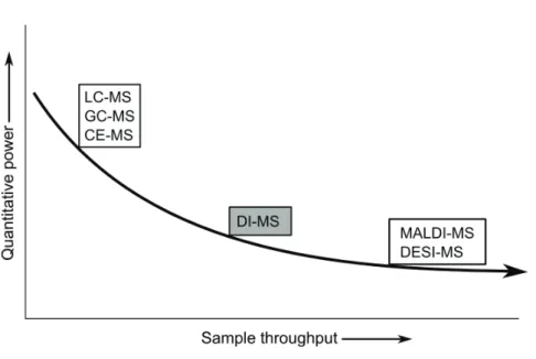
For the profiling of small molecules there are different techniques available. There is often an inverse relation between assay comprehensiveness (analyte coverage) and the throughput of the assay (see Figure 1). Whereas a single analyte high-throughput screen (HTS), such as microscopy assays, enzyme-linked immunosorbent essays (ELISA) or fluorescence-activated cell sorting (FACS) can screen over a million compounds per week, comprehensive mass spectrometry (MS)-based assays which can potentially screen for hundreds of analytes, are generally much slower [2]. As a consequence, comprehensive profiling assays have the potential to provide more in-depth information about the phenotype of a specific biological system than a single read-out biochemical assay. Therefore, in choosing the appropriate profiling technology, the balance between assay throughput and analytical comprehensiveness should be considered carefully with probably in many cases preferably maximal depth at high sample throughput.
Many conventional HTS approaches rely on fluorescent- or radio-labeling since these labels can be rapidly detected with high sensitivity [3]. Label-free strategies, including MS, have gained wide interest, since they can be applied to a broader range of assays and target classes. Moreover, MS-based screening is a comprehensive, selective and sensitive analytical tool which is not susceptible to the limitations imposed by labelling and coupling enzymes[4]. As a consequence, profiling of small molecules by MS has the potential to be a very powerfull technique.
Mass Spectrometry
Chapt
er 1
Figure 1: Schematic picture representing the comprehensiveness of the small molecule assay versus the sample throughput of the assay. Assays with potential hundreds of read-out variables such as MS are oft en much slower than other single variable read-out assays, such as microscopy assays, enzyme-linked immunosorbent assays (ELISA) or fl uorescence-activated cell sorting (FACS).
Chapt
er 1
distribution patterns of the analytes. IMS techniques can in principle be very fast, especially when only one spot per sample needs to be analyzed. Moreover, when employing e.g. DESI, limited or even no sample pretreatment is necessary. As a consequence, IMS has become a rapid and valuable tool across many fields, yet its major challenge is to acquire quantitative data regarding the surface concentrations of the analytes[5].
The developments in (HR)MS instruments have drastically increased ion selectivity for obtaining high resolution and moreover, enabled the detection of minute amounts of analyte (femtograms)[6]. However, one of the disadvantages of a MS is its limited dynamic range which is at best five orders of magnitude. More importantly, when using ESI, potential analyte competition during ionisation in the ion source, called ion suppression, imposes challenges in order to obtain quantitative results[7]. The same is true for most other ionization techniques. As a consequence, prior to MS detection, several separation techniques are often employed such as liquid chromatography (LC)-MS, gas chromatography (GC)-MS or capillary electrophoresis (CE)-MS in order to reduce the amount of potential co-eluting ions in the ion source and therefore reduce potential ion suppression effects. These hyphenated platforms can provide quantitative data, however at the expense of sample throughput (Figure 2). Direct infusion-MS (DI-MS) is a fast and comprehensive analytical method in which a sample is introduced into the MS without prior separation, possibly after sample pretreatment. In DI-MS, samples can be introduced into the MS in several ways: (i) manually, by means of a syringe pump, or (ii) automated. Flow injection analysis-MS is a commonly used automated DI-MS approach which involves the injection of an analyte plug (typically a few microliters) into a flowing solvent stream by employing e.g. a LC autosampler and pump, omitting a chromatographic column[8]. Additionally, automated nanoESI robots are available which can directly introduce a sample into the MS through a small nanoESI microchip-nozzle (typically 5 µm), in a ‘carryover-free’ manner[9]. However, although DI-MS can be fast and enables high-throughput analysis, it is often prone to ion suppression due to co-eluting ions such as (endogenous) salts and it is often not possible to differentiate between isomers. By employing nanoelectrospray (nanoESI) instead of conventional ESI, ionization efficiency can be increased and ion suppression can be reduced [10]. As a consequence, effective sample pretreatment is a key step prior to DI-MS analysis in order to obtain quantitative data[6]. When samples are effectively purified and the extracted analyte is within the dynamic range of the MS, analytical performance of DI-MS should be comparable with hyphenated MS platforms. As discussed, MALDI and DESI-MS are in principle faster per sample point than DI-MS as they can faster acquire MS spectra per sample. When e.g. using chip-based nanoESI-DI-MS, after every sample the nozzle has to be changed in order to prevent carry over, which will usually take a few seconds.
Sample pretreatment
Chapt
er 1
subsequent analysis with the analytical instrument. However, MS-based methods have the drawback that they often require tedious protocols for sample pretreatment that decrease the throughput of the assay. Therefore, to achieve high-throughput automated and integrated analytical systems have to be developed which include the sample pretreatment procedure. Although these goals can partly be met by traditional methods, new concepts are required which simplify sample pretreatment and maintain robustness and a high-throughput capacity. Additionally, when sample volume is limited, miniaturization of the sampling as well as the sample pretreatment procedure is needed which can also potentially lead to the concept of massive parallelization.
Metabolomics
The metabolome is the whole set of small molecules (typically <1000 Da) in a given biological sample. It can provide a functional readout of cellular biochemistry and therefore has the strongest correlation with the phenotype[1]. Therefore, metabolomics offers a promising phenotyping platform which has the potential to e.g. discover biomarkers for the diagnosis and prognosis of diseases and the prediction of the efficacy of drug interventions[11] [12]. Metabolites are a highly diverse range of molecules with vast differences in chemical structure, physicochemical properties and dynamic range (up to nine decades[13][14]). As a consequence, metabolomics may be the most challenging phenotyping omics science. Whereas genomics and transcriptomics can amplify DNA sequences using polymerase chain reaction (PCR), in metabolomics there is no effective method which can ‘amplify’ all low-abundant metabolites in an unbiased manner.
The metabolome can be considered as to the largest part a regulated dynamic phenotype in which changes in metabolite levels can e.g. reflect a transition from a healthy to a diseased state or the resilience of a system, in the context of systems homeostasis and allostasis. For the discovery of metabolic profiles to predict disease progression or the effect of interventions at an individual level, performing longitudinal studies is key in monitoring individuals which may result in personalized biology and eventually personalized medicine. For this strategy, following individuals over years will require high-throughput metabolomics approaches[1]. In addition, due to the vast biological variability of individuals, many individual samples may be analysed in a metabolomics study in order to ensure statistically meaningful results. As a consequence, such metabolomics studies should be performed preferably in a HT manner. In addition, each study should not only be carried out to analyse more samples within a period of time, but also to quantify as many relevant metabolites as possible in a single analysis, for which MS is in principle very suitable[15].
Aim and outline of the thesis
Chapt
er 1
as well as sample pretreatment are needed. In addition, miniaturization of the experimental setup can play an important aspect with regards to the potential of massive parallelization and, consequently, can tremendously increase sample throughput.
In Chapter 2 an overview of recent developments in sample pretreatment procedures for MS-based metabolomics is given. Deproteinization, removal of interfering molecules, liquid-liquid extraction (LLE), solid-phase extraction (SPE), electromigration-based extraction methods and possibly emerging sample pretreatment techniques for metabolomics are described and discussed. Advantages and limitations of these techniques for metabolomics are given, and aspects such as potential for automation and high-throughput analysis are evaluated
In Chapter 3 the potential of nanoESI-DI-High Resolution-MS for the metabolic phenotyping of early zebrafish embryogenesis is investigated. It is studied whether efficient but limited sample pretreatment is sufficient to obtain metabolic profiles to distinguish early developmental stages during zebrafish embryos embryogenesis. Reproducible and high-quality MS data is generated by implementing an automated data-processing tool, which includes data clean-up steps and a dedicated normalization-optimization algorithm. Principle component analysis reveals that periods of 1 hour time shifts post fertilization can be differentiated from each other.
In Chapter 4, a new and fast and selective electromigration-based sample enrichment and purification technique called 3-phase electroextraction (3-phase EE) is presented and coupled to nanoESI-DI-MS. The electromigration of analytes from an aqueous sample through an immiscible organic filter phase into a small-volume aqueous acceptor phase is demonstrated. It is shown that selectivity of 3-phase EE can be tuned by changing the composition of the organic filter phase which also prevents proteins from migrating into the acceptor phase. Proof of principle towards online 3-phase EE-nanoESI-DI-MS is demonstrated and thus the compatibility of 3-phase EE with HTS is shown.
In Chapter 5 a proof of principle of a new miniaturized LLE technique in a 384-well plate is presented, based on gas pressure mixing followed by passive phase separation. This fully-automated approach is integrated online into a nanoESI-DI-MS robot. It is shown that this high-throughput platform is suitable for screening drugs from human plasma with similar or better analytical performance compared to a conventional LLE procedure. Finally, the micro LLE method applied to dried blood spots is demonstrated.
In Chapter 6, a miniaturized sampling technique based on microneedle sampling is
developed in order to monitor drug uptake in the (small-volume) yolk of zebrafish larvae using MS analysis. It is demonstrated that conventional whole zebrafish embryo lysis is not suitable for measuring drug uptake, due to potential adherence of drugs to the skin. The in vivo uptake MS-data is correlated to the results of in vitro and in vivo high-throughput drug screening platforms and it was shown that this new approach can give relevant information for interpretation of drug efficacy data.
REFERENCES
[1] R. Ramautar, R. Berger, J. van der Greef, and T. Hankemeier, “Human metabolomics: strategies to understand biology.,” Curr. Opin. Chem. Biol., vol. 17, no. 5, pp. 841–6, Oct. 2013. [2] Y. Feng, T. J. Mitchison, A. Bender, D. W. Young, and J. A. Tallarico, “Multi-parameter
phenotypic profiling: using cellular effects to characterize small-molecule compounds.,” Nat. Rev. Drug Discov., vol. 8, no. 7, pp. 567–78, Jul. 2009.
Chapt
er 1
screening of small molecule libraries using SAMDI mass spectrometry.,” ACS Comb. Sci., vol. 13, no. 4, pp. 347–50, Jul. 2011.
[4] T. P. Roddy, C. R. Horvath, S. J. Stout, K. L. Kenney, P.-I. Ho, J.-H. Zhang, C. Vickers, V. Kaushik, B. Hubbard, and Y. K. Wang, “Mass spectrometric techniques for label-free high- throughput screening in drug discovery.,” Anal. Chem., vol. 79, no. 21, pp. 8207–13, Nov. 2007.
[5] S. R. Ellis, A. L. Bruinen, and R. M. A. Heeren, “A critical evaluation of the current state-of- the-art in quantitative imaging mass spectrometry.,” Anal. Bioanal. Chem., vol. 406, no. 5, pp. 1275–89, Feb. 2014.
[6] A. Namera and T. Saito, “Recent advances in unique sample preparation techniques for bioanalysis.,” Bioanalysis, vol. 5, no. 8, pp. 915–32, Apr. 2013.
[7] A. Furey, M. Moriarty, V. Bane, B. Kinsella, and M. Lehane, “Ion suppression; a critical review on causes, evaluation, prevention and applications.,” Talanta, vol. 115, pp.
104–22, Oct. 2013.
[8] T. Fuhrer, D. Heer, B. Begemann, and N. Zamboni, “High-throughput, accurate mass metabolome profiling of cellular extracts by flow injection-time-of-flight mass spectrometry.,” Anal. Chem., vol. 83, no. 18, pp. 7074–80, Sep. 2011.
[9] R.-J. Raterink, F. M. Kloet, J. Li, N. A. Wattel, M. J. M. Schaaf, H. P. Spaink, R. Berger, R. J. Vreeken, and T. Hankemeier, “Rapid metabolic screening of early zebrafish embryogenesis based on direct infusion-nanoESI-FTMS,” Metabolomics, vol. 9, no. 4, pp. 864–873, Jan. 2013.
[10] A. Schmidt, M. Karas, and T. Dülcks, “Effect of different solution flow rates on analyte ion signals in nano-ESI MS, or: when does ESI turn into nano-ESI?,” J. Am. Soc. Mass Spectrom., vol. 14, no. 5, pp. 492–500, May 2003.
[11] R. Kaddurah-Daouk, M. B. Bogdanov, W. R. Wikoff, H. Zhu, S. H. Boyle, E. Churchill, Z. Wang, A. J. Rush, R. R. Krishnan, E. Pickering, M. Delnomdedieu, and O. Fiehn,
“Pharmacometabolomic mapping of early biochemical changes induced by sertraline and placebo.,” Transl. Psychiatry, vol. 3, p. e223, Jan. 2013.
[12] M. Mamas, W. B. Dunn, L. Neyses, and R. Goodacre, “The role of metabolites and metabolo mics in clinically applicable biomarkers of disease.,” Arch. Toxicol., vol. 85, no. 1, pp. 5–17, Jan. 2011.
[13] N. Psychogios, D. D. Hau, J. Peng, A. C. Guo, R. Mandal, S. Bouatra, I. Sinelnikov, R. Krishnamurthy, R. Eisner, B. Gautam, N. Young, J. Xia, C. Knox, E. Dong, P. Huang, Z. Hollander, T. L. Pedersen, S. R. Smith, F. Bamforth, R. Greiner, B. McManus,
J. W. Newman, T. Goodfriend, and D. S. Wishart, “The human serum metabolome.,” PLoS One, vol. 6, no. 2, p. e16957, Jan. 2011.
[14] S. Krug, G. Kastenmüller, F. Stückler, M. J. Rist, T. Skurk, M. Sailer, J. Raffler, W. Römisch- Margl, J. Adamski, C. Prehn, T. Frank, K.-H. Engel, T. Hofmann, B. Luy, R. Zimmermann, F. Moritz, P. Schmitt-Kopplin, J. Krumsiek, W. Kremer, F. Huber, U. Oeh, F. J.
Theis, W. Szymczak, H. Hauner, K. Suhre, and H. Daniel, “The dynamic range of the human metabolome revealed by challenges.,” FASEB J., vol. 26, no. 6, pp. 2607–19, Jun. 2012. [15] J. Han, R. Datla, S. Chan, and C. H. Borchers, “Mass spectrometry-based technologies for
Chapter 2
Recent Developments in Sample
Pretreatment Techniques for Mass
Spectrometry-based Metabolomics
Based on
R.J. Raterink, P. W. Lindenburg, R. J. Vreeken, R. Ramautar and T. Hankemeier, “Recent developments in sample pretreatment techniques for mass spectrometry-based metabolomics”
Accepted for publication in Trends in Analytical Chemistry. ABSTRACT
Chapt
er 2
INTRODUCTION
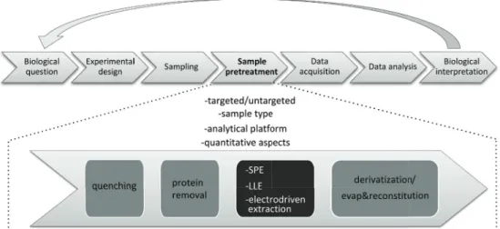
Metabolites are key regulators of system homeostasis and provide a functional readout of cellular biochemistry. Metabolomics can be defi ned as a comprehensive analytical approach for the study of the complete set of metabolites (metabolome), present in a biological system[1]. Interest in metabolomics is rapidly expanding in various research fi elds, such as cancer research[2], drug discovery and development[3], toxicological, biomedical and clinical research and nutritional research. Th e major aim of metabolomics is to obtain an answer or insight to a biological question. An analytical workfl ow comprised of experimental design, sampling, sample pretreatment, data acquisition and data analysis is generally used to address a biological question by a metabolomics approach (Figure 1).
Within the analytical workfl ow for metabolomics studies, sample pretreatment is a key step as it has a major infl uence on both the overall coverage and quality of the obtained metabolic profi les and biological interpretation of the data. As such, it may be considered the most error-prone and time-consuming step of metabolomics studies and is maybe the major source of inconsistencies between laboratories[4].
Although technical advances have been made in the fi elds of bioanalysis and metabolomics in terms of automation and high-throughput analysis, a signifi cant part of the sample pretreatment procedure is still carried out manually[5]. Th e main challenges of sample pretreatment in metabolomics is the great diversity of chemical structures and physicochemical properties as well as vast diff erences in dynamic range (up to 9 decades) of the metabolites present in a biological sample[6],[7] and at the same time the lack of universal analytical comprehensive techniques with a suffi cient dynamic range and physicochemical coverage. To ensure that the measured metabolic profi les are representative, the sample pretreatment procedures used should allow the reproducible enrichment of all metabolites of interest, and remove those compounds that interfere with the subsequent analytical method in order to preserve the integrity of the analytical system. Moreover, MS analysis can be disturbed by interfering, co-eluting matrix components in the samples resulting in ion suppression eff ects, especially when using electrospray ionization (ESI)[8]. Sample pretreatment is also required to release metabolites from the sample matrix and transport the analytes to a medium compatible with the analytical instrument. Th erefore, there is an urgent need for the development of
Chapt
er 2
reproducible, standardized and quantitative sample pretreatment approaches for biological samples critical for large-scale metabolomics studies and for minimizing inter-laboratory inconsistencies[9].
The purpose of this review is to present the state-of-the-art sample pretreatment methodologies for MS-based metabolomics, not focusing on lipodomics as this is covered including sample pretreatment in another review article by Fiehn et al. in this issue of Trends in Analytical Chemistry. In addition, some of the new emerging sample pretreatment techniques, which have not yet been fully evaluated for metabolomics studies, will also be covered. Recent developments in sample pretreatment techniques such as protein removal, liquid-liquid extraction (LLE), solid-phase extraction (SPE) and electromigration-based extraction will be highlighted, and aspects such as automation and high-throughput analysis will be considered. Subsequently, sample pretreatment issues relevant for obtaining reliable quantitative metabolomics data are shortly discussed. Finally, some general conclusions and perspectives are given. As plants represent a particular complex case as plant cell walls are difficult to break and they comprise special biochemical classes, sample pretreatment procedures for plant metabolomics studies are not discussed in this review.
SAMPLE PRETREATMENT FOR METABOLOMICS: CRITICAL
ASPECTS
Sample pretreatment is an essential part of a metabolomics study as it has a major effect on the metabolite coverage and the quality of the results obtained. Prior to sample pretreatment, experimental design and pre-analytical steps such as sample collection/storage and stabilization need to be carefully examined and chosen in each metabolomics study (Figure 1); however, these aspects are not covered in this review, but are discussed elsewhere [10]– [14]. Sample pretreatment strategies used in metabolomics prior to separation and detection can include ultrafiltration, protein precipitation, LLE, SPE, derivatization, and evaporation followed by reconstitution[15]. Sample pretreatment finally results in a fraction of a sample that can be analyzed by a subsequent analysis platform. In this section important aspects with regard to the choice of a sample pretreatment technique for metabolomics are considered. First, the difference between targeted and untargeted metabolomics is outlined, followed by a discussion concerning the influence of the sample type and the subsequent analysis platform on the sample pretreatment strategy.
General aspects: targeted versus untargeted metabolomics
In metabolomics, two different approaches may be distinguished, i.e. the targeted and the untargeted detection of metabolites. The targeted approach is focused on the quantitative analysis of preselected metabolites in a biological sample. Targeted metabolomics approaches are commonly driven by a specific biochemical hypothesis, typically focusing on one or more related pathways of interest[16][17]. The untargeted approach involves the profiling of biological samples without having a priori knowledge on the nature and the identity of the measured metabolites. The goal is to obtain qualitative and (semi)-quantitative information, which can be used to compare patterns or fingerprints of metabolites that change in response to normal and abnormal biological processes, genetic alterations and to external stimuli, for example drug exposure[18]. As a consequence, untargeted metabolomics can be used to reveal the involvement of metabolic pathways that may not have been predicted and is therefore often hypothesis-generating or hypothesis-refining.
Chapt
er 2
metabolomics studies may be quite different. For untargeted metabolomics studies, the biological sample should preferably be analyzed with minimal pretreatment in order to prevent the potential loss of metabolites, or at least, within a certain wide biochemical window, metabolites should not be removed during pretreatment. The sample pretreatment steps can be quite straightforward, separating low molecular weight (LMW) compounds from proteins and/or lipids using deproteinization and/or delipidation techniques[15]. Therefore in untargeted metabolomics studies of biofluids, non-selective sample pretreatment methods such as “dilute-and-shoot” and solvent protein precipitation are often used, since they enable broad metabolite coverage and high-throughput analysis[18]. Untargeted metabolomics approaches can also be applied to a fractionated sample obtained after e.g., a LLE and/or SPE step.
In contrast to untargeted metabolomics studies, development and optimization of sample pretreatment procedures for targeted metabolomics are often relatively straightforward, as it is known a priori on which metabolite class(es) to focus. Sample pretreatment includes the separation of metabolites from proteins, often followed by LLE and/or off-line/online SPE for the selective isolation and enrichment of the target compounds, and removal of interfering matrix components.
Evaporation and reconstitution is often the final step during sample pretreatment in order to concentrate the analytes by reconstitution in a smaller volume and/or convert the solvent to an analysis-compatible solvent. During such a step, attention should be paid to analyte solubility, and potential oxidation of analytes such as thiol-containing molecules[19].
Sample type
The sample pretreatment strategy also largely depends on the nature of the sample to be analyzed. The protein content of biological samples is an important aspect to consider, as most subsequent analysis methods are not compatible with protein-rich samples such as, plasma, serum, cells and tissue. In contrast, urine, particularly from healthy human individuals, contains relatively low protein amounts, and centrifugation followed by dilution with water is often all that is required prior to analysis[18].
Chapt
er 2
transportation at ambient conditions. Therefore, the use of DBS may have advantages for large-scale epidemiological studies where the pretreatment of blood and the subsequent storage and transport poses logistical challenges. So far only a few studies on untargeted metabolomics have been performed with the use of DBS. Analyte coverage for DBS seem to be comparable with solvent precipitation of whole blood and plasma[25]–[27]. Moreover, the result of stability studies seem to indicate that stability for most metabolites does not seem to be critical when analysis is done within a week of collection[25]. Lately, instrumental manufacturers have developed automated systems which can handle, extract and introduce the DBS extract into the analytical system, thus enabling the use of DBS for large-scale high-throughput profiling studies[28].However, some challenges related to DBS need to be examined, such as the influence of the hematocrit level and the non-uniform distribution of the blood spot on the sampling area[29].
Saliva is another readily available biofluid that may contain metabolites of interest for diagnosis and prognosis of diseases. Nevertheless, there are only a few reports concerning metabolomics. The major challenge in saliva metabolomics is the relatively low abundance of metabolites[30]–[32]. Therefore, in this field there is a need for more sensitive analytical tools which may include sample pretreatment methods that allow analyte enrichment.
Subsequent analytical platform
One major aim of metabolomics is to obtain a comprehensive view of the metabolites present in a biological sample. As no single analytical technique covers the entire spectrum of the human metabolome, various complementary analytical platforms (e.g. gas chromatography-mass spectrometry (GC-MS), liquid chromatography-MS (LC-MS), capillary electrophoresis-MS (CE-electrophoresis-MS) and/or direct infusion-electrophoresis-MS (DI-electrophoresis-MS) should be employed in order to improve metabolite coverage and identification power[33]–[36]. Each of these analytical platforms has different requirements with regard to sample pretreatment. In general, deproteinization is required for each analytical platform, as the presence of proteins can seriously influence precision, accuracy and instrument component lifetime (e.g. LC and GC columns). Moreover, proteins can induce severe matrix effects, may cause clogging of nanoESI emitters and can modify the inner surface of capillaries in CE.
In GC-MS, sample pretreatment involves a number of steps generally including deproteinization, lyophilization and chemical derivatization. Chemical derivatization is often required to decrease the boiling point of many endogenous metabolites. There are a multitude of different chemical derivatization reagents used, although a two-stage process of oximation followed by trimethylsilylation is mostly applied[37]. However, the derivatization process may add more error-prone complex steps in the sample pretreatment method. Furthermore in general, derivatization can impose added requirements to the sample pretreatment procedure in order to clean up compounds that can interfere with the derivatization reactions.
In DI-MS, “dilute and shoot” after deproteinization is the most common approach, including optimization of the dilution factor in order to minimize ion suppression and detector saturation effects. Additionally, prior to DI-MS, a desalting step should be included, because salts can significantly contribute to ion suppression effects[38]. However, to date no general straightforward method is available which separates (endogenous) inorganic ions from the wide range of polar and often ionic metabolites.
SAMPLE PRETREATMENT TECHNIQUES
Chapt
er 2
electromigration-driven sample extraction will be discussed. Furthermore, throughout this section delipidation techniques will be discussed.
Protein removal
In metabolomics, the most commonly used methods for protein removal are organic solvent-based protein precipitation (PPT) followed by centrifugation, or membrane-solvent-based techniques such as ultrafiltration[4]. It is preferable to use an organic solvent for PPT, since protein denaturation using heat or inorganic acids have been shown to result in lower metabolite coverage[39] and the organic solvent also disrupts binding between many metabolites and proteins. Actually, the latter is also true for many metabolites using inorganic acids, and some inorganic acids extracts metabolites more efficient than organic solvents. However, organic solvent PPT is able to extract both hydrophilic and hydrophobic compounds. Therefore, further clean-up of the extracted samples is often required as the supernatant still contains many components such as lipids which may disturb subsequent MS analysis (i.e. ion suppression) and may reduce lifetime of e.g. LC columns. As a consequence, the combination of protein and lipid removal is often used as an effective strategy to improve metabolite coverage and reproducibility. Moreover, metabolite losses may occur due to co-precipitation with proteins and/or poor solubility in the selected extraction solvent[40].
Over the past few years, the performance of various PPT procedures for metabolomics have been evaluated in terms of protein removal efficiency, metabolite coverage, and precision[20], [37], [39], [41]–[44]. The diverging practices used in these studies reveal that there is still no general consensus on the best PPT procedure that should be used for metabolomics. However, some trends are apparent: precipitation with acetonitrile or acetone seems to perform better in terms of protein removal, whereas PPT with methanol, ethanol or a mixture of both results in improved metabolic coverage and method precision. Therefore, the latter solvents are more often applied[28][15].
In conventional solvent PPT however, it is costly to include centrifugation into robotic automation solutions. Recent innovations have become available overcoming the need of centrifugation after PPT. Surface-functionalized magnetic beads have been recently suggested for sample pretreatment in several fields, addressing large and small molecules. König et al. reported a serum protein removal method based on surface-modified MagSi magnetic beads. Proteins were denatured using a proprietary reagent followed by the immobilization of the protein-adsorbing magnetic beads, by applying a magnetic field[45]. Neither centrifugation nor application of pressure is required, but only a time-controlled application of a magnetic field making this technique promising for automated high-throughput applications.
Also recently, solutions have become available for automated solvent PPT in a well-plate format such as the Sirocco™ (Waters), Impact™ (Phenomenex) or HyperSep™ (Thermoscientific). Using these PPT plates the solvent precipitated sample is filtered through a well plate by using a vacuum or pressure. Cao et al. demonstrated the automated removal of proteins in 50 µL dog plasma followed by LC-MS analysis, using the Sirocco™ plate[46]. These available solutions could be promising for metabolomics studies, but have not been demonstrated yet. In section 3.3 other recently available well plates are discussed which simultaneously can remove proteins and lipids from a complex sample.
Chapt
er 2
using stationary phase functionality. After the sample is injected into a turbo flow column the high flow-rate of 1.5-5.0 mL/min generates turbulent flow conditions inside the column. Small analyte molecules are retained via diffusion into the pores, while proteins are washed to waste. In 2010, Michopoulos et al. demonstrated the use of TFC for the protein removal in a metabolomics study of plasma, followed by LC-MS analysis. A comparison between TFC and methanol-based PPT was made and resulted in similar numbers of molecular features (2900), with a somewhat poorer repeatability for TFC. TFC reduced the concentration of phospholipids 10-60 fold, probably because the protein binding compounds (such as certain lipids) were washed away[47]. The principle of TFC has shown potential for the direct analysis of crude serum samples in automated, high-throughput metabolomics studies, however at the price of very high flow-rates and substantial consumption of solvents.
Liquid-Liquid-Extraction
LLE enables the extraction of metabolites into two fractions (aqueous and organic phase) that separately contain polar and apolar compounds and which can be then independently analyzed. In LLE it is challenging to extract polar analytes into an organic phase, therefore in metabolomics LLE is mostly used for sample cleanup purposes, predominantly for the removal of lipids. According to Folch et al.[48] and Bligh and Dyer[49], chloroform extraction can be used to recover all major lipid classes, but the use of dichloromethane has been suggested as a less toxic alternative[50]. Recently Matyash et al. reported that substituting chloroform with methyl tert-butyl ether (MTBE) also recovered the major lipid classes with the same or better recovery, but with reduced health risks[51]. Moreover, MTBE has a more advantageous phase configuration in which the MTBE phase is at the top and where the precipitated proteins are at the bottom (instead of between the phases using chloroform-based LLE), therefore simplifying online coupling. For these reasons, in the last years several papers have been published comprising LLE with MTBE for metabolomics studies. Chen et al. presented an untargeted approach to simultaneously perform metabolomics as well as lipidomics from a small piece of tissue sample (10 mg) based on a MTBE LLE. After the MTBE-based extraction, a mix of the resulting polar and apolar fraction was pooled, evaporated and reconstituted and analyzed with ultra performance (UP)LC-MS. This approach was comparable or superior in yield (3429 vs. 2641 features) and reproducibility (0.3-9.9% vs. 0.5-15.4%) to a standard methanol extraction for the profiling of polar and apolar metabolites[52].
Recently the group of Barbas and co-workers developed a MTBE-based extraction method by performing the whole sample preparation and analysis within and from a single LC-vial, which they coined as in vial dual extraction (IVDE)[53]. The upper MTBE and lower methanol-water phases were successively injected onto an reversed-phase (RP)LC-MS system directly from the vial by the adjustment of the instrument needle height in two separate runs. This way, pretreatment time and analytical variation was reduced. To date, this approach was tested for the direct LC-MS analysis of 20 µL plasma resulting in over 4500 reproducible features, as well as DBS samples for untargeted metabolomics[54], see Figure 2, and also for the application of a biological case [55].
Chapt
er 2
Figure 2: Schematic illustration of the IVDE method comprising the (from bottom to top) precipitated proteins, aqueous and organic MTBE layer. Both organic and aqueous layer are injected on a LC separately for apolar and polar metabolite analysis respectively. Figure adapted with permission from [55].
Chapt
er 2
metabolites, see Figure 3[56].
LLE can also be used for cleaning up samples in order to obtain effi cient labeling in metabolomics. Recently, Peng et al. reported an improved method for detecting organic acids based on LLE followed by diff erential isotope p-dimethylaminophenacyl (DmPA) labeling of the acidic metabolites[57]. It is shown that this strategy off ers superior performance (3-fold increase in putative detected metabolites) over the method of direct labeling of metabolites in biofl uids such as human urine.
Still, conventional LLE as described above has drawbacks being time-consuming and diffi cult to automate (e.g. in a multi-well format), due to the necessary mixing and oft en required centrifugation steps.
New LLE approaches have emerged in overcoming automation diffi culties, as discussed below. Additionally, these approaches may use less organic solvent than conventional LLE. Supported Liquid Extraction (SLE), or solid-supported liquid extraction, is an emerging LLE-based technique for metabolite analysis in which an aqueous sample phase is absorbed into a chemically inert, porous, high-surface area diatomaceous earth support. Th e organic phase is passed through the cartridge and a highly effi cient extraction is obtained, therefore bypassing cumbersome mixing and centrifugation steps (Figure 4). As a consequence, the SLE is easier to automate and uses less organic solvent than conventional LLE[58]. Several commercial 96-well plate SLE are available, compatible with automated high-throughput analysis. Although, to date, the analysis of drugs in biofl uids is one of the most popular application of SLE, it may also be promising for high-throughput sample clean-up in metabolomics[59][60].
Figure 4: Schematic picture of a typical SLE procedure, modifi ed from[84].
Miniaturization of LLE is another trend such as liquid-phase microextraction (LPME) tech-niques [61]. In LPME, extraction generally takes place from an aqueous sample phase into a small amount of a water-immiscible organic acceptor phase, and reducing the acceptor-to-donor ratio. Even though this miniaturized approach might be promising in metabolomics studies where limited sample amounts are available, no recent metabolomics applications of LPME techniques have been reported so far.
Chapt
er 2
of metabolites which increases sample throughput. So far, the potential of this approach has been only demonstrated for a few metabolomics studies [62].
Solid Phase Extraction
SPE is an eff ective sample pretreatment method for the removal of interfering substances and for the enrichment of analytes. A variety of extraction sorbents is available for SPE; examples are: reversed-phase materials including phenyl groups or polymeric material, weak and strong ion exchange materials including amino groups, mixed mode materials covering for example reversed- phase and weak ion exchange and stationary phases for HILIC separations. Th e-refore, SPE can address more specifi c molecular characteristics of target analytes and allows the design of protocols which can be more selective than LLE[5]. However, due to sorbent selectivity, obtaining high metabolite coverage may be challenging, especially in untargeted metabolomics. Th erefore, within targeted metabolomics, the combination of SPE and LC sy-stems using reversed-phase sorbents are most oft en employed for clean-up (desalting and de-lipidation) and enrichment of relatively hydrophobic analytes. Th is approach can also be used for analysis of more polar compounds if employing alternative phases (e.g. C30) or ion-pairing reagents[63]. However complementary principles may be more appropriate, such as mixed-mode or HILIC phases. RP SPE-and LC columns are used in most online SPE-LC systems and are now widely commercially available in various dimensions[64].
Recently, Yang et al. reported a combined untargeted MTBE-based LLE and SPE (NH2) me-thod to improve the coverage of the metabolome in plasma caused by eff ective delipidation. [65]. Th ey separated the sample into fi ve fractions including aqueous species, lipids, fatty acids, neutral lipids and hydrophobic lipids prior to RPLC-MS and HILIC-MS analysis, and detected over 3806 versus 1851 molecular features using methanol extraction only (Figure 5). Improved reproducibility was shown with CV’s below 15% for the combined LLE-SPE method, compared to 30% using the methanol method. However, this LLE-SPE method is time-consuming and therefore may be less suitable for high-throughput applications.
Chapt
er 2
Fully automated SPE has clear advantages over conventional LLE and can outperform manual protocols in terms of reproducibility and sample throughput. Off-line automated SPE sam-ple pretreatment using robotics is available, as well as SPE platforms cousam-pled on-line to MS such as the Agilent Rapidfire system which is a robotic high-throughput SPE-MS system or the Spark Symbiosis™ system which combines SPE and LC in parallel for high sample throu-ghput. These systems could be suitable for rapidly tailoring an efficient targeted metabolite extraction protocol, by employing the various SPE materials that are currently available and optimizing organic solvents, pH or ion strength.
As discussed, delipidation of the sample has gained increased interest, since it can reduce ion suppression effects and can increase column lifetime. Recently, several commercial available lipid depletion plates have become available such as the Hybrid SPE™ (Sigma Aldrich), Ostro™ (Waters), Captiva™ ND (Agilent) and the Phree™ (Phenomenex) 96 well plates which can re-move lipids as well as proteins from a sample in a single step. These recent innovations are ef-fective for removing phospholipids and compatible with automation and high sample throu-ghput, but their protocols dilute samples and do not easily allow sample concentration[66]. In SPE recent promising innovations regarding miniaturization, sorbent material and sorbent functionality has appeared in literature and is discussed below.
Also in SPE there is a trend towards miniaturization. One such approach is solid-phase mi-croextraction (SPME) in which a fiber is coated with a thin layer of sorbent material. SPME can integrate sampling, extraction, concentration and sample introduction into a single step[61]. For more details the reader is referred to the review article by Pawliszyn et al. in this issue of Trends in Analytical Chemistry. Microextraction by packed sorbent (MEPS) is a more recent miniaturized SPE approach that integrates the sorbent cartridge in a microliter syringe which can be used in e.g. an autosampler. Sample and solvent volume has been scaled down to the microliter level and is therefore very compatible with volume-limited samples. Several MEPS sorbents are available, including reversed- and normal phase, HILIC, mixed mode and ion exchange functionalities[67]. This technique could be very promising in online automa-ted, high-throughput metabolomics studies.
The use of alternative SPE sorbent chemistry based on aptamers could also bring potential to highly selective targeted metabolomics approaches, for example by employing RNA oli-gomers that have high affinity to small molecule targets including ATP and several amino acids[68]. Another interesting alternative sorbent is the use of immobilized ionic liquids in sample pretreatment for the extraction of polar compounds[69]. However, the potential of these alternative sorbents for metabolomics has not been demonstrated yet.
The use of monolithic material in SPE columns has gained increased interest. This material has several advantages over the use of particle sorbents including low back pressure, high efficiency and a low dead volume[70]. Recently, Abe et al. used a spin column, which was pac-ked with a monolithic silica disk, for the extraction of drugs from human urine. In this spin column, the monolithic silica works as a frit, the surface to volume ratio is larger than the use of silica particles and it requires a small volume of extraction solvent [71]. As a consequence this material could be promising for metabolomics, but this has not been demonstrated yet. However, sample loading, washing and elution were accomplished by centrifugation of the spin column, which makes it less suitable for robotic automation.
Chapt
er 2
CNT could be promising for metabolomics, however it has not been demonstrated yet.
Electromigration-based Extraction Techniques
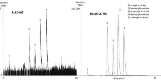
Electromigration-based extraction utilizes an electric field to induce selective migration of charged compounds. A major part of the metabolome can be targeted with this approach, as many metabolites are charged in solution or can be made charged by adjusting the pH. Elec-tromigration-based sample extraction has received increased attention in the past 5 years[73] [74]. In this section, we outline recent developments in two techniques that may be promising for metabolite analysis: electromembrane extraction (EME) and electroextraction (EE). EME is based on an the application of an electric field between a donor phase and an acceptor phase which are separated by a membrane filled with an organic solvent in its pores[75]. Via the membrane, which consists of a polymeric material, analytes are extracted from a sample to an acceptor compartment. The electric field enhances the extraction rate of the analytes and as a consequence it is fast and typically enriches analytes (expressed in concentration units) by one order of magnitude. Strieglerová et al. demonstrated the potential of EME for the targe-ted extraction of endogenous amino acids from human plasma and urine[76]. Here, a simple EME device was coupled with capillary electrophoresis with capacitively coupled contactless conductivity detection. In total, 12 endogenous physiological amino acids were analyzed in human plasma, whole blood, serum and urine with peak area RSDs ranging from 1 to 13%. However, not all endogenous 20 physiological amino acids were recovered[76]. Further op-timization of the composition of the liquid phase of the membrane may result in an EME protocol that is capable of extracting a wider physicochemical range of analytes in one run. EE is an electromigration-based sample enrichment technique in which analytes are concen-trated from a large donor phase, through one or more liquid-liquid interfaces, into a small acceptor phase[77]. In the most simple set-up, the donor phase is an organic solvent, which may contain some water to dissolve ions as has been done for ethyl acetate, and the adjacent acceptor phase is an aqueous phase; both phases should be immiscible. It has been shown that EE allows the fast enrichment of metabolites and is promising for high-throughput ap-plications and analysis of low-abundant metabolites [78][79]. The driving force of EE is an electric field that is applied over the liquid-liquid system: in the donor phase a very high field strength is present. When the metabolites enter the aqueous acceptor phase, they enter a very low electric field, their migration speed diminishes and they are enriched in a small volume. In 2012, Lindenburg et al. presented EE of acylcarnitines from human urine. EE took place in a wide-bore PTFE capillary, was coupled online with LC-MS via a switching valve and was automated. Within a few minutes, acylcarnitines at the nM level in urine were enriched up to 1000 times with excellent repeatability (below 12%) in comparison with conventional LC-MS injection, see Figure 6 [78].
Chapt
er 2
Until now, it has been shown that electromigration-driven sample extraction methods are in principle promising for metabolomics. Its approach can off er speed, selectivity and excellent potential for both miniaturization and high-throughput analysis. Th erefore, we expect that electromigration-based sample extraction will be used as complementary tools for metabolo-mics studies in the upcoming years. However, an important aspect to study is the applicability of these methods to a wider range of metabolites.
QUANTITATION AND VALIDATION
Th e systematic evaluation and validation of an extraction procedure for a given biological sample matrix is necessary in order to obtain reliable and meaningful metabolomics data. In this section, attention is paid to some issues that should be considered during sample extrac-tion to obtain reliable quantitative metabolite data.
Th e performance (i.e. recovery, repeatability, enrichment factors etc.) of the extraction me-thod should be critically examined by using proper internal standards. In untargeted metabo-lomics, the performance of an extraction method is still oft en only evaluated in terms of the number of molecular features observed[15]. Th is strategy may provide useful information, however specifi c information concerning metabolite enrichment and recovery, or metabolite losses due to e.g. protein binding, is not provided. In this context, we suggest using multiple stable isotopically labeled internal standards, each representing a metabolite (sub)class, du-ring sample extraction in order to evaluate the performance of the method[80][81]. Recently Strassburg et al. developed a quantitative (precision RSD ranged from 4% - 15%) and sensitive (down to nM levels) LC-MS/MS method for the profi ling of approximately 100 oxylipins in human plasma by using 11 deuterated internal standards which were considered represen-tative for the diff erent compound classes[82]. Method validation in untargeted metabolo-mics is more challenging than in targeted metabolometabolo-mics regarding the choice of the internal standard. Another elegant approach to tackle these challenges is the use of 13C-labeled cell extracts, which has been recently used for the quantifi cation of 91 metabolites representing central carbon and energy metabolism[33]. For targeted metabolites, isotopically-labelled
Chapt
er 2
metabolites can be used to correct for potential matrix effects (ion suppression/enhancement) and batch-to-batch effects of the corresponding endogenous metabolites.
At present, there is no consensus yet on the most suited type of sample extraction method that should be used for a certain biological sample matrix or question. Standardization and inter-laboratory comparisons are crucial in order to demonstrate the validity of a sample extraction procedure for metabolic profiling of a particular biological sample, and this would help to compare different metabolomics studies published or even accessible in data repositories. For inter-laboratory comparisons, reference material[83] should be used and preferably absolute concentrations should be provided for metabolites. Especially when reporting relative con-centrations, as is often the case in untargeted metabolomics studies, it is crucial to use pooled samples and/or internal standards as quality controls and for correction of variations and possible biases in the overall analytical procedure during studies[81].
CONCLUSIONS AND OUTLOOK
Sample pretreatment is a key step in the metabolomics workflow as it has a major influence on the overall quality of the obtained metabolic profile, metabolite coverage and sample through-put. This review highlights that the development of sample pretreatment for metabolomics is still an active area of research. Although no universal sample pretreatment technique is avai-lable for the comprehensive analysis of all metabolites in a given biological sample, the combi-nation of protein and lipid removal is often used as an effective strategy to improve metabolite coverage and reproducibility. In LLE several developments are made towards the compati-bility with automation, of which supported liquid extraction is one of the most promising techniques, readily available in a well-plate format. As a consequence, deproteinization and delipidation can be performed by using LLE as well as SPE techniques in a fully automated, high-throughput manner. The recent innovations in automated offline well-plate extraction (including protein precipitation, LLE and SPE) or online extraction (including turbulent flow chromatography) have allowed fast sample cleanup and partly removed the bottlenecks asso-ciated with sample pretreatment. All these techniques are very promising for metabolomics, however the effective extraction of polar metabolites is still challenging and the separation of polar analytes from interfering salts is a hurdle for some analytical methods which still needs to be overcome. New emerging electromigration-based sample pretreatment techniques are promising and their compatibility with automation has been demonstrated. Still, more re-search is needed in order to exploit the full potential of these techniques for metabolomics. To improve quantitative aspects during sample pretreatment, more research is needed in the use of internal standards, such as 13C-labeled cell extracts. Proper internal standards are cru-cial to correct for matrix and batch effects and to obtain insights in metabolite enrichment and recovery. In the future, sample pretreatment procedures should not only be automated and fast, but also quantitative and standardized so that metabolomics data of different studies are comparable within and between labs, which would be needed for breakthroughs in bio-medical and pharmacological research.
ACKNOWLEDGEMENTS
Chapt
er 2
REFERENCES
[1] O. Fiehn, “Metabolomics--the link between genotypes and phenotypes.,” Plant Mol. Biol., vol. 48, no. 1–2, pp. 155–71, Jan. 2002.
[2] R. Beger, “A Review of Applications of Metabolomics in Cancer,” Metabolites, vol. 3, no. 3, pp. 552–574, Jul. 2013.
[3] J. D. Rabinowitz, J. G. Purdy, L. Vastag, T. Shenk, and E. Koyuncu, “Metabolomics in drug target discovery.,” Cold Spring Harb. Symp. Quant. Biol., vol. 76, pp. 235–46, Jan. 2011. [4] T. Hyötyläinen, “Critical evaluation of sample pretreatment techniques.,” Anal. Bioanal.
Chem., vol. 394, no. 3, pp. 743–58, Jun. 2009.
[5] M. Vogeser and F. Kirchhoff, “Progress in automation of LC-MS in laboratory medicine.,” Clin. Biochem., vol. 44, no. 1, pp. 4–13, Jan. 2011.
[6] N. Psychogios, D. D. Hau, J. Peng, A. C. Guo, R. Mandal, S. Bouatra, I. Sinelnikov,
R. Krishnamurthy, R. Eisner, B. Gautam, N. Young, J. Xia, C. Knox, E. Dong, P. Huang, Z. Hollander, T. L. Pedersen, S. R. Smith, F. Bamforth, R. Greiner, B. McManus, J. W. Newman, T. Goodfriend, and D. S. Wishart, “The human serum metabolome.,” PLoS One, vol. 6, no. 2, p. e16957, Jan. 2011.
[7] S. Krug, G. Kastenmüller, F. Stückler, M. J. Rist, T. Skurk, M. Sailer, J. Raffler, W. Römisch- Margl, J. Adamski, C. Prehn, T. Frank, K.-H. Engel, T. Hofmann, B. Luy, R. Zimmermann, F. Moritz, P. Schmitt-Kopplin, J. Krumsiek, W. Kremer, F. Huber, U. Oeh, F. J. Theis, W. Szymczak, H. Hauner, K. Suhre, and H. Daniel, “The dynamic range of
the human metabolome revealed by challenges.,” FASEB J., vol. 26, no. 6, pp. 2607–19, Jun. 2012.
[8] A. Furey, M. Moriarty, V. Bane, B. Kinsella, and M. Lehane, “Ion suppression; a critical review on causes, evaluation, prevention and applications.,” Talanta, vol. 115, pp. 104–22, Oct. 2013.
[9] C. Simó, C. Ibáñez, A. Gómez-Martínez, J. A. Ferragut, and A. Cifuentes, “Is metabolomics reachable? Different purification strategies of human colon cancer cells provide different CE- MS metabolite profiles.,” Electrophoresis, vol. 32, no. 13, pp. 1765–77, Jun. 2011.
[10] T. Rosenling, M. P. Stoop, A. Smolinska, B. Muilwijk, L. Coulier, S. Shi, A. Dane, C. Christin, F. Suits, P. L. Horvatovich, S. S. Wijmenga, L. M. C. Buydens, R. Vreeken, T. Hankemeier, A. J. van Gool, T. M. Luider, and R. Bischoff, “The impact of delayed storage on the measured proteome and metabolome of human cerebrospinal fluid.,” Clin. Chem., vol. 57, no. 12, pp. 1703–11, Dec. 2011.
[11] D. Siegel, H. Permentier, D.-J. Reijngoud, and R. Bischoff, “Chemical and technical challenges in the analysis of central carbon metabolites by liquid-chromatography mass spectrometry.,” J. Chromatogr. B. Analyt. Technol. Biomed. Life Sci., Nov. 2013. [12] B. Kamlage, S. G. Maldonado, B. Bethan, E. Peter, O. Schmitz, V. Liebenberg, and P. Schatz,
“Quality markers addressing preanalytical variations of blood and plasma processing identi fied by broad and targeted metabolite profiling.,” Clin. Chem., vol. 60, no. 2, pp. 399–412, Mar. 2014.
[13] J. LaBaer, “Improving international research with clinical specimens: 5 achievable objectives.,” J. Proteome Res., vol. 11, no. 12, pp. 5592–601, Dec. 2012.
[14] V. Gonzalez-Covarrubias, A. Dane, T. Hankemeier, and R. J. Vreeken, “The influence of ci trate, EDTA, and heparin anticoagulants to human plasma LC–MS lipidomic profiling,” Metabolomics, vol. 9, no. 2, pp. 337–348, Jul. 2012.
[15] D. Vuckovic, “Current trends and challenges in sample preparation for global metabolomics using liquid chromatography-mass spectrometry.,” Anal. Bioanal. Chem., vol. 403, no. 6, pp. 1523–48, Jun. 2012.
[16] B. Álvarez-Sánchez, F. Priego-Capote, and M. D. Luque de Castro, “Metabolomics analysis I. Selection of biological samples and practical aspects preceding sample preparation,” TrAC Trends Anal. Chem., vol. 29, no. 2, pp. 111–119, 2010.
Chapt
er 2
spectrometry.,” Adv. Protein Chem. Struct. Biol., vol. 80, pp. 45–83, Jan. 2010.
[18] E. J. Want, I. D. Wilson, H. Gika, G. Theodoridis, R. S. Plumb, J. Shockcor, E. Holmes, and J. K. Nicholson, “Global metabolic profiling procedures for urine using UPLC-MS.,” Nat. Protoc., vol. 5, no. 6, pp. 1005–18, Jun. 2010.
[19] L. A. D’Agostino, K. P. Lam, R. Lee, and P. Britz-McKibbin, “Comprehensive plasma thiol redox status determination for metabolomics.,” J. Proteome Res., vol. 10, no. 2, pp. 592–603, Feb. 2011.
[20] W. Römisch-Margl, C. Prehn, R. Bogumil, C. Röhring, K. Suhre, and J. Adamski, “Procedure for tissue sample preparation and metabolite extraction for high-throughput targeted meta bolomics,” Metabolomics, vol. 8, no. 1, pp. 133–142, Mar. 2011.
[21] C. A. Sellick, R. Hansen, G. M. Stephens, R. Goodacre, and A. J. Dickson, “Metabolite extraction from suspension-cultured mammalian cells for global metabolite profiling.,” Nat. Protoc., vol. 6, no. 8, pp. 1241–9, Aug. 2011.
[22] J. J. Ellinger, D. C. Miller, I. a Lewis, and J. L. Markley, “Semiautomated device for batch extraction of metabolites from tissue samples.,” Anal. Chem., vol. 84, no. 4, pp. 1809–12, Feb. 2012.
[23] E. J. Want, P. Masson, F. Michopoulos, I. D. Wilson, G. Theodoridis, R. S. Plumb, J. Shockcor, N. Loftus, E. Holmes, and J. K. Nicholson, “Global metabolic profiling of animal and human tissues via UPLC-MS.,” Nat. Protoc., vol. 8, no. 1, pp. 17–32, Jan. 2013.
[24] F. M. Geier, E. J. Want, A. M. Leroi, and J. G. Bundy, “Cross-platform comparison of Caenor habditis elegans tissue extraction strategies for comprehensive metabolome coverage.,” Anal. Chem., vol. 83, no. 10, pp. 3730–6, May 2011.
[25] I. Wilson, “Global metabolic profiling ( metabonomics / metabolomics ) using dried blood spots : advantages and pitfalls,” pp. 2255–2257, 2011.
[26] F. Michopoulos, G. Theodoridis, C. J. Smith, and I. D. Wilson, “Metabolite profiles from dried biofluid spots for metabonomic studies using UPLC combined with oaToF-MS.,” J. Proteome Res., vol. 9, no. 6, pp. 3328–34, Jun. 2010.
[27] J. Dénes, E. Szabó, S. L. Robinette, I. Szatmári, L. Szőnyi, J. G. Kreuder, E. W. Rauterberg, and Z. Takáts, “Metabonomics of newborn screening dried blood spot samples: a novel approach in the screening and diagnostics of inborn errors of metabolism.,” Anal. Chem., vol. 84, no. 22, pp. 10113–20, Nov. 2012.
[28] H. Gika and G. Theodoridis, “Sample preparation prior to the LC-MS-based metabolomics/ metabonomics of blood-derived samples.,” Bioanalysis, vol. 3, no. 14, pp. 1647–61, Jul. 2011. [29] M. O’Mara, B. Hudson-Curtis, K. Olson, Y. Yueh, J. Dunn, and N. Spooner, “The effect of
hematocrit and punch location on assay bias during quantitative bioanalysis of dried blood spot samples.,” Bioanalysis, vol. 3, no. 20, pp. 2335–47, Oct. 2011.
[30] J. Zheng, R. A. Dixon, and L. Li, “Development of isotope labeling LC-MS for human salivary metabolomics and application to profiling metabolome changes associated with mild cognitive impairment.,” Anal. Chem., vol. 84, no. 24, pp. 10802–11, Dec. 2012. [31] B. Cuevas-Córdoba and J. Santiago-García, “Saliva: A Fluid of Study for OMICS.,” OMICS,
vol. 18, no. 2, pp. 87–97, Feb. 2014.
[32] S. Medina, R. Domínguez-Perles, J. I. Gil, F. Ferreres, and A. Gil-Izquierdo, “Metabolomics And The Diagnosis Of Human Diseases -A Guide To The Markers And Pathophysiological Pathways Affected.,” Curr. Med. Chem., Nov. 2013.
[33] J. M. Büscher, D. Czernik, J. C. Ewald, U. Sauer, and N. Zamboni, “Cross-platform comparison of methods for quantitative metabolomics of primary metabolism.,” Anal. Chem., vol. 81, no. 6, pp. 2135–43, Mar. 2009.
[34] C. Ibáñez, C. Simó, V. García-Cañas, A. Gómez-Martínez, J. A. Ferragut, and A. Cifuentes, “CE/LC-MS multiplatform for broad metabolomic analysis of dietary polyphenols effect on colon cancer cells proliferation.,” Electrophoresis, vol. 33, no. 15, pp. 2328–36, Aug. 2012. [35] H. G. Gika, G. A. Theodoridis, and I. D. Wilson, “Hydrophilic interaction and reversed-
Chapt
er 2
Zucker rat urine.,” J. Sep. Sci., vol. 31, no. 9, pp. 1598–608, May 2008.
[36] R. Mohamed, E. Varesio, G. Ivosev, L. Burton, R. Bonner, and G. Hopfgartner, “Comprehen- sive analytical strategy for biomarker identification based on liquid chromatography coupled to mass spectrometry and new candidate confirmation tools.,” Anal. Chem., vol. 81, no. 18, pp. 7677–94, Sep. 2009.
[37] W. B. Dunn, D. Broadhurst, P. Begley, E. Zelena, S. Francis-McIntyre, N. Anderson, M. Brown, J. D. Knowles, A. Halsall, J. N. Haselden, A. W. Nicholls, I. D. Wilson, D. B. Kell, and R. Goodacre, “Procedures for large-scale metabolic profiling of serum and plasma using gas chromatography and liquid chromatography coupled to mass spectrometry.,” Nat. Protoc., vol. 6, no. 7, pp. 1060–83, Jul. 2011.
[38] A. Schmidt, M. Karas, and T. Dülcks, “Effect of different solution flow rates on analyte ion signals in nano-ESI MS, or: when does ESI turn into nano-ESI?,” J. Am. Soc. Mass Spectrom. , vol. 14, no. 5, pp. 492–500, May 2003.
[39] E. J. Want, G. O’Maille, C. A. Smith, T. R. Brandon, W. Uritboonthai, C. Qin, S. A. Trauger, and G. Siuzdak, “Solvent-dependent metabolite distribution, clustering, and protein extraction for serum profiling with mass spectrometry.,” Anal. Chem., vol. 78, no. 3, pp. 743–52, Feb. 2006.
[40] K. O. Boernsen, S. Gatzek, and G. Imbert, “Controlled protein precipitation in combination with chip-based nanospray infusion mass spectrometry. An approach for metabolomics profiling of plasma.,” Anal. Chem., vol. 77, no. 22, pp. 7255–64, Nov. 2005.
[41] F. Michopoulos, L. Lai, H. Gika, G. Theodoridis, and I. Wilson, “UPLC-MS-based analysis of human plasma for metabonomics using solvent precipitation or solid phase extraction.,” J. Proteome Res., vol. 8, no. 4, pp. 2114–21, Apr. 2009.
[42] S. J. Bruce, I. Tavazzi, V. Parisod, S. Rezzi, S. Kochhar, and P. a Guy, “Investigation of human blood plasma sample preparation for performing metabolomics using ultrahigh
performance liquid chromatography/mass spectrometry.,” Anal. Chem., vol. 81, no. 9, pp. 3285–96, May 2009.
[43] M. a Lorenz, C. F. Burant, and R. T. Kennedy, “Reducing time and increasing sensitivity in sample preparation for adherent mammalian cell metabolomics.,” Anal. Chem., vol. 83, no. 9, pp. 3406–14, May 2011.
[44] O. Yanes, R. Tautenhahn, G. J. Patti, and G. Siuzdak, “Expanding coverage of the
metabolome for global metabolite profiling.,” Anal. Chem., vol. 83, no. 6, pp. 2152–61, Mar. 2011.
[45] K. König, S. F. Goethel, V. M. Rusu, and M. Vogeser, “Deproteination of serum samples for LC-MS/MS analyses by applying magnetic micro-particles.,” Clin. Biochem., vol. 46, no. 7–8, pp. 652–5, May 2013.
[46] D. Cao, W. Li, X. Zhao, X. Ye, F. Sun, J. Li, F. Song, and G. Fan, “Development and validation of a rapid and high-sensitivity liquid chromatography-tandem mass spectrometry assay for the determination of neostigmine in small-volume beagle dog plasma and its application to a pharmacokinetic study.,” Biomed. Chromatogr., vol. 28, no. 3, pp. 354–61, Mar. 2014. [47] F. Michopoulos, A. M. Edge, G. Theodoridis, and I. D. Wilson, “Application of turbulent flow
chromatography to the metabonomic analysis of human plasma: comparison with protein precipitation.,” J. Sep. Sci., vol. 33, no. 10, pp. 1472–9, Jun. 2010.
[48] J. Folch, M. Lees, and G. H. Sloane Stanley, “A simple method for the isolation and
purification of total lipides from animal tissues.,” J. Biol. Chem., vol. 226, no. 1, pp. 497–509, May 1957.
[49] E. G. Bligh and W. J. Dyer, “A rapid method of total lipid extraction and purification.,” Can. J. Biochem. Physiol., vol. 37, no. 8, pp. 911–7, Aug. 1959.
[50] E. Cequier-Sánchez, C. Rodríguez, A. G. Ravelo, and R. Zárate, “Dichloromethane as a solvent for lipid extraction and assessment of lipid classes and fatty acids from samples of different natures.,” J. Agric. Food Chem., vol. 56, no. 12, pp. 4297–303, Jun. 2008.
Chapt
er 2
extraction by methyl-tert-butyl ether for high-throughput lipidomics.,” J. Lipid Res., vol. 49, no. 5, pp. 1137–46, May 2008.
[52] S. Chen, M. Hoene, J. Li, Y. Li, X. Zhao, H.-U. Häring, E. D. Schleicher, C. Weigert, G. Xu, and R. Lehmann, “Simultaneous extraction of metabolome and lipidome with methyl tert- butyl ether from a single small tissue sample for ultra-high performance liquid chromato graphy/mass spectrometry,” J. Chromatogr. A, vol. 1298, pp. 9–16, 2013.
[53] L. Whiley, J. Godzien, F. J. Ruperez, C. Legido-Quigley, and C. Barbas, “In-vial dual extraction for direct LC-MS analysis of plasma for comprehensive and highly reproducible metabolic fingerprinting.,” Anal. Chem., vol. 84, no. 14, pp. 5992–9, Jul. 2012.
[54] A. Sen, Y. Wang, K. Chiu, L. Whiley, D. Cowan, R. C.-C. Chang, and C. Legido-Quigley, “Metabolic phenotype of the healthy rodent model using in-vial extraction of dried serum, urine, and cerebrospinal fluid spots.,” Anal. Chem., vol. 85, no. 15, pp. 7257–63, Aug. 2013. [55] J. Godzien, M. Ciborowski, L. Whiley, C. Legido-Quigley, F. J. Ruperez, and C. Barbas, “In-
vial dual extraction liquid chromatography coupled to mass spectrometry applied to strepto zotocin-treated diabetic rats. Tips and pitfalls of the method.,” J. Chromatogr. A, vol. 1304, pp. 52–60, Aug. 2013.
[56] J. Saric, E. J. Want, U. Duthaler, M. Lewis, J. Keiser, J. P. Shockcor, G. A. Ross, J. K. Nicholson, E. Holmes, and M. F. M. Tavares, “Systematic evaluation of extraction methods for multiplatform-based metabotyping: application to the Fasciola hepatica metabolome.,” Anal. Chem., vol. 84, no. 16, pp. 6963–72, Aug. 2012.
[57] J. Peng and L. Li, “Liquid-liquid extraction combined with differential isotope dimethylami- nophenacyl labeling for improved metabolomic profiling of organic acids.,” Anal. Chim. Acta, vol. 803, pp. 97–105, Nov. 2013.
[58] J. A. Tweed, J. Walton, and Z. Gu, “Automated supported liquid extraction using 2D barcode processing for routine toxicokinetic portfolio support.,” Bioanalysis, vol. 4, no. 3, pp. 249–62, Feb. 2012.
[59] H. Jiang, H. Cao, Y. Zhang, and D. M. Fast, “Systematic evaluation of supported liquid extraction in reducing matrix effect and improving extraction efficiency in LC-MS/ MS based bioanalysis for 10 model pharmaceutical compounds.,” J. Chromatogr. B. Analyt. Technol. Biomed. Life Sci., vol. 891–892, pp. 71–80, Apr. 2012.
[60] R. N. Rao, K. G. Prasad, K. V. S. Kumar, and B. Ramesh, “Diatomaceous earth supported liquid extraction and LC-MS/MS determination of elvitegravir and ritonavir in rat plasma: application to a pharmacokinetic study,” Anal. Methods, vol. 5, no. 23, p. 6693, Nov. 2013. [61] A. Sarafraz-Yazdi and A. Amiri, “Liquid-phase microextraction,” TrAC Trends Anal. Chem.,
vol. 29, no. 1, pp. 1–14, 2010.
[62] M. D. Luque de Castro and M. M. Delgado-Povedano, “Ultrasound: A subexploited tool for sample preparation in metabolomics,” Anal. Chim. Acta, vol. 806, pp. 74–84, Jan. 2014. [63] H. Yoshida, T. Mizukoshi, K. Hirayama, and H. Miyano, “On-line desalting-mass
spectrometry system for the structural determination of hydrophilic metabolites, using a column switching technique and a volatile ion-pairing reagent.,” J. Chromatogr. A, vol. 1119, no. 1–2, pp. 315–21, Jun. 2006.
[64] M. Rogeberg, H. Malerod, H. Roberg-Larsen, C. Aass, and S. R. Wilson, “On-line solid phase extraction–liquid chromatography, with emphasis on modern bioanalysis and miniaturized systems,” J. Pharm. Biomed. Anal., 2013.
[65] Y. Yang, C. Cruickshank, M. Armstrong, S. Mahaffey, R. Reisdorph, and N. Reisdorph, “New sample preparation approach for mass spectrometry-based profiling of plasma results in improved coverage of metabolome.,” J. Chromatogr. A, vol. 1300, pp. 217–26, Jul. 2013. [66] D. Neville, R. Houghton, and S. Garrett, “Efficacy of plasma phospholipid removal
during sample preparation and subsequent retention under typical UHPLC conditions.,” Bioanalysis, vol. 4, no. 7, pp. 795–807, Apr. 2012.
[67] J. Pereira, C. L. Silva, R. Perestrelo, J. Gonçalves, V. Alves, and J. S. Câmara, “Re-
![Figure 3: Venn diagram depicting 142 metabolites identifi ed across fi ve analytical platforms aft er using a single optimized LLE procedure[56].](https://thumb-us.123doks.com/thumbv2/123dok_us/8237210.2183331/21.722.124.602.427.827/figure-depicting-metabolites-identifi-analytical-platforms-optimized-procedure.webp)
![Figure 4: Schematic picture of a typical SLE procedure, modifi ed from[84].](https://thumb-us.123doks.com/thumbv2/123dok_us/8237210.2183331/22.722.113.607.507.744/figure-schematic-picture-typical-sle-procedure-modifi-ed.webp)