Develop Small Molecule Regulators of GTPase-activating Proteins of ADP-ribosylation Factors (ARFGAPs)
Wei Sun
A dissertation submitted to the faculty of the University of North Carolina at Chapel Hill in partial fulfillment of the requirements for the degree of Doctor of Philosophy in the UNC Eshelman School of Pharmacy (Division of Chemical Biology and Medicinal Chemistry).
Chapel Hill 2012
Approved by:
Qisheng Zhang, Ph.D. Jian Liu, Ph.D.
ii © 2012 Wei Sun
iii Abstract
WEI SUN: Develop Small Molecule Regulators of GTPase-activating Proteins of ADP-ribosylation Factors (ARFGAPs)
(Under the direction of Qisheng Zhang)
GTPase-activating proteins of ADP-ribosylation factors (ARFGAPs) play essential roles in cell growth and migration, tumor invasion and neuronal development. Increasing evidence also implicates that ARFGAPs are involved in cancer, Alzheimer’s disease, and autism. However, the precise mechanisms whereby ARFGAPs regulate different diseases are yet to be elucidated. Consequently, direct and efficient regulators of ARFGAPs are urgently needed. In this thesis, I describe our efforts in developing small molecule ARFGAP inhibitors.
It has been reported that a small molecule, QS11, potentially inhibits multiple ARFGAPs and is able to activate the Wnt/β-catenin signaling pathway. However, the mechanism of how QS11 inhibits different ARFGAPs is not well known. To define the molecular basis of the regulation of ARFGAP1 by QS11, we demonstrate that QS11 binds to the lipid packing sensor (ALPS) motifs of ARFGAP1 instead of its GAP domain. This interaction also contributes to the inhibition of ARFGAP1 by QS11 (IC50 = 4.0 μM).
iv
To identify novel small molecule inhibitors of the catalytic GAP domain of ARFGAPs, a fluorescence polarization-based ARFGAP assay has been developed. The Z’ factor of the assay is 0.75 in 384-well format. When applied to a pilot screen of the LOPAC library of 1,280 compounds, the assay demonstrated high reproducibility, reasonable hit rates, high tolerance with DMSO, and suitability for automation. Compared to the traditional assays for ARFGAP activity, this new assay is more user and environmentally friendly, and represents the first assay of ARFGAP enzymatic activity that allows the large-scale screening of compound libraries to identify inhibitors of ARFGAPs.
v Dedication
I dedicate this work to my parents Mr. Shouming Sun and Ms. Yuzheng Cao, and my wife Ms. Chia-wen Hsu, who are always there to support and inspire me through the
vi
Acknowledgements
I give my most sincere thanks to my advisor, Dr. Qisheng Zhang, for his scientific guidance throughout my graduate study and research. Dr. Zhang has been a tremendous advisor and has polished my rough edges, teaching me how to critically think about science and how to effectively present my discoveries. I was his first graduate student at the University of North Carolina and I have been fortunate to take on the lead role in his exciting and rewarding projects.
vii
I give my special thanks to Drs. Jonathan Goldberg (Memorial Sloan-Kettering Cancer Center), Paul Randazzo (National Cancer Institute), Richard Premont (Duke University), Bruno Antonny (Université de Nice Sophia Antipolis et CNRS) and Masanobu Satake (Tohoku University) for generously providing protein constructs.
I have enjoyed the Zhang research group members past and present: Dr. Zhiquan Song, Dr. Weigang Huang, Dr. Xiaoyang Wang, Mr. Pavan Denduluri, Dr. Ju Youn Beak, and Dr. Manish Singh. Dr Zhiquan Song helped me synthesize nine QS11 analogs and collaborated with me on the project of generating novel myristoylated ARF1s.
viii Preface
Chapter 2 and Chapter 4 represent unpublished research. Part of Chapter 3 was published previous to writing this dissertation with the following citation:
Sun W, Vanhooke JL, Sondek J, Zhang Q. High-throughput Fluorescence Polarization Assay for the Enzymatic Activity of GTPase-activating Protein of ADP-ribosylation Factor (ARFGAP). J Biomol Screen. 2011 Aug; 16(7):717-23.
Permission to include the article in its entirety in a Ph.D dissertation was retained from Society for Laboratory Automation and Screening as explained at
http://s100.copyright.com/AppDispatchServlet
ix
Table of Contents
Abstract ... iii
Dedication ... v
Acknowledgements ... vi
Preface ...viii
Table of Contents ... ix
List of Tables ...xiii
List of Schemes ... xiv
List of Figures ... xv
List of Abbreviations ... xviii
Chapter 1. GTPase-activating Protein of ADP-ribosylation Factor (ARFGAP) ... 1.1INTRODUCTION TO ARFGAP ... 1
1.1.1 ARF family proteins ... 1
1.1.2 ARFGAP family proteins ... 3
1.1.3 Domain structures of ARFGAP family proteins ... 4
1.2ROLES OF ARFGAPS IN CELLULAR PROCESSES ... 6
1.2.1 Membrane traffic ... 7
1.2.2 Cellular signaling ... 7
1.2.3 Cytoskeleton reorganization ... 8
1.3MODELS OF ARFGAPFUNCTIONS IN CELL SIGNALING ... 9
1.3.1 Membrane curvature sensing ... 9
1.3.2 Cargo sorting and coatomer ... 10
x
1.5REGULATION OF ARFGAPACTIVITY ... 15
1.5.1 Crystal structures of ARFGAPs ... 15
1.5.2 Biochemical studies ... 16
1.6PEPTIDE AND SMALL MOLECULE REGULATORS OF ARFGAPS ... 17
Chapter 2. Inhibition of ARFGAP1 Activity by QS11: Mechanism of Action and Analog Synthesis. ... 18
2.1INTRODUCTION ... 18
2.2RESULTS AND DISCUSSION ... 20
2.2.1 Generation of purified ARFGAP1 and ARF1... 20
2.2.2 QS11 inhibits the activity of full-length ARFGAP1 but not that of [1-136]ARFGAP1 ... 22
2.2.3 Characterization of binding affinities between QS11 and different forms of ARFGAP1... 26
2.2.4 QS11 inhibited GAP activity of ARFGAP1 ... 30
2.2.5 Investigation of secondary structures of ARFGAP1 ... 34
2.2.6 Localization of ARFGAP1 in the presence of QS11 ... 36
2.2.7 Working model on how QS11 inhibits the GAP activity of ARFGAP1... 37
2.2.8 Synthesis of a small library of QS11 analogs ... 38
2.2.9 Inhibition studies of QS11 analogs ... 43
2.2.10 Binding studies of QS11 analogs ... 45
2.3EXPERIMENTAL SECTION ... 47
2.3.1 Expression and purification of ARFs and ARFGAPs ... 47
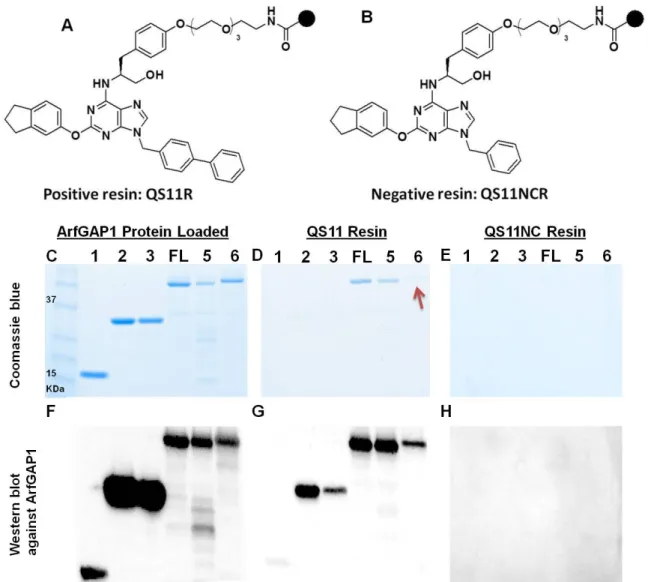
2.3.2 Small molecule pulldown assay and western blot ... 47
2.3.3 Measurement of binding affinities by SPR ... 48
2.3.4 Liposome ... 48
2.3.5 Radio active GTP hydrolysis assay ... 49
2.3.6 Secondary structure measurements by circular dichroism ... 49
2.3.7 Golgi localization of ARFGAP1 in the presence of QS11 ... 50
2.3.8 Chemical synthesis ... 50
xi
2.3.10 NMR and mass analysis ... 51
2.4CONCLUSION ... 60
2.5FUTURE PLAN ... 61
Chapter 3. High Throughput Fluorescence Polarization Assay for the Enzymatic Activity of ARFGAP ... 63
3.1INTRODUCTION ... 63
3.2RESULTS AND DISCUSSION ... 65
3.2.1 Development of a high throughput fluorescence polarization assay for the enzymatic activity of ARFGAP ... 65
3.2.2 Scope of the fluorescence polarization assay ... 70
3.2.3 Assay development towards high throughput screening ... 72
3.2.4 Screen of the Prestwick and LOPAC1280 Collection ... 74
3.2.5 Screen of 5, 000 kinase inhibitors ... 75
3.3EXPERIMENTAL SECTION ... 80
3.3.1 Expression and purification of ARF1 and ARFGAPs ... 80
3.3.2 Native gel assay ... 80
3.3.3 Fluorescence polarization assay ... 81
3.3.4 Enzyme kinetics ... 81
3.3.5 Screening of the Prestwick chemical library ... 81
3.3.6 Screening of the LOPAC1280 chemical library ... 82
3.3.7 Screening of 5, 000 Kinase Inhibitors ... 83
3.3.8 Validation of hit compounds in radio active GTP hydrolysis assays ... 83
3.3.9 ITC binding assays ... 83
3.3.10 SPR Analysis ... 83
3.4CONCLUSION ... 84
3.5FUTURE PLAN ... 85
xii
4.1INTRODUCTION ... 86
4.2RESULTS AND DISCUSSION ... 89
4.2.1 Modified myristic acid ... 89
4.2.2 Characterization of novel ARFs with modified myristic acids ... 90
4.2.3 Fluorophore labeling assays ... 95
4.3EXPERIMENTAL SECTION ... 97
4.3.1 Expression and purification of modified myristoylated ARFs ... 97
4.3.2 GTP loading and GTP hydrolysis in intrinsic fluorescence assay ... 97
4.3.3 In Vitro fluorescence labeling... 98
4.3.4 Sample preparation for mass spectra ... 98
4.4CONCLUSION ... 99
4.5FUTURE PLAN ... 100
APPENDIX ... 101
xiii List of Tables
xiv List of Schemes
xv List of Figures
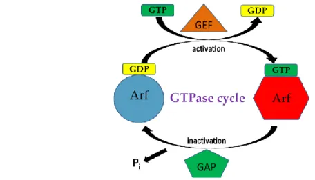
Figure 1.1. General regulation of ARF activation and inactivation. ... 1
Figure 1.2. The common hydrophobic area of the ARF family proteins ... 2
Figure 1.3. Domain organization of human ARFGAP subfamilies and structure of GAP domain of the ARFGAP1... 5
Figure 1.4. Selected ARFGAPs protein complexes involved in receptor trafficking, cell migration and invasion... 6
Figure 1.5. GTP hydrolysis and COP dynamics: a complex issue ... 10
Figure 1.6. Scheme of the dual role of ARFGAPs ... 11
Figure. 1.7. Model for the role of the PH domain in autoinhibition of ASAP1 GAP activity ... 12
Figure 1.8. Disease relevance of ARFGAP family proteins ... 13
Figure 1.9. Overall structure and superimpositions... 16
Figure 2.1. ARFGAP1 contains one GAP domain and two ALPS motifs. ... 21
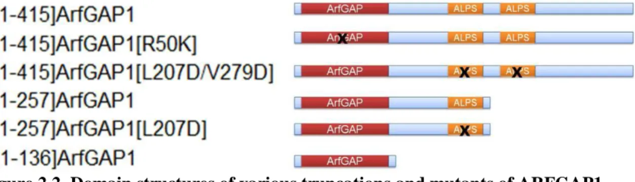
Figure 2.2. Domain structures of various truncations and mutants of ARFGAP1. .. 22
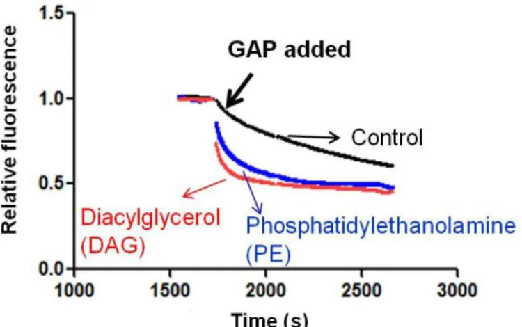
Figure 2.3. Stimulation of ARFGAP1 activity by DAG or PE ... 24
Figure 2.4. Inhibition of 1-415ARFGAP1 by QS11 ... 24
Figure 2.5. No inhibition of [1-136]ARFGAP1 by QS11. ... 25
Figure 2.6. Binding affinity of [1-136]ARFGAP1 to QS11 as measured by isothermal titration calorimetry (ITC). ... 25
Figure 2.7. ARFGAP1 pull-down experiments with small molecule matrices. ... 28
Figure 2.8. Binding affinities between different ARFGAP1 proteins and QS11 as measured by surface plasmon resonance (SPR). ... 30
xvi
proteins by QS11 ... 32
Figure 2.11. Non-competitive inhibition of ARFGAP1 by QS11 to ARF-GTP binding sites. ... 33
Figure 2.12. Far-UV CD spectra of [1-415]ARFGAP1 with liposome or with both liposome and QS11 ... 35
Figure 2.13. Effects of QS11 in localization of ARFGAP1 ... 36
Figure 2.14. Chemical structure of QS11 ... 39
Figure 2.15. Synthetic schemes for the focused library of 2,6,9-trisubstituted purines ... 39
Figure 2.16. Building blocks selected for analog synthesis ... 40
Figure 2.17. Chemical structures of 31 analogs ... 41
Figure 2.18. IC50 curve of analog-B0C1 ... 44
Figure 3.1. Schematic illustration of the assay. ... 66
Figure 3.2. GTPase-activating protein (GAP) domain of ARFGAP1 effectively catalyzes the conversion of ARF1-GTP to ARF1-GDP. ... 67
Figure 3.3. Time course reaction of ARF-GTP hydrolysis in FP assay. ... 67
Figure 3.4. The ARFGAP1-catalyzed GTP hydrolysis with different concentrations of ARF1-GTP ... 69
Figure 3.5. Standard curve for guanosine triphosphate (GTP) hydrolysis. ... 70
Figure 3.6. Highly purified human His6-[325-724]ASAP1 and human GST-[1-163]SMAP2 ... 71
Figure 3.7. Fluorescence polarization assay monitoring the GTPase-activating protein (GAP) activity of SMAP2 and ASAP1. ... 72
Figure 3.8. Assay development toward high-throughput screen. ... 73
xvii
Figure 3.10. Pilot screen of the Prestwick library ... 74
Figure 3.11. Pilot screen of the LOPAC1280 library. ... 75
Figure 3.12. Screen of 5, 000 kinase inhibitors. ... 76
Figure 3.13. Chemical structures and IC50 of E13, I3 and L3... 78
Figure 3.14. Effecs of E13, I3, L3 and DMSO on GAP activity of ARF-GTP hydrolysis. ... 79
Figure 4.1. Mass spectra of full length Keto-ARF1. ... 91
Figure 4.2. Mass spectra of full-length myristoylated ARF1. ... 92
Figure 4.3. GTP exchange assays of ARFs. ... 93
Figure 4.4. GTP hydrolysis on ARFs catalyzed by ARFGAP1. ... 94
xviii
List of Abbreviations
ARFGAP – ADP-ribosylation factors directed GTPase activating proteins
ALPS – ARFGAP1 lipid packing sensor
SAR – Structure-activity relationship
ARF – ADP-ribosylation factor
GEF – Guanine nucleotide-exchange factor
GAP – GTPase-activating protein
ER – Endoplasmic reticulum
CHA – Common hydrophobic area
CB – Calm binding Domain
CALM – Clathrin-box
FG – Phenylalanine-glycine repeats
SHD – Spa-homology domain
CC – coiled-coil
PBS – Paxillin binding sites
BAR – Bin/Amphiphysin/Rvs
PH – Pleckstrin homology
xix PxxP – Proline-rich
D/ELPPKP – Tandem Proline rich
SH3 – Src homology 3
GLD – GTP-binding protein-like domain
SAM – Sterile α-motif
RA – Ras association motif
COPI – Coat protein complex I
TfR – Transferrin receptor
PLC-γ – Phospholipase C gamma
PIP2 – Phosphatidylinositol 4,5-bisphosphate
IP3 – Inositol 1,4,5-trisphosphate
PI3K – Phosphatidylinositol 3-kinases
SHIP2 – Phosphatidylinositol 5-phosphatase
PAK – P21-activated kinase
PIX – PAK-interacting exchange factor
GPCR – G-protein-coupled receptor
xx MHC – Histocompatibility complex
DAG – Diacylglycerol
PIP3 – Phosphatidylinositol 3,4,5-trisphosphate
PE – Phosphatidylethanolamine
PS – Phosphatidylserine
PI – Phosphatidylinositol
PC – Phosphatidylcholine
ITC – Isothermal Titration Calorimetry
SPR – Surface plasmon resonance
CD – Circular Dichroism
HTS – High throughput screening
FP – Fluorescence polarization
SD – Standard deviation
LOPAC – Library of Pharmacologically-Active Compounds
SMAP2 – Stromal membrane-associated GTPase-activating protein 2
NMT – N-myristoyl transferase
xxi
LC-MS/MS – Liquid chromatography-tandem mass spectrometry
PBS – Phosphate buffered saline
SDS-PAGE – Sodium dodecyl sulfate polyacrylamide gel electrophoresis
DMF – Dimethylformamide
RFU – Relative fluorescence units
DIAD – Diisopropyl azodicarboxylate
NMR – Nuclear magnetic resonance
Log P – The logarithm of the ratio of the concentrations of the un-ionized solute in the solvents
Chapter 1.
GTPase-activating Protein of ADP-ribosylation Factor (ARFGAP)
1.1 Introduction to ARFGAP
1.1.1 ARF family proteins ARF family proteins are small GTPases that regulate membrane traffic and organelle structures.(1,2) They function through cycling between active GTP-bound forms and inactive GDP-bound forms (Fig. 1.1).(3) The activation of ARF-GDP is promoted by guanine nucleotide-exchange factors (GEFs) whereas the hydrolysis of ARF-GTP is catalyzed by GTPase-activating proteins (GAP). (1,4-6) Unlike other GTPases within the Ras super family, the nucleotide exchange rates and the intrinsic GTP hydrolysis rates for ARFs are slow. (7-10) Consequently, ARFGEFs and ARFGAPs are essential for the regulation of ARF activity.
2
exchange catalyzed by a GEF. The GTP molecule bound to ARF is then hydrolyzed to GDP with the aid of a GAP.
Six conserved members of ARFs have been identified in mammalian cells. They are classified into three subfamilies based on structure similarities: Class I (ARF1, ARF2 and ARF3), Class II (ARF4 and ARF5) and Class III (ARF6). ARFs localize both on the lipid membranes and in the cytosol.(11) ARF1 and ARF3 bind to the plasma membrane when in GTP-bound form and are released to cytosol when in GDP-bound form. ARF4, ARF5 and ARF6 bind to the plasma membrane when in either GTP- or GDP-bound form.(12,13) The terminal amphipathic helix of ARFs and the myristoylation at the N-terminus are critical elements for their membrane binding (Fig. 1.2).(10,14)
ARF1 and ARF6 have been extensively studied among the six ARFs. ARF1 regulates vesicle trafficking from endoplasmic reticulum (ER) to Golgi, as well as function and morphology of Golgi.(15) ARF6 is involved in endocytosis, phagocytosis, receptor recycling and actin-cytoskeletal remodeling.(14,16) Importantly, overexpression of ARF6 has been found in multiple invasive breast cancer cells.(17-19) Knock-down of the level of ARF6 effectively reduced tumor invasion in these breast cancer cells. (17)
3
region in colors (switch 1 part, in blue; interswitch part, in green; switch 2 part, in red). A detailed view (right) of the CHA region is shown in ribbons, with the residues forming this region indicated in sticks. (This figure was reprinted from (4). Chavrier, P., and Ménétrey, J. (2010) Structure 18, 1552-1558 © 2010 Elsevier Science, used with permission).
In humans, 15 ARFGEFs have been discovered and they share a conserved catalytic Sec7 domain of approximately 200 amino acids,(5) while 31 discovered human ARFGAPs are categorized into 10 subfamilies based on the sequence similarity of GAP and other functional domains (Fig 1.3).(20) All ARFGAPs share a catalytic GAP domain of approximately 130 amino acids in which a characteristic zinc finger motif (CX2CX16CX2CX4R) and an arginine residue are highly conserved.(21)
4
5
6
1.2 Roles of ARFGAPs in Cellular Processes
ARFGAPs are primarily considered as negative regulators of ARFs before numerous studies have shown that most ARFGAPs also served as effectors for other proteins and lipids due to their multi-domain structures.(1,2,4,29-32) ARFGAPs containing catalytic domains other than ARFGAP could also regulate other protein family through their enzymatic activity. For instance, ARAPs containing Rho GAP domains catalyze the hydrolysis of GTP that bind to the RhoA GTPase. (28)
ARFGAPs have a variety of interacting partners due to their multi-domain structures. The functions of interacting proteins dictate the roles of ARFGAPs for membrane traffic, cellular signaling and cytoskeleton reorganization (Fig 1.4).
7
colored in green. (This figure was reprinted from from (33). Inoue, H., and Randazzo, P. A. (2007) Traffic 8, 1465-1475. © 2007 Blackwell Publishing Ltd, used with permission) 1.2.1 Membrane traffic Several ARFGAPs are involved in regulating membrane traffic. For example, ERD2 is a transmembrane receptor that mediates retrograde transport of ER-resident proteins from the Golgi to the ER. ARFGAP1 binds to p24 cargo proteins and to ERD2 to regulate cargo sorting.(34-36) ARFGAP1 was also indicated to bind to coatomer and clathrin AP1 to control membrane traffic.(37,38) The binding to the coatmer was reported to stimulate the activity of ARFGAP1 by 10-1,000 fold. (21) (39)Another ARFGAP, ACAP1, binds to transferrin receptor (TfR), cellubrevin, and integrin-β1 to serve as novel coat or adaptor protein in the recycling compartments.(40,41) In addition, SMAP proteins interact with clathrin to drive the formation of transport intermediates from both the plasma membrane and the trans Golgi network.(42,43) Furthermore, AGAP1 and AGAP2 interact with clathrin adaptor proteins, AP3 and AP1, respectively, to regulate the endocytic compartments.(34) Finally, ASAP1 interacts with CIN85 to accelerate the recycling of EGF and EGFR,(44) and also coordinates with POB1 and RalBP to regulate actin cytoskeleton and membrane traffic.(45,46) ASAP2 has also been shown to bind to the SH3 domain of amphiphysin IIm to function in synaptic vesicle endocytosis.(47) It was suggested that the ARFGAPs function as a subunit of a vesicle coat protein similar to the role of Sec23 in ER to Golgi transport mediated by COPII vesicle coats.(48)
1.2.2 Cellular signaling GIT1, AGAP2 and ARAP3 interact with related enzymes to control the levels of important phosphoinositol lipids in cellular signaling. GIT1 binds to phospholipase C gamma (PLC-γ), which catalyzes the hydrolysis of PIP2
8
could prevent neuronal apoptosis.(47,49,50) ARAP3 binds to phosphatidylinositol 5-phosphatase (SHIP2) to negatively regulate PI3K signaling.(51) In addition, GIT1 interacts with Rac1, Cdc42, p21-activated kinase (PAK), PAK-interacting exchange factor (PIX), MEK1, and paxillin.(52) The interaction between GIT1 and PAK are shown to regulate cytoskeletal dynamics by inhibiting Rac1 and Cdc42. GRK2 recruits and binds to both GIT1 and GIT2 to mediate internalization of the G-protein-coupled receptor (GPCR).(52,53) Fyn, a Src family kinase, phosphorylates AGAP2 and prevents degradation of AGAP2 during programmed cell death in the anti-apoptotic signaling pathway.(54) The interaction between AGAP2 and Akt is essential to this pathway as well.(55,56) ASAP1 binds to focal adhesion kinase (FAK) to mediate the localization of paxillin and affect cell motility.(57) Src and Pyk2 bind to and phosphorylate ASAP1 to inhibit the GAP activity of ASAP1.(58) ARAP1, ARAP2 and ARAP3 containing Rho GAP domains interact with RhoA, a Rho GTPase to regulate actin and actin-associated structures.(51,59) ARAP3 also regulates peripheral actin ruffles by binding to Rap1 GTPase.(51)
9
1.3 Models of ARFGAP Functions in Cell Signaling
ARFGAP1 and ARFGAP2/3 are the simplest ARFGAPs in domain structures and studies on them form the basis for many models of the ARFGAP functions. Initially, ARFGAPs were proposed to only function as negative regulators of ARF signaling. In the prevailing model, the cycle of active ARF1 and inactive ARF1 is linked to coat association and dissociation from membranes.(2,34,64-67) Although a variety of modifications to this model have been proposed, the central hypothesis is that active ARF1-GTP is critical for recruiting coat proteins on the membrane while its hydrolysis would release the coat proteins from the membranes. In this model, the function of ARFGAP1 is to catalyze hydrolysis of ARF1-GTP to induce coat dissociation. The GTP hydrolysis is also dependent on the assembling of coat promoters into a vesicle coat.
10
Figure 1.5 GTP hydrolysis and COP dynamics: a complex issue (A) The typical G-protein activation cycle. The GEF and GAP regulate the functions of the G G-protein. (B) The assembly–disassembly cycle of the protein coats is not necessarily in phase with the GTPase cycle. The first coatbuilding unit is a recently formed 1:1 complex between Arf-GTP and a COP complex wandering at the membrane surface by lateral diffusion. The second is an older unit, which has been incorporated in the coat lattice. (This figure was reprinted from (67), Antonny, B., Bigay, J., Casella, J. F., Drin, G., Mesmin, B., and Gounon, P. (2005) Biochem Soc Trans 33, 619-622. © 2005 Biochemical Society, used with permission)
The model of membrane curvature sensing is attractive and has been extensively demonstrated in numerous experiments.(67,75,77-79) However, the model has its own limitations. First, although multiple ARFGAPs are involved in membrane traffic, the ALPS motifs are only present in ARFGAP1.(74) Therefore, expanding this model to other ARFGAPs is conceptually challenging, especially for other large ARFGAPs with additional domains. Second, ASAPs with BAR domains are not as sensitive as ARFGAP1 to curvature changing, although the primary role of BAR domain is reported to sense membrane curvature.(30,74,80,81) Third, cargo sorting is not involved in this model.(30)
11
ARFGAPs (Gcs1p, Glo3p, Age1p and Age2p) expressed from a high copy plasmid suppressed a loss-of-function allele of ARF1(2,83) suggesting that ARFGAPs function as downstream effectors of ARFs. Structure evidences suggested that both the yeast ARFGAP1 Gcs1p and the ARFGAP2/3 homologue Glo3p interact with SNARE proteins to induce the recruitment of ARF1p and coatomer to the SNAREs.(84-86) It is proposed that the formation of the primer complexes is required for vesicle transportation. In other studies, the most intriguing finding was that COPI vesicles only contain ARFGAP1 instead of complex of ARFs and ARFGAPs. In vivo data confirmed that COPI persists on membrane after the dissociation of ARF.(87) In addition, ARF was not detected in proteomic analysis of the COPI coated vesicles(66,88-91) further indicating that ARFGAP1 plays a critical role in this type of cargo sorting.
12
Randazzo, P. A. (2010) FEBS Lett 584, 2646-2651. © 2010 Federation of European Biochemical Societies, used with permission)
Figure. 1.7. Model for the role of the PH domain in autoinhibition of ASAP1 GAP activity. (1) The PH domain interacts with the GAP domain in the absence of PIP2. (2)
PIP2 binds to the PH domain, leading to the exposure of the GAP domain. (3) The GAP
domain interacts with ARF-GTP and catalyzes GTP hydrolysis. (This Figure was reprinted from (92), Randazzo, P. A., and Hirsch, D. S. (2004) Cell Signal 16, 401-413. © 2003 Elsevier Inc, used with permission)
The function of ASAP1 has been examined extensively. PIP2 and PA enhance
GAP activity approximately 10,000 fold.(93,94) It is proposed that PIP2 binds to the PH
domain and induces a conformational change in the ARFGAP domain.(93) In this model, the PH domain binds to the catalytic GAP domain and blocks the interaction of ASAP1 with ARF-GTP (Fig 1.7).(95) The binding of PIP2 to the PH domain releases the catalytic
13
1.4 Disease Relevance of ARFGAPs
ARFGAPs are involved in various diseases (Fig 1.8). For example, ASAP1 is amplified and overexpressed in uveal melanomas and in colorectal, prostate, and breast carcinomas.(16,96-99) Overexpression of ASAP1 causes increased cell motility in low-grade melanoma cells, while siRNAs against ASAP1 reduce cell migration in ASAP1-overexpessing cells.(16) Furthermore, overexpression of ASAP1 correlates with poor metastasis-free survival and prognosis in colorectal cancer patients. (100)
Figure 1.8. Disease relevance of ARFGAP family proteins
14
GIT proteins were also involved in HIV infectivity. The inactivation of ARF6 by GITs are likely to mediate downregulation of major histocompatibility complex (MHC) class I on the host cell after infection by the viral protein Nef.(105)
15
1.5 Regulation of ARFGAP Activity
The regulation mechanism of ARFGAP is not completely resolved. The catalytic mechanism of other GAPs, such as Ras and Rho, has been elucidated.(112,113) Both structural and biochemical studies lead to an “arginine finger” model for catalysis of GTP hydrolysis.(112-115) In this model, GAPs of Ras and Rho GTPases supply a critical arginine residue, which is missing in the GTPases. Mutation of the arginine residue caused a reduction of GAP activity. (112-115)
16
Figure 1.9. Overall structure and superimpositions. (A) Ribbon representation of the ARF6-GDPAlF3ASAP3 structure, with the ASAP3 GAP domain in cyan, the ankyrin
domain in blue, and ARF6 in green, and with its switch I in yellow and switch II in gray.(B) Comparison of ARF6-GDPAlFxASAP3 with ARF1-GDPARFGAP1 reported previously (118), obtained by superimposition of ARF1 with ARF6, with ARFGAP1 in dark pink, and ARF6GDP-AlF3-ASAP3 as in Figure 1.9 A, leaving out the ankyrin
repeats for clarity.(This Figure was reprinted from (117). Ismail, S. A., Vetter, I. R., Sot, B., and Wittinghofer, A. (2010) Cell 141, 812-821. © 2010 Elsevier Inc, used with permission)
1.5.2 Biochemical studies Besides the structural studies on ARFGAPs, extensive biochemical experiments have been carried out to understand other lipid-based regulators of ARFGAP activity.(8,68,75,77,81,93,94,119,120) PIP2 was shown to stimulate the
17
1.6 Peptide and Small Molecule Regulators of ARFGAPs
Currently, there are no effective small molecule inhibitors of ARFGAPs. Because cargo proteins bind to coatomer and ARFGAP to form a complex, peptides from the cytoplasmic tail of p24 cargo proteins were synthesized and found to inhibit the GAP activity of ARFGAP1 and ARFGAP2.(64,121) However, the inhibitory effect was nonspecific and coatomer independent. Later, peptides from several p24 family members p23 and p25 were also found to enhance GAP activity of ARFGAP1 and ARFGAP2.(68) These results are in conflict with the current regulator mechanism in which activation of ARFGAP occurs after vesicle assembly.
Chapter 2.
Inhibition of ARFGAP1 Activity by QS11: Mechanism of Action and Analog Synthesis
2.1 Introduction
Small molecule regulators of ARFGAPs will be useful tools to study the functions of ARFGAPs in cellular processes, yet only a peptide inhibitor of ARFGAP1 have been characterized in biochemical assays. (122) Recently, a small molecule, QS11, has been shown to synergize with the Wnt proteins to activate the Wnt/β-catenin signaling pathway. (123) In the subsequent target identification process, ARFGAP1 was identified as one of the primary targets of QS11. In the presence of QS11, both ARF1-GTP and ARF6-GTP showed accumulated levels in NIH 3T3 cells indicating that the GAP activities in these cells were inhibited. In addition, in ASAP1-overexpressing MDA-MB-231 cells, QS11 inhibited cell migration in a dose-dependent manner. Overexpression of ARFGAP1 in HEK293 cells abolished synergistic effect of QS11. These results implicate that QS11 is an inhibitor of ARFGAPs in vivo. However, whether QS11 inhibits the activities of ARFGAPs in in vitro biochemical assays and what is the detailed mechanism of this inhibition are not well understood.
(p-19
trifluoromethyl)phenyl yields more than 10-fold decrease in activity. Replacement of the aryloxy group at the C2 position with amino groups also leads to reduced activity. Furthermore, the stereochemistry of the substituent at the C6 position is important as the enantiomer of QS11 does not show synergistic activation with the Wnt proteins.
20
2.2 Results and Discussion
21
Figure 2.1. ARFGAP1 contains one GAP domain and two ALPS motifs. A. Location of the GAP domain and ALPS motifs in ARFGAP1. B. Three critical amino acids-R50, L207, and V279 are highlighted in sequences of the GAP domain and two ALPS motifs.
22
Figure 2.2. Domain structures of various truncations and mutants of ARFGAP1. We chose ARF1 as the substrate for ARFGAP1 because the crystal structure of ARF1 and GAP domain of ARFGAP1 has been solved and the detailed kinetic studies on ARFGAP1-catalyzed hydrolysis of ARF1-GTP have been carried out.(21,68) Under physiological conditions, ARF1 is myristoylated and localized at the Golgi membrane when it is GTP-bound. A truncated ARF1 with the N-terminal 17 amino acid residues deleted has also been widely used in enzymatic assays because it is soluble and technically less challenging to prepare in large quantities. Consequently, we have purified both mysritoylated ARF1 and [△17]ARF1.(21,122,128) The expression construct for myristoylated ARF1 was obtained from Dr. Paul Randazzo (National Cancer Institute) while that for soluble ARF1 was obtained from Dr. Jonathan Goldberg (Memorial Sloan-Kettering Cancer Center).
2.2.2 QS11 inhibits the activity of full-length ARFGAP1 but not that of [1-136]ARFGAP1 Varieties of phosphoinositol lipids stimulate GAP activity.(8,75,82,94,119) For instance, phosphatidylinositol 3,4,5-trisphosphate (PIP3)
23
24
Figure 2.3. Stimulation of ARFGAP1 activity by DAG or PE. Time course of [1– 415]ARFGAP1-catalyzed hydrolysis of ARF1-GTP with liposome containing phosphatidylserine (PS) (5%), phosphatidylinositol (PI) (10%), cholesterol (16%) and where indicated DAG (15%) or PE (19%). The remaining lipid is phosphatidylcholine (PC). Myristoylated ARF1-GDP (0.4 μM) was mixed with liposome (total lipid concentration, 400 μM) and exchanged to ARF1-GTP state by adding GTP (40 μM )and chelating free Mg2+ with EDTA. The concentration of Mg2+ was then adjusted to 3 mM and [1-415]ARFGAP1 (40 nM) was added to initiate the GTP hydrolysis on ARF.
25
Figure 2.5. No inhibition of [1-136]ARFGAP1 by QS11. QS11 (20 μM) or DMSO were incubated with [1-136]ARFGAP1 for 10 min. [△17]ARF1 (10 μM) was exchanged with [γ32P] GTP This mixture containing compounds (20 μM) and [1-136]ARFGAP1 (5
μM) were then added into [△17]ARF-[γ32P] GTP (10 μM) as a ratio of 1:1. The reactions were stopped, sperated and record. GAP activity in the presence of DMSO was normalized to 100%
26
2.2.3 Characterization of binding affinities between QS11 and different forms of ARFGAP1 To understand the mechanism of action, we then studied the molecular basis of the interaction between ARFGAP1 and QS11. We have generated six truncated or mutated ARFGAP1 proteins and used them for the binding studies (Figs 2.2 and 2.7). Small molecule affinity pull-down and MS analysis were used to identify ARFGAP1 as the potential target of QS11 in the previous study. (123) This pull-down approach measures the relative levels of the QS11-bound proteins in cell lysates through the small molecule matrix. In analogy, we used this method to distinguish the relative binding affinities between QS11/QS11NC and different forms of ARFGAP1 (Fig 2.7). Compared with the traditional binding assays such as SPR analysis and ITC, this pull-down experiment is more efficient since multiple proteins and small molecules can be evaluated at the same time.
27
28
29
We further validated this interaction in surface plasmon resonance (SPR) assays. Purified full-length ARFGAP1, ARFGAP1[R50K], [1-136]ARFGAP1, [1-257]ARFGAP1, [1-257]ARFGAP1[L207D] or [1-415]ARFGAP1[L207D/V279D] was covalently immobilized to a CM5 chip surface. The control channel was treated in the same way but without protein immobilization. Ethanolamine was subsequently injected to block the unreacted surface. QS11 was then injected for at increasing concentrations (0, 156, 312, 625, 1,250, 2,500, 5,000, 8,000 and 10,000 nM) in HBS-EP buffer and the dissociation of ARFGAP1-QS11 complex was followed for 10 min. SPR analysis afforded Kd values of 1.3 ± 0.3 μM for both full-length ARFGAP1 and ARFGAP1[R50K]
30
Figure 2.8. Binding affinities between different ARFGAP1 proteins and QS11 as measured by surface plasmon resonance (SPR). The proteins were immobilized on surfaces of CM5 chips. Equilibrium responses of the binding of QS11 to ARFGAP1 proteins were plotted against concentrations of QS11. The data were fitted using a 1:1 binding model to calculate the binding constant Kd.
2.2.4 QS11 inhibited GAP activity of ARFGAP1 To quantify the inhibition of GAP activity by QS11, we first compared the relative capacity of the six ARFGAP1 proteins as described in the binding experiments in catalyzing the hydrolysis of ARF1-GTP. We used [γ32P] GTP hydrolysis assay to detect the loss of [γ32P] GTP that was bound to myristoylated ARF1 in liposome, which can indirectly report the GAP activity. Myristoylated ARF1 was activated in the presence of GTP and [γ32P] GTP, EDTA and liposome. Then, MgCl2 was added to stabilize myristoylated ARF-[γ32P] GTP. The GTP
31
ARFGAP1[L207D/V279D] dramatically reduced the GAP activity probably due to its poor affinity to the liposome, which was also observed in another reported intrinsic fluorescence ARF-GTP hydrolysis assay.(77) In addition, [1-257]ARFGAP1 showed a slightly reduced GAP activity compared to full-length ARFGAP1(77) while [1-257]ARFGAP1[L207D] dramatically reduced the GAP activity due to the reduced interactions with liposome. Finally, the catalytic domain of ARFGAP1 did not exhibit good activity under current reaction conditions, possibly due to its weak interaction with liposome.
Figure 2.9. Activities of different forms of ARFGAP1. A. Determination of GAP activity of ARFGAP1 on GTP hydrolysis. ARFGAP1 was titrated into a reaction containing myristoylated ARF-[γ32P] GTP and measured as described under “Experimental Procedures”
Next, dose-dependent inhibition of the GAP activity of each ARFGAP1 was evaluated under conditions that the rate of GTP hydrolysis was within the linear range (Fig. 2.10). The IC50 for inhibiting the GAP activity of full-length ARFGAP1 by QS11
32
ARF1-GTP varied. QS11 inhibited the GAP activity of full length ARFGAP1 across a wide range of concentrations of ARF-GTP (Fig 2.11) indicating that QS11 likely inhibited ARFGAP1 non-competitively with ARF1-GTP.
33
Figure 2.11. Non-competitive inhibition of ARFGAP1 by QS11 to ARF-GTP binding sites. QS11 was at the IC50 concentration (4.0 μM) while the concentrations of
substrate ARF1-[γ32P] GTP varied from 100 nM to 6.4 μM.
-7.5 -7.0 -6.5 -6.0 -5.5 -5.0 -4.5 0
20 40 60 80 100
Log ARF-GTP [M]
%
I
n
h
ib
it
io
34
2.2.5 Investigation of secondary structures of ARFGAP1 The ALPS motifs in ARFGAP1 are random coils in solution and rearrange to α-helix structures when they interact with lipid membranes.(67,77,120) The Circular Dichroism (CD) spectra showed that adding liposome to the full length ARFGAP1 led to increased absorbance at 208 and 222 nm. More detailed analysis indicated that the major contribution of this increase in absorbance was from the ALPS motifs, especially the first ALPS motif.(77) Mutations in the ALPS motifs dramatically decreased or disrupted the increase of the α-helix structures.
35
36
2.2.6 Localization of ARFGAP1 in the presence of QS11 The ALPS motifs are required for proper localization of ARFGAP1 in Golgi membrane(77,129,130): ARFGAP1 majorly localize in Golgi membrane(76,129,130) while mutations in the ALPS motifs cause diffused localization of ARFGAP1.(77,129) Consequently, QS11 should disrupt the Golgi localization of ARFGAP1 to regulate its catalytic action on hydrolysis of ARF1-GTP. Dr. Juyoun Beak in our lab thus transfected NIH3T3 cells with YFP-ARFGAP1 and treated the resulting cells with QS11 or QS11NC. Preliminary data indeed suggested that the Golgi structures were disrupted upon QS11 treatment (Fig 2.13). We are optimizing the condition and testing the effects of QS11 in these cells.
37
2.2.7 Working model on how QS11 inhibits the GAP activity of ARFGAP1 Based on our data, we propose a working model where QS11 regulates the activity of ARFGAP1 through hydrophobic interaction with the ALPS motifs which disrupts the Golgi localization of ARFGAP1 in the endogenous systems (Scheme 2.1). QS11 non competitively inhibites ARFGAP1 activity through the ALPS motifs. Consistent with this model, QS11 is a highly hydrophobic compound partly due to the biphenyl rings. In addition, four hydrophobic amino acids in the second ALPS region of ARFGAP1 are essential for the interaction of ARFGAP1 with lipid membrane and subsequent formation
of α-helix structures by non-structured ALPS motifs.
38
39 Figure 2.14. Chemical structure of QS11
40
41 Figure 2.17. Chemical structures of 31 analogs
N N N N O O O B0C1 N N N N O B0C2 N N N N O B0C3
HN OH HN OH HN OH
N N N
N O F3C
B0C4 HN OH Total: 31 QS11 analogs
N N N N O O B0C5 HN OH N N N N O HN H N O N N N N O O O HN H N O B1C0 B1C1 N N N N O HN H N O B1C2 O O O N N N N O O HN H N O B1C5 O N N N N HN O B2C0 N N N N HN O O O B2C1 N N N N HN O B2C2 N N N N HN O B2C3 N N N N HN O F3C
B2C4 N N N N HN O O B2C5 N N N N HN O B3C0 N N N N HN O O O B3C1 N N N N HN O B3C2 N N N N HN O B3C3 N N N N HN O F3C
42
43
2.2.9 Inhibition studies of QS11 analogs The synthetic analogs were first evaluated in ARF-[γ32P] GTP hydrolysis assays where their capacities to inhibit the catalytic activity of the full-length ARFGAP1 were tested. Myristoylated ARF1 was loaded with [γ32P] GTP in the presence of EDTA and liposome. Then, MgCl2 was added
to quench the nucleotide exchange reaction. All analogs were first dissolved in DMSO to make a stock solution at 10 mM and then diluted into aqueous solution to make the final concentration at 20 μM. In a representative reaction, one QS11 analog was pre-incubated with ARFGAP1 (16 nM) at room temperature for 10 min and the resulting mixture was added to myristoylated ARF-[γ32P] GTP in liposome to initiate the hydrolysis. As fixeded time, the reaction was stopped, free GTP and phosphate were separated and the remaining membrane-bound [γ32P]ATP was measured by scintillation counting (Table 2.1).
Replacing the biphenyl group in QS11 with a phenyl group (QS11NC) dramatically abolished the inhibition. This is consistent with the fact that the removal of one of the benzene groups of QS11 abolished the synergist effect of QS11 in the Wnt reporter assays.(123) Replacing the 5-methyl-2,3-dihydro-1H-indene moiety (QS11) with 5-methylbenzo[d][1,3]dioxole (B0C1) did not affect the inhibition effect (IC50 of B0C1 is
44 Figure 2.18 IC50 curve of analog-B0C1
The water solubility of QS11 is a concern when the compound is used in aqueous solution. QS11 begins to precipitate beyond 10 μM in aqueous solution. It was shown that when Kd and IC50/EC50 of the compound is around low micromolar or high
nanomolar range, in a number of biochemical and biophysical assays, the concentrations of small molecules need to go beyond 10 μM to get sufficient signal for accurate activity measurement.(131-133) It is well known that PEGylation can provide water solubility to hydrophobic drugs and proteins.(134-137) In order to improve the water solubility of QS11, PEG like structure, 2-(2-aminoethylamino)ethanol (B1) was utilized to modify the C6 position of QS11. This set of four analogs (B1C0, B1C1, B1C2, and B1C5) indeed showed a better solubility in assay buffer (50 mM Hepes pH 7.2, 120 mM KAc, 3 mM MgCl2, 2 mM EDTA, 1 mM DTT). No precipitations were observed when the
45
Substitution of the C6 position of QS11 with naphthalen-1-ylmethanamine (B2C0) significantly decreased the inhibition of the molecule (Table 2.1). The bulky naphthalene moiety is not favored at this position. Interestingly, the analog B2C3, with naphthalene groups on both C2 and C6 positions, showed moderate inhibition. It is not clear whether the moderate inhibition is due to the increased steric effects or aromatic effects provided by the substitutions at the C6 position of QS11.
The removal of the methoxy group in amine part (B3C0) dramatically decreased the inhibition effect (Table 2.1). This hydroxyl group is likely to be important to form a hydrogen bond with ARFGAP1 for its activity. This hypothesis was further confirmed by replacing the hydroxyl group with a methyl formate group in B5C0. This analog B5C0 and hydroxyl-free analog B3C0 showed similar decreased activity against ARFGAP1. Surprisingly, by replacing the cyclopentane moiety (C0) with 1,3-dioxolane (C1), the new analog B5C1 almost rescued the inhibition of this molecule. The two oxygen atoms in B5C1 are suspected to form hydrogen bonds with the ARFGAP1 which contribute to the inhibition.
The benzene group in the amine part is necessary for maintaining the inhibition of this molecule. After removal of this benzene group, all the analogs (B4C0, B4C1, B4C2, B4C3, and B4C5) showed dramatically decreased inhibition of ARFGAP1.
46
QS11NC lacking one benzene ring in the biphenyl group did not bind to ARFGAP1. This is consistent with previous results that QS11NC did not inhibit the GAP activity in the in vitro GTP hydrolysis assays, did not increase ARF-GTP levels in NIH 3T3 cells and did not activate the Wnt signaling pathway.
47
2.3 Experimental Section
2.3.1 Expression and purification of ARFs and ARFGAPs Rat ARFGAP1 and human myristoylated ARF1 are used in this work. Full-length ARFGAP1 and its R50K mutant are expressed in SF9 cells by Dr. Richard Premont (Duke Univesity) (125,138). The other constructs are expressed in bacterial systems.The expression constructs of [1-257]ARFGAP1, [1-257]ARFGAP1[L207D] and [1-415]ARFGAP1[L207D/V279D] are from Dr. Bruno Antonny (Institut de Pharmacologie Moleculaire et Cellulaire). (77,79) The expression constructs of [△17]ARF1 and [1-136]ARFGAP1 are from Dr. Jonathan Goldberg (Memorial Sloan-Kettering Cancer Center). (21) The single colonies of mysritoylated ARF1 are from Dr. Paul Randazzo (National Cancer Institute). (139,140) All the proteins were expressed and purified according to literature protocols.(21,77,125,139)
48
2.3.3 Measurement of binding affinities by SPR BIACore 3000 instrument (BIACore, GE) was used in this study. The CM5 sensor chip was activated by running through a 1:1 mixture of N-hydroxysuccinimide (0.05 M) and 1-ethyl-3-(3-dimethylaminopropyl) carbodiimide (0.2 M) at 10 μl/min for 7 min. Purified full-length ARFGAP1, ARFGAP1[R50K], [1-136]ARFGAP1, [1-257]ARFGAP1, or 257]ARFGAP1[L207D] [100 μg/ml in 10 mM KAc (pH 5.0)] or [1-415]ARFGAP1[L207D/V279D] [100 μg/ml in 10 mM KAc (pH 4.0)] was then injected for 10 min at 10 μl/min to covalently immobilize it to the CM5 chip surface. The control channel was treated in the same way but without protein immobilized. Ethanolamine (1.0 M, pH 8.5) was injected for 7 min to block the unreacted surface. The CM5 chip surface was equilibrated in HBS-EP buffer (0.01 M Hepes, 0.15 M NaCl, 3 mM EDTA, and 0.005% surfactant P20, pH 7.4). QS11 was then injected for 3 min at increasing concentrations (0, 156, 312, 625, 1,250, 2,500, 5,000, 8,000 and 10,000 nM) in HBS-EP buffer with a flow rate of 20 μl/min, and dissociation of ARFGAP1-QS11 complexes was followed for 10 min. The surface was regenerated with 10 mM glycine (pH 2.5). Data from at least two independent titration experiments were averaged. Data in the control channel were subtracted from that in the corresponding protein channel. Data resulted from DMSO injection were further subtracted from those derived from QS11 injections. The data were analyzed by fitting into a one-site specific binding mode to calculate the binding affinities using Graphpad Prism 5.
49
argon steam for 1 h and followed by drying under vacuum for 1 h. The film was resuspended in buffer [50 mM Hepes (pH 7.5), 120 mM KAc, 3 mM MgCl2, 2 mM
EDTA and 1 mM DTT] and hydrated at for 0.5 h. (77,79) The mixtures were freezed and thawed in ethanol/dry ice and warm water batch five times, and then extrude through (pore size) 0.03 µm polycarbonate filters using a hand extruder (Avanti). (Note: mole percent is the ratio of the moles of a substance in a mixture to the moles of the mixture).
2.3.5 Radio active GTP hydrolysis assay Myristoylated ARF1 (800 nM) is activated in buffer [50 mM Hepes (pH 7.5), 120 mM KAc, 1 mM MgCl2, 1 mM DTT, 2.4
μM GTP, 800 nM [γ32
P] GTP (specific activity = 6, 000-3, 000 Ci/mmol), 2 μM EDTA and 0.2 mM liposome] for 40 min at room temperature. Then, 2 mM MgCl2 was added to
stabilize myristoylated ARF-GTP. The so-formed myristoylated ARF-GTP is stable on ice for up to two days. The GTP hydrolysis was initiated by adding ARFGAP1, stopped by diluting with ice cold buffer [50 mM Hepes (pH 7.5), 120 mM KAc, 3 mM MgCl2, 3
mM EDTA, 1 mM DTT), and separated by filtration through BA 85 with pore size at 0.45 µm (Millipore). The radioactivity of the membrane-bound [γ32P] GTP was measured by a scintillation counter. For IC50 measurement, ARFGAP1 was pretreated with QS11 or
DMSO in buffer [50 mM Hepes (pH 7.5), 120 mM KAc, 3 mM MgCl2, 3 mM EDTA, 1
mM DTT, 50 μg/ml BSA). After incubation at room temperature for 10 min, ARFGAP1 and QS11 mixture was added to myristoylated ARF-GTP at a 1:1 ratio to initiate the hydrolysis. The reactions were incubated at indicated time, stopped and analyzed as described above.
50
Photophysics). The proteins were dialyzed against buffer [10 mM Tris pH 7.5, 150 mM KCl and 1 mM DTT] at 4 °C overnight. The experiments were performed at room temperature in a HELLMA quartz cell with an optical path length of 0.1 cm. Each spectrum was recorded from 200 to 260 nm with a bandwidth of 1 nm and a speed of 1.25 s per point. QS11 (10 μM) was added to a mixture of ARFGAP1 and liposome and the spectrum was recorded after incubation for 10 min. Control spectra of liposome or QS11 in buffer were subtracted from the protein spectra.
2.3.7 Golgi localization of ARFGAP1 in the presence of QS11 The procedures were similar as previously described (18). Briefly, GFP-[1-415]ARFGAP1 was transfected into NIH3T3 cells. The cells were cultured at 37 oC with 5% CO2 for 24 h
before QS11 (2 μM) or QS11NC (2 μM) were added. After another 24 h, the cells were fixed for confocal microscopy.
2.3.8 Chemical synthesis DIAD (2.58 ml, 26.25 mmol) was added dropwise to a mixture of biphenyl-4-ylmethanol (2.42 g, 26.25 mmol ), 2,6-dichloro-9H-purine (2.36 g, 25 mmol) and PPh3 (3.36 g, 26.25 mmol) in THF (100 ml). The resulting mixture was
heated to 75 °C (oil bath temperature) for overnight before it was cooled and diluted with CHCl3 (50 ml). The layers were separated and the organic fraction was washed with H2O
and brine, dried over Na2SO4, and concentrated under vacuum. The residue was purified
by flash column chromatography over silica gel to give 9-[(1,1'-biphenyl)-4-ylmethyl]-2,6-dichloro-9H-purine (5.68 g, 16 mmol, 64%).
51
room temperature. After stirring at 80 °C overnight, the reaction mixture was cooled and diluted with CHCl3 (10 ml). The layers were separated and the organic fraction was
washed with H2O and brine, dried over Na2SO4, and concentrated under vacuum. The
residue was purified by flash column chromatography over silica gel to give compound 3. Next, compound 3 (0.05 mmol) was mixted with commercial available phenol derivatives (0.18 mmol) in the presence of tris(dibenzylideneacetone)dipalladium (2.3 mg, 0.002 mmol) and di-tert-butyl(phenyl)phosphine (2 mg, 0.0045 mmol) under reflux conditions for 2 h. The reaction mixture was diluted with CHCl3 (100 ml) and the layers
were separated. The organic layer was washed with H2O and brine, dried over Na2SO4
and concentrated under vacuum. The residue was purified by flash column chromatography over silica gel to generate the final analog 4.
2.3.9 Calculation of ClogP Chemdraw software was employed to estimate the ClogP value of each analog.
52
9-(biphenyl-4-ylmethyl)-2,6-dichloro-9H-purine (2): (5.68 g, 16 mmol, 64%). 1H NMR (400 MHz, CDCl3) δ 8.09 (s, 1H), 7.62-7.57 (m, 4H), 7.55-7.38 (m, 5H), 5.45 (s, 2H).
(S)-2-(9-(biphenyl-4-ylmethyl)-2-chloro-9H-purin-6-ylamino)-3-phenylpropan-1-ol (B0): (144.75 mg, 0.31 mmol, 55%). 1H NMR (400 MHz, CDCl3) δ 7.64-7.54 (m, 5H),
7.44-7.18 (m, 10H), 7.00-6.97 (m, 1H), 5.38-5.28 (m, 2H), 4.58 (br. s, 1H), 3.90-3.88 (m, 1H), 3.74-3.71 (m, 1H), 3.06-2.96 (m, 2H).
2-(2-(9-(biphenyl-4-ylmethyl)-2-chloro-9H-purin-6-ylamino)ethylamino)ethyl
acetate (B1): (97.44 mg, 0.21 mmol, 36%). 1H NMR (400 MHz, CDCl3) δ 7.89-7.84 (m,
1H), 7.56-7.54 (m, 4H), 7.44-7.28 (m, 5H), 5.35 (s, 2H), 4.25 (s, 2H), 3.79 (s, 2H), 3.68-3.65 (m, 4H), 2.63 (s, 1H), 2.19-2.06 (m, 4H), 2.01 (m, 3H), 1.26 (m, 2H).
9-(biphenyl-4-ylmethyl)-2-chloro-N-(naphthalen-1-ylmethyl)-9H-purin-6-amine (B2): (261.25 mg, 0.55 mmol, 99%). 1H NMR (400 MHz, CDCl3) δ 8.08-8.07 (m, 1H),
7.85-7.82 (m, 2H), 7.56-7.27 (m, 14H), 6.62 (br. s, 1H), 5.28-5.22 (m, 2H).
9-(biphenyl-4-ylmethyl)-2-chloro-N-phenethyl-9H-purin-6-amine (B3): (237.06 mg, 0.54 mmol, 97%). 1H NMR (400 MHz, CDCl3) δ 7.59-7.55 (m, 5H), 7.46-7.42 (m, 2H),
7.38-7.22 (m, 9H), 6.01 (br. s, 1H), 5.35 (s, 2H), 3.91 (br. s, 1H), 3.01-2.97 (m, 2H). (S)-2-(9-(biphenyl-4-ylmethyl)-2-chloro-9H-purin-6-ylamino)butan-1-ol (B4): (219.78 mg, 0.54 mmol, 96%). 1H NMR (400 MHz, CDCl3) δ 7.66-7.54 (m, 4H),
7.45-7.41 (m, 2H), 7.37-7.33 (m, 3H), 7.07 (br. s, 1H), 5.39-5.23 (m, 2H), 4.33 (br. s, 1H), 4.00-3.96(m, 1H), 3.75-3.71 (m, 1H), 1.76-1.62 (m, 2H), 1.01-0.97 (m, 3H).
53
7.70 (s, 1H), 7.59-7.55 (m, 4H), 7.46-7.41 (m, 2H), 7.37-7.34 (m, 3H), 7.30-7.15 (m, 7H), 6.39 (br. s, 1H), 5.34 (s, 2H), 4.12-4.09 (m, 1H), 3.74 (s, 3H), 3.32-3.22 (m, 2H).
(S)-2-(2-(benzo[d ][1,3]dioxol-5-yloxy)-9-(biphenyl-4-ylmethyl)-9H-purin-6-ylamino)-3-phenylpropan-1-ol (B0C1): (26.28 mg, 0.046 mmol, 92%). 1H NMR (400 MHz, CDCl3) δ 7.60-7.54 (m, 5H), 7.43 (t, J = 7.4 Hz, 2H), 7.38-7.32 (m, 3H), 7.11-7.22 (m,
3H), 7.01 (s, 1H), 6.90 (s, 1H), 6.81-6.83 (d, J = 8.0 Hz, 1H), 6.74 (s, 1H), 6.88 (dd, J = 8.0, 4.0 Hz, 1H), 5.95 (d, J = 20 Hz, 2H), 5.25 (t, J = 18 Hz, 2H), 4.31 (br. s, 1H), 3.78-3.59 (m, 2H), 2.96 (dd, J = 14.0, 6.0 Hz, 1H). MALDI-MS: m/z 572.2 (M + H)+.
(S)-2-(9-(biphenyl-4-ylmethyl)-2-(naphthalen-2-yloxy)-9H-purin-6-ylamino)-3-phenylpropan-1-ol (B0C2): (25.98 mg, 0.045 mmol, 90%).1H NMR (400 MHz, CDCl3)
δ 7.87-7.80 (m, 3H), 7.67 (s, 1H), 7.59-7.33 (m, 13H), 7.11 (s, 1H), 7.00 (s, 1H), 6.82 (s, 2H), 6.68 (s, 2H), 5.32-5.23 (m, 2H), 4.68 (s, 1H), 4.13 (s, 1H), 3.64-3.52 (m, 2H), 2.91-2.61 (m, 2H). MALDI-MS: m/z 578.3 (M + H)+.
(S)-2-(9-(biphenyl-4-ylmethyl)-2-(naphthalen-1-yloxy)-9H-purin-6-ylamino)-3-phenylpropan-1-ol (B0C3): (25.98 mg, 0.045 mmol, 90%). 1H NMR (400 MHz, CDCl3)
δ 8.02-7.77 (m, 3H), 7.51-7.36 (m, 15H), 7.15 (s, 3H), 6.83 (s, 2H), 6.47 (s, 1H), 5.22 (s, 2H), 4.04 (br. s, 1H), 3.41 (m, 1H), 2.75 (s, 2H). MALDI-MS: m/z 578.3 (M + H)+. (S)-2-(9-(biphenyl-4-ylmethyl)-2-(3-(trifluoromethyl)phenoxy)-9H-purin-6-ylamino)-3-phenylpropan-1-ol (B0C4): (23.80 mg, 0.040 mmol, 80%). 1H NMR (400 MHz, CDCl3) δ 7.67 (s, 1H), 7.55-7.51 (m, 7H), 7.46-7.33 (m, 6H), 7.22-7.18 (m, 2H), 7.05 (s,
54
(S)-2-(9-(biphenyl-4-ylmethyl)-2-(3-methoxyphenoxy)-9H-purin-6-ylamino)-3-phenylpropan-1-ol (B0C5): (25.08 mg, 0.045 mmol, 90%). 1H NMR (400 MHz, CDCl3)
δ 7.62-7.56 (m, 5H), 7.46-7.43 (m, 2H), 7.37-7.26 (m, 4H), 7.22-7.15 (m, 3H), 7.03 (s, 2H), 5.35-5.20 (m, 2H), 4.30 (br. s, 1H), 3.77-3.71 (m, 4H), 3.63-3.59 (m, 1H), 2.96 (dd, J = 12.0, 8.0 Hz, 1H), 2.89-2.84 (m, 1H). MALDI-MS: m/z 558.2 (M + H)+.
2-(2-(9-(biphenyl-4-ylmethyl)-2-(2,3-dihydro-1H-inden-5-yloxy)-9H-purin-6-ylamino)ethylamino)ethanol (B1C0): (18.20 mg, 0.035 mmol, 70%). 1H NMR (400 MHz, CDCl3) δ 7.56-7.54 (m, 1H), 7.44-7.42 (m, 4H), 7.37-7.35 (m, 2H), 7.35-7.34 (m,
3H), 7.22-7.20 (m, 1H), 7.09-7.07 (m, 1H), 7.00-6.96 (m, 1H), 5.26-5.22 (m, 2H), 3.82-3.68 (m, 4H), 3.51-3.40 (m, 4H), 2.92 (s, 4H). MALDI-MS: m/z 521.3 (M + H)+.
2-(2-(2-(benzo[d][1,3]dioxol-5-yloxy)-9-(biphenyl-4-ylmethyl)-9H-purin-6-ylamino)ethylamino)ethanol (B1C1): (17.29 mg, 0.033 mmol, 65%). 1H NMR (400 MHz, CDCl3) δ 7.68-7.67 (m, 1H), 7.57-7.55 (m, 4H), 7.46-7.42 (m, 2H), 7.37-7.35 (m,
3H), 6.78-6.73 (m, 2H), 6.66-6.58 (m, 1H), 6.01 (s, 2H), 5.25-5.22 (m, 2H), 3.80-3.67 (m, 5H), 3.54-3.44 (m, 4H), 2.16 (s, 2H), 2.02 (s, 2H). MALDI-MS: m/z 525.5 (M + H)+.
2-(2-(9-(biphenyl-4-ylmethyl)-2-(naphthalen-2-yloxy)-9H-purin-6-ylamino)ethylamino)ethanol (B1C2): (18.03 mg, 0.034 mmol, 68%). 1H NMR (400 MHz, CDCl3) δ 7.85-7.80 (m, 3H), 7.70-7.63 (m, 2H), 7.56-7.26 (m, 12H), 5.25 (s, 2H),
3.99 (s, 1H), 3.77 (s, 1H), 3.64-3.44 (m, 4H), 3.27-3.08 (m, 2H), 2.02-1.98 (m, 4H).
55
4H), 6.85-6.76 (m, 3H), 5.26-6.23 (m, 2H), 3.79-3.68 (m, 7H), 3.51-3.38 (m, 4H). MALDI-MS: m/z 511.2 (M + H)+.
9-(biphenyl-4-ylmethyl)-2-(2,3-dihydro-1H-inden-5-yloxy)-N-(naphthalen-1-ylmethyl)-9H-purin-6-amine (B2C0): (27.52 mg, 0.048 mmol, 95%). 1H NMR (400 MHz, CDCl3) δ 8.02-8.00 (m, 1H), 7.85-7.77 (m, 2H), 7.56-7.27 (m, 15H), 7.18-7.09 (m,
2H), 7.03-7.01 (m, 1H), 6.21 (s, 1H), 5.28-5.09 (m, 4H), 2.89-2.85 (m, 4H), 2.09-2.04 (m, 2H). MALDI-MS: m/z 574.3 (M + H)+.
2-(benzo[d][1,3]dioxol-5-yloxy)-9-(biphenyl-4-ylmethyl)-N-(naphthalen-1-ylmethyl)-9H-purin-6-amine (B2C1): (27.71 mg, 0.048 mmol, 96%). 1H NMR (400 MHz, CDCl3)
δ 8.02-8.00 (m, 1H), 7.86-7.78 (m, 2H), 7.55-7.27 (m, 16H), 6.80-6.76 (m, 3H), 6.35 (s, 1H), 5.98-5.95 (m, 2H), 5.27-5.21 (m, 2H), 5.10 (s, 2H). MALDI-MS: m/z 578.2 (M + H)+.
9-(biphenyl-4-ylmethyl)-N-(naphthalen-1-ylmethyl)-2-(naphthalen-2-yloxy)-9H-purin-6-amine (B2C2) (28.59 mg, 0.049 mmol, 98%).: 1H NMR (400 MHz, CDCl3) δ
7.96-7.94 (m, 1H), 7.85-7.69 (m, 6H), 7.55-7.20 (m, 17H), 6.42 (s, 1H), 5.18 (s, 2H), 5.05 (s, 2H). MALDI-MS: m/z 584.2 (M + H)+.
9-(biphenyl-4-ylmethyl)-N-(naphthalen-1-ylmethyl)-2-(naphthalen-1-yloxy)-9H-purin-6-amine (B2C3): (28.59 mg, 0.049 mmol, 97%). 1H NMR (400 MHz, CDCl3) δ
8.09-8.07 (m, 1H), 7.91-7.73 (m, 5H), 7.52-7.34 (m, 16H), 7.20-7.15 (m, 4H), 6.22 (s, 1H), 5.15 (s, 2H), 4.93 (s, 2H). MALDI-MS: m/z 584.2 (M + H)+.
56
δ 8.01-7.98 (m, 1H), 7.88-7.80 (m, 2H), 7.56-7.26 (m, 17H), 6.26 (s, 1H), 5.23 (s, 2H), 5.08 (s, 2H). MALDI-MS: m/z 602.2 (M + H)+.
9-(biphenyl-4-ylmethyl)-2-(3-methoxyphenoxy)-N-(naphthalen-1-ylmethyl)-9H-purin-6-amine (B2C5): (26.47 mg, 0.047 mmol, 93%). 1H NMR (400 MHz, CDCl3) δ
8.01-7.99 (m, 1H), 7.86-7.77 (m, 2H), 7.54-7.24 (m, 14H), 6.89-6.74 (m, 3H), 6.35 (s, 1H), 5.24 (s, 2H), 5.09 (s, 2H), 3.75 (s, 3H). MALDI-MS: m/z 564.2 (M + H)+.
9-(biphenyl-4-ylmethyl)-2-(2,3-dihydro-1H-inden-5-yloxy)-N-phenethyl-9H-purin-6-amine (B3C0): (25.79 mg, 0.048 mmol, 96%). 1H NMR (400 MHz, CDCl3) δ 7.63 (s,
1H), 7.57-7.55 (m, 4H), 7.46-7.42 (m, 2H), 7.37-7.34 (m, 3H), 7.28-7.18 (m, 3H), 7.11-7.08 (m, 2H), 7.02-7.00 (m, 2H), 6.07 (br. s, 1H), 5.26 (s, 2H), 3.69 (br. s, 2H), 2.95-2.87 (m, 6H), 2.13-2.04 (m, 2H). MALDI-MS: m/z 538.3 (M + H)+.
2-(benzo[d][1,3]dioxol-5-yloxy)-9-(biphenyl-4-ylmethyl)-N-phenethyl-9H-purin-6-amine (B3C1): (25.44 mg, 0.047 mmol, 93%). 1H NMR (400 MHz, CDCl3) δ 7.63 (s,
1H), 7.57-7.55 (m, 3H), 7.46-7.42 (m, 2H), 7.37-7.30 (m, 3H), 7.28-7.13 (m, 3H), 6.83-6.81 (m, 2H), 6.79-6.72 (m, 1H), 5.26 (m, 2H), 3.72 (br. s, 2H), 2.89 (s, 2H). MALDI-MS: m/z 542.2 (M + H)+.
9-(biphenyl-4-ylmethyl)-2-(naphthalen-2-yloxy)-N-phenethyl-9H-purin-6-amine (B3C2): (26.27 mg, 0.048 mmol, 96%). 1H NMR (400 MHz, CDCl3) δ 7.90-7.81 (m, 3H),
7.70 (s, 1H), 7.64 (s, 1H), 7.55-7.32 (m, 12H), 7.12 (m, 3H), 6.91 (br. s, 2H), 5.25 (s, 2H), 3.64 (s, 2H), 2.82 (s, 2H). MALDI-MS: m/z 548.2 (M + H)+.
9-(biphenyl-4-ylmethyl)-2-(naphthalen-1-yloxy)-N-phenethyl-9H-purin-6-amine (B3C3): (26.27 mg, 0.048 mmol, 96%). 1H NMR (400 MHz, CDCl3) δ 8.05-8.03 (m, 1H),
57
2H), 5.94 (br. s, 1H), 5.17 (s, 2H), 3.54 (s, 2H), 2.67 (s, 2H). MALDI-MS: m/z 548.2 (M + H)+.
9-(biphenyl-4-ylmethyl)-N-phenethyl-2-(3-(trifluoromethyl)phenoxy)-9H-purin-6-amine (B3C4): (25.43 mg, 0.045 mmol, 90%). 1H NMR (400 MHz, CDCl3) δ 7.62-7.42
(m, 11H), 7.37-7.32 (m, 3H), 7.24-7.18 (m, 2H), 7.09 (br. s, 2H), 5.25 (s, 2H), 3.70 (s, 2H), 2.86 (s, 2H). MALDI-MS: m/z 566.2 (M + H)+.
9-(biphenyl-4-ylmethyl)-2-(3-methoxyphenoxy)-N-phenethyl-9H-purin-6-amine (B3C5): (24.25 mg, 0.046 mmol, 92%). 1H NMR (400 MHz, CDCl3) δ 7.61-7.54 (m, 5H),
7.45-7.42 (m, 2H), 7.35-7.32 (m, 3H), 7.28-7.25 (m, 4H), 7.21-7.18 (m, 1H), 7.11 (br. s, 2H), 6.87-6.77 (m, 3H), 5.95 (br. s, 1H), 5.25 (s, 2H), 3.80 (s, 3H), 3.72-3.70 (m, 2H), 2.87 (s, 2H). MALDI-MS: m/z 528.2 (M + H)+.
(S)-2-(9-(biphenyl-4-ylmethyl)-2-(2,3-dihydro-1H-inden-5-yloxy)-9H-purin-6-ylamino)butan-1-ol (B4C0): (22.74 mg, 0.045 mmol, 90%). 1H NMR (400 MHz, CDCl3)
δ 7.58-7.53 (m, 4H), 7.43-7.31 (m, 5H), 7.21-7.19 (m, 1H), 7.05 (s, 1H), 6.97-6.95 (m, 1H), 6.21 (br. s, 1H), 5.21 (s, 2H), 4.03 (br. s, 1H), 3.76-3.64 (m, 3H), 2.91 (s, 4H), 2.11-2.09 (m, 2H), 1.62-1.58 (m, 2H), 0.94-0.88 (m, 3H). MALDI-MS: m/z 506.3 (M + H)+.
(S)-2-(2-(benzo[d][1,3]dioxol-5-yloxy)-9-(biphenyl-4-ylmethyl)-9H-purin-6-ylamino)butan-1-ol (B4C1): (22.91 mg, 0.045 mmol, 91%). 1H NMR (400 MHz, CDCl3)
δ 7.60-7.54 (m, 5H), 7.45-7.41 (m, 2H), 7.36-7.32 (m, 3H), 6.81-6.79 (m, 1H), 6.74 (m, 1H), 6.67-6.65 (m, 1H), 6.34 (br. s, 1H), 5.99 (s, 2H), 5.21 (s, 2H), 4.06 (br. s, 1H), 3.77-3.63 (m, 2H), 1.61 (m, 2H), 0.87-0.83 (m, 3H). MALDI-MS: m/z 510.2 (M + H)+.
58
2H), 7.80-7.78 (m, 1H), 7.63-7.61 (m, 2H), 7.53-7.34 (m, 10H), 7.30-7.28 (m, 2H), 6.20 (br. s, 1H), 5.19 (s, 2H), 3.76 (br. s 1H), 3.62-3.58 (m, 2H), 1.69 (m, 2H), 0.91 (m, 3H). MALDI-MS: m/z 516.2 (M + H)+.
(S)-2-(9-(biphenyl-4-ylmethyl)-2-(naphthalen-1-yloxy)-9H-purin-6-ylamino)butan-1-ol (B4C3): (24.73 mg, 0.048 mm(S)-2-(9-(biphenyl-4-ylmethyl)-2-(naphthalen-1-yloxy)-9H-purin-6-ylamino)butan-1-ol, 95%). 1H NMR (400 MHz, CDCl3) δ 7.36 (s, 1H),
7.56-7.54 (m, 4H), 7.45-7.42 (m, 2H), 7.35-7.26 (m, 4H), 6.83-6.77 (m, 3H), 6.24 (br. s, 1H), 5.23 (s, 2H), 4.04 (br. s, 1H), 3.78 (s, 5H), 3.66-3.61 (m, 2H), 2.63 (s, 1H), 1.63 (m, 2H), 0.93 (m, 3H). MALDI-MS: m/z 516.2 (M + H)+.
(S)-2-(9-(biphenyl-4-ylmethyl)-2-(3-methoxyphenoxy)-9H-purin-6-ylamino)butan-1-ol (B4C5): (22.28 mg, 0.045 mm(S)-2-(9-(biphenyl-4-ylmethyl)-2-(3-methoxyphenoxy)-9H-purin-6-ylamino)butan-1-ol, 90%). 1H NMR (400 MHz, CDCl3) δ 7.99-7.97 (m,
1H), 7.90-7.80 (m, 1H), 7.75-7.73 (m, 1H), 7.63-7.26 (m, 13H), 7.25-7.17 (m, 2H), 6.25 (br. s, 1H), 5.09 (s, 2H), 3.78-3.48 (m, 3H), 2.63 (s, 1H), 1.77 (m, 2H), 0.93 (m, 3H). MALDI-MS: m/z 496.2 (M + H)+.
(S)-methyl 2-(9-(biphenyl-4-ylmethyl)-2-(2,3-dihydro-1H-inden-5-yloxy)-9H-purin-6-ylamino)-3-phenylpropanoate (B5C0): (23.80 mg, 0.040 mmol, 80%). 1H NMR (400 MHz, CDCl3) δ 7.65 (s, 1H), 7.57-7.55 (m, 4H), 7.46-7.45 (m, 2H), 7.44-7.43 (m, 3H),
7.24-7.22 (m, 4H), 7.08-7.05 (m, 3H), 6.99-6.97 (m, 1H), 6.26 (br. s, 1H), 5.27 (s, 2H), 5.09 (br. s, 1H), 3.64 (s, 3H), 3.15 (s, 2H), 2.94 (s, 4H), 2.15-2.09 (m, 2H). MALDI-MS: m/z 596.3 (M + H)+.
59
7.24-7.22 (m, 5H), 7.08 (s, 2H), 6.75-6.66 (m, 3H), 6.22 (br. s, 1H), 5.99 (m, 2H), 5.26 (s, 2H), 5.03 (br. s, 1H), 3.66 (s, 3H), 3.16 (s, 2H). MALDI-MS: m/z 600.2 (M + H)+.
(S)-methyl 2-(9-(biphenyl-4-ylmethyl)-2-(naphthalen-2-yloxy)-9H-purin-6-ylamino)-3-phenylpropanoate (B5C2): (24.20 mg, 0.040 mmol, 80%). 1H NMR (400 MHz, CDCl3) δ 7.88-7.86 (m, 2H), 7.82-7.81 (m, 1H), 7.61-7.29 (m, 14H), 6.96-6.84 (m, 3H),
6.84 (s, 2H), 6.82 (m, 1H), 6.33 (br. s, 1H), 5.25 (s, 2H), 4.96 (s, 1H), 3.98 (s, 1H), 3.54 (s, 3H), 3.09 (s, 2H). MALDI-MS: m/z 606.2 (M + H)+.
(S)-methyl 2-(9-(biphenyl-4-ylmethyl)-2-(naphthalen-1-yloxy)-9H-purin-6-ylamino)-3-phenylpropanoate (B5C3): (25.11 mg, 0.041 mmol, 83%). 1H NMR (400 MHz, CDCl3) δ 8.01-7.99 (m, 1H), 7.92-7.90 (m, 1H), 7.78-7.76 (m, 1H), 7.52-7.31 (m, 13H),
7.18-7.10 (m, 3H), 6.91 (s, 2H), 6.19 (br. s, 1H), 5.19 (s, 2H), 4.78 (s, 1H), 3.52 (s, 3H), 2.93 (s, 2H). MALDI-MS: m/z 606.2 (M + H)+.
(S)-methyl 2-(9-(biphenyl-4-ylmethyl)-2-(3-methoxyphenoxy)-9H-purin-6-ylamino)-3-phenylpropanoate (B5C5): (23.99 mg, 0.041 mmol, 82%). 1H NMR (400 MHz, CDCl3) δ 7.55 (s, 1H), 7.46-7.44 (m, 4H), 7.37-7.34 (m, 2H), 7.32-7.28 (m, 8H), 7.07 (s,
60 2.4 Conclusion
Small GTPases ARFs and their effectors ARFGAPs and ARFGEFs have emerged to be therapeutic targets for many cancers and neurological diseases. (5,138,141,142) However, there are limited small molecule regulators of these families of proteins. In this study, we report the characterization of one small molecule ARFGAP inhibitor, QS11, and the molecular basis of its inhibition. QS11 non-competitively inhibits ARFGAP1 activity in ARF-GTP hydrolysis assay (IC50 = 4.0 ± 0.5 μM). To our knowledge, this is


![Figure 2.6. Binding affinity of [1-136]ARFGAP1 to QS11 as measured by isothermal titration calorimetry (ITC)](https://thumb-us.123doks.com/thumbv2/123dok_us/8251182.2186259/46.918.295.668.541.809/figure-binding-affinity-arfgap-measured-isothermal-titration-calorimetry.webp)