CHARACTERIZATION OF ALPHAVIRUS REPLICON PARTICLE BASED VACCINES AND ADJUVANTS IN A NEONATAL MURINE MODEL
Syed Muaz Khalil
A dissertation submitted to the faculty at the University of North Carolina at Chapel Hill in particial fulfillment of the requirements for the degree of Doctor of Philosophy in the
Department of Microbiology and Immunology.
Chapel Hill 2014
Approved by Laura J. White Blossom Damania Mark Heise
ABSTRACT
Syed Muaz Khalil: Characterization of alphavirus replicon based vaccines and adjuvants in a neonatal murine model
(Under the direction of Laura J. White)
Infectious diseases are the leading cause of childhood morbidity and mortality worldwide. Not only is there a lack of vaccines for many deadly diseases, early life
immunization with the available vaccines is frequently ineffective and requires multiple doses to induce protective immune response. Additionally, the neonatal immune responses to infection and vaccination are biased towards TH2 at the cost of pro-inflammatory TH1 responses needed to combat intracellular pathogens. However, upon appropriate stimulation, the neonatal immune system can induce adult-like TH1 responses. We hypothesized that non-propagating Venezuelan equine encephalitis virus replicon (VEE)-based vaccine platforms (VRP) are good candidates for safe early life immunization, based on their ability to (1) target dendritic cells (DC) in the
draining lymph node and (2) induction of robust innate immune response through intracellular amplification of replicon RNA.
In the VRP expression vector, the same replicon particle functions to deliver the antigen and to serve as innate immune-stimulant. On the other hand, in the VRP as adjuvant (GVI3000), the replicon particle only provides the danger signal function, while the antigen is not expressed from the replicon particle, but co-delivered as purified protein or inactivated virions. By
able to demonstrate the role of the adjuvant function in the VRP, and to start to understand how it mediates the changes in the quality and magnitude of the immune response in the neonate.
In this thesis, using a neonatal mouse model, we showed that both platforms induced effective and protective immunity against two different infectious agents, influenza virus and dengue virus. Furthermore, VRP replicon particles served as useful tools to better understand the induction of the neonatal immune response.
I dedicate this work to my parents, Mozaffar and Farzana, my wife, Sarah, my brothers,
Musab and Muasir, my sister-in-laws, Sumaiya and Aiysha, my kids, Ahmed Zakareya and
Rayhana, my nieces and nephew, Zara, Isra, and Yusuf, and last but not the least, my father- and
mother-in-law, John and Nancy.
Thank you for all your love and support. I couldn’t have been more fortunate than to
ACKNOWLEDGMENTS
First and foremost, I would like to thank my advisor and mentor (and the scientific
grandmother of my kids) Laura White for the opportunity to work with her. She is an outstanding scientist and even better person. I am forever grateful for her guidance and support during my time as a PhD student. Bob Johnston at the Global Vaccines Inc. provided me with guidance and support. He has also been a great resource of knowledge and I learned a great deal about science from him. I would also like to acknowledge the tremendous support as well as words of advice and encouragement by Mark Heise. I would like to thank each member of my committee
Blossom Damania, Kristina Abel, Jason Whitmire, and Ron Swanstrom for their time, guidance, and patience. Finally, at UNC, Bob Bourret has been a great program advisor and Dixie Flannery has provided tremendous amount of support.
Life outside the lab was fun because of the great group of dear friends: Aimen, Shaad, Mohammed, and Madih. I am also grateful to the brothers at Masjid Tawheed wa Sunnah, Durham. My soccer and basketball teammates in Chapel Hill also deserve my gratitude.
TABLE OF CONTENTS
LIST OF TABLES ... xi
LIST OF FIGURES ... xii
LIST OF COMMONLY USED ABBREVIATIONS ... xiv
CHAPTER 1: Introduction ... 1
1.1. Vaccination and Immunology ... 1
1.1.1. Brief History ... 1
1.1.2. Immune Response to Vaccination ... 2
1.1.2.1. Antibody-response ... 3
1.1.2.2. Mucosal response ... 4
1.1.2.3. T-cell response ... 5
1.1.2.4. Innate immune response ... 6
1.1.2.5. Herd immunity ... 7
1.1.3. Early Life Immunology and Challenges ... 7
1.1.3.1. Adaptive immunity ... 8
1.1.3.2. Innate immunity ... 11
1.2. Need for Early Life Vaccines ... 13
1.2.1. Influenza vaccines ... 13
1.2.2. Dengue vaccines ... 15
1.3.1. Live-attenuated vaccines ... 17
1.3.2. Inactivated vaccines ... 18
1.3.3. Subunit vaccines ... 18
1.3.4. DNA vaccines ... 18
1.3.5. Viral expression vectors ... 19
1.3.5.1. Poxviruses ... 20
1.3.5.2. Adenoviruses ... 21
1.3.5.3. Alphaviruses ... 22
1.3.6. Adjuvants ... 23
1.3.6.1. Mineral salts (Alum) ... 24
1.3.6.2. Emulsion (Freund’s Adjuvants) ... 25
1.3.6.3. Microbial derivative (CpG) and other TLR ligands ... 26
1.4. Venezuelan Equine Encephalitis Virus (VEE) ... 27
1.4.1. VEE Genome and Replication ... 28
1.4.2. VEE pathogenesis ... 30
1.4.3. VEE-based vaccine vectors ... 32
1.4.4. VEE-based adjuvants ... 34
CHAPTER 2: A tetravalent alphavirus-vector based dengue vaccine provides effective immunity in an early life mouse model ... 36
2.1. OVERVIEW ... 36
2.2. INTRODUCTION ... 37
2.3. MATERIALS & METHODS ... 39
2.4. RESULTS ... 41
CHAPTER 3: An Alphavirus-based Adjuvant Enhances Serum and Mucosal Antibodies, T cells and Protective Immunity to Influenza
Virus in Neonatal Mice ... 59
3.1. OVERVIEW ... 59
3.2. INTRODUCTION ... 60
3.3. MATERIALS & METHODS ... 65
3.4. RESULTS ... 73
3.5. DISCUSSION ... 83
CHAPTER 4: DISCUSSION & FUTURE DIRECTIONS ... 103
4.1. Contributions and impact to the field ... 103
4.2. Immune induction by alphavirus-based vaccines and adjuvants in neonatal mice ... 106
4.2.1. Humoral immune response ... 106
4.2.2. Cell-mediated immune response ... 108
4.2.3. Innate immune responses ... 108
4.2.4. Mucosal immune response ... 110
4.3. Current challenges and future directions ... 111
4.3.1. Autoimmunity ... 111
4.3.2. Anti-VRP immunity ... 112
4.3.3. Maternal antibodies issues ... 114
4.3.4. Further improvement in VRP vaccines ... 115
4.3.5. NHP models and clinical trials ... 116
LIST OF TABLES
LIST OF FIGURES
Figure 2.1. DENV3-VRP vaccine induces neutralizing antibody
response after a single immunization in neonates and adult mice. ... 49
Figure 2.2: Serotype-specific Nab titers induced by each MV and TV vaccine in adult and neonatal mice. (A-D) ... 50
Figure 2.3: Induction of DENV E-specific cell-mediated immunity (CMI) after monovalent and tetravalent VRP immunization in adult and neonatal mice. ... 52
Figure 2.4: Neonatal immunization with VRP vaccine primes robust neutralizing antibody and T-cell responses. ... 54
Figure 2.5: VRP expression vector immunization is protective in mice after a single immunization on day 7. ... 56
Supplementary Figure 2.1: Monovalent DV2-VRP immunization is protective in mice after a single immunization on day 21. ... 57
Supplementary Figure 2.2: TV-prME-VRP vaccine induces long-term neutralizing antibody response to each component of the vaccine in adult mice. .... 58
Figure 3.1: GVI3000 is an effective adjuvant to iFlu after a single dose in mice immunized as neonates. ... 91
Figure 3.2: GVI3000 adjuvant promotes flu-specific cellular immune response in neonatal mice DLN after a single neonatal immunization. ... 93
Figure 3.3: GVI3000-adjuvanted iFlu protects neonatally immunized mice from a lethal influenza challenge. ... 94
Figure 3.4: Viral titers in nasal tissues and lungs of challenged mice. ... 96
Figure 3.5: GVI3000 enhances inflammation in the DLNs of neonatal mice. ... 97
Figure 3.6: Dendritic cells are activated in VRP-GFP adjuvanted mice. ... 99
Figure 3.7: GVI3000 induces systemic Type I interferon immune response neonatal mice. .... 100
Figure 4.1. VRP expression vector and adjuvant particle can overcome
LIST OF COMMONLY USED ABBREVIATIONS
Ab Antibody
Ad Adenovirus
APC Antigen presenting cell ASC Antibody-secreting cell AST Average survival time BMDC Bone marrow dendritic cell cDC Conventional dendritic cell CFA Complete Freund’s adjuvant CHIK Chikungunya virus
CNS Central nervous system CRBC Chicken red blood cell CT Cholera toxin
CTL Cytotoxic T lymphocyte CSE Conserved sequence element DC Dendritic cell
DENV Dengue virus
DHF Dengue Hemorrhagic Fever DLN Draining lymph node DNA Deoxyribonucleic acid DSS Dengue Shock Syndrome EID Egg infectious dose
ELISPOT Enzyme-linked immunospot assay ER Endoplasmic reticulum
FDA Food and Drug Administration GALT Gut-associated lymphoid tissue GFP Green fluorescent protein
GVI3000 VRP adjuvant with V3000 envelope
GVI3A VRP adjuvant with mutations in the nt3 of 5’ UTR HA Hemagglutinin
HAI Hemagglutinin inhibition assay HEV High endothelial venules Hib Haemophilus influenzae type b HIV Human immunodeficiency virus HLA Human leukocyte antigen hpi Hours post infection HS Heparin sulfate
iDC Inflammatory dendritic cell iDENV Inactivated dengue virus iFlu Inactivated influenza virus IFA Incomplete Freund’s adjuvant IFN Interferon
IPV Inactivated polio vaccine IRF Interferon regulatory factor ISCOM Immune stimulating complex ISG Interferon stimulated gene IU Infectious units
kb Kilobase
KO Knockout
LAV Live-attenuated vaccine LC Langerhans cell
LOD Limit of detection LRT Lower respiratory tract
MAdCAM-1 Mucosal addressin cell adhesion molecule-1 MALT Mucosal-associated lymphoid tissue
MHC I Major histocompatibility complex I MHC II Major histocompatibility complex II MLN Mesenteric lymph node
MV Monovalent
MVA Modified vaccinia Ankara NAb Neutralizing antibody NHP Non-human primate NK cells Natural Killer cells
nt Nucleotide
NTR Non-translated region ONN O’nyong-nyong virus OPV Oral polio vaccine
PAMP Pathogen-associated molecular pattern PBMC Peripheral blood mononuclear cell PBS Phosphate buffered saline
PCR Polymerase chain reaction pDC Plasmacytoid dendritic cell PRR Pattern recognition receptor RKO Receptor knockout
RNA Ribonucleic acid RRV Ross River virus RT Room temperature
SEM Standard error of the mean SFV Semliki Forest virus
SHIV Simian human immunodeficiency virus SIgA Secretory IgA
SIN Sindbis virus
TH1 T-helper 1 TH2 T-helper 2
TNF Tumor necrosis factor TLR Toll-like receptor TV Tetravalent
URT Upper respiratory tract
VEE Venezuelan equine encephalitis virus VRP VEE replicon particles
CHAPTER 1: INTRODUCTION
1.1. Vaccination and Immunology
1.1.1. Brief History
The earliest recorded use of vaccination can be dated back to 1000 A.D. in ancient China, where individuals who were naïve to smallpox vaccination were inoculated with the content from smallpox vesicules, pustules or scabs (1, 2). Later records show this practice to be
widespread in parts of Asia and North Africa (1, 3). The introduction of vaccination in England is credited to Lady Mary Wortley Montagu—the wife of English ambassador to the Ottoman Empire—who observed its practice in Constantinople in early eighteenth century (1). During that period, smallpox was the cause of 8-20% of all deaths in both rural and urban areas (4).
However, it was well-known at the time that milkmaids who had shown signs of pox-like infection acquired from cows they milked, were well-protected from the worst symptoms of smallpox disease (1, 5). Edward Jenner, a rural physician in the late eighteenth century, used this knowledge to draw a link between the mild cowpox infection and protection from smallpox infection.
vaccine (1). The work started by Jenner has led to smallpox being the only communicable disease to be completely eradicated from its natural environment (5).
Following the example of Jenner, in 1881, Louis Pasteur observed that culture plates with
Pasteurella septica, which causes a lethal form of cholera in chicken, left on the lab bench for two weeks, did not lead to illness in chickens (2, 3). In fact, the inoculated chickens were protected after infection with fresh P. septica cultures. This observation led to Pasteur’s conclusion that external insults can cause changes in bacteria leading to its attenuation but not any loss of immunogenicity (2, 3). Following these results, Pasteur replicated his success with vaccination against anthrax and rabies (1-3). In the twentieth century several pathogens were attenuated for vaccination, including Mycobacterium bovis by Calmette and Guerin in 1921 (6) and yellow fever virus by Max Theiler (7). It is a remarkable feat achieved by the pioneers of vaccination to have developed several highly effective live, attenuated vaccines given that the knowledge of pathogen and host immunity was so limited.
1.1.2. Immune Response to Vaccination
especially evident in early life as it is estimated that early life immunization is saving 3 million children a year worldwide (4). It is estimated that approximately 80% of children were
immunized at birth in 1990, up from 5% estimate in 1974 (4). However, many more lives could be saved if existing immunization programs were implemented properly (4).
Immune response to an effective vaccine is multi-faceted and complex. Our knowledge of how vaccines work is growing but still limited. Vaccines that are currently available were, for the most part, developed empirically with little understanding of the host immune response (11). The primary effector of vaccine-mediated immune response is antigen-specific antibody.
However, several factors are important during an antibody response, including the quality, location, persistence etc. of antibody response. Persistence of antibody response underscores the important role B-cells play in effective immunity. Furthermore, T-cells also play an important role in the induction of high-affinity antibodies and immune memory, in addition to being direct effectors themselves.
1.1.2.1. Antibody-response
paralytic poliomyelitis virus. Sera from a hyperimmune NHP was able to prevent virus spread to the CNS and reduce viremia in passively immunized animals, even though viral replication in the gut was no different from control animals (15). Several other studies also demonstrate the
protective effect of systemic serum IgG including to pertussis (12), yellow fever virus infection (16), rabies virus infection (17), meningococcal polysaccharides (18), pneumococcal
polysaccharides (19), and typhoid polysaccharides (20). Serum IgG against bacterial toxins, such as diphtheria and tetanus, have also been demonstrated to provide protection (21, 22).
Antibodies can also have a pathogenic effect as seen after dengue infection.
Sub-neutralizing antibody concentrations against an infecting dengue virus serotype can enhance the risk of severe dengue disease, such as dengue hemorrhagic fever or dengue shock syndrome (23-25). Therefore, any live-attenuated vaccine design has to overcome this challenge by inducing protective antibody response against all four dengue serotypes simultaneously. Issues related to dengue vaccines are addressed in more detail below.
1.1.2.2. Mucosal response
Mucosal surface includes all of body’s mucus-covered epithelia that is exposed to the external world, such as those of respiratory, gastrointestinal, and urogenital tract as well as the exposed cornea/conjunctiva (26). These surfaces play a crucial role in preventing infections while simultaneously facilitating in the uptake of nutritional components and respiratory exchanges (27). In terms of surface area, the combined mucosal surface is the largest
practical and effective ways of mucosal immunization are through oral and nasal routes (28). An example of an effective oral vaccine is OPV, which replicates in the gut and delivers antigen to the gut-associated lymphoid tissues (GALT), inducing a potent intestinal immune response. Immunization through nasal route has also been efficient, such as for cold-adapted influenza virus vaccine (29).
Although all antibody isotypes are present at the mucosal surfaces, IgA antibodies are the predominant isotypes in the mucosal secretion (30-32). The adaptive immune response at the mucosa initiates at the mucosal-associated lymphoid tissues (MALT), including the production of IgA-secreting B-lymphocytes (27). Furthermore, antigen transport to the MALT also starts the process of lymphocyte activation with effector functions at the mucosal surface (27).
1.1.2.3. T-cell response
Most current vaccines mediate protection through highly specific serum IgG antibodies (11). However, for some diseases such as tuberculosis, a passive antibody-mediated immune response is inefficient and T-cell induction by BCG vaccination is the primary effector of protection (33). There is evidence in other cases where vaccine induced T-cells contribute in mediating protection. Long-term protection in children immunized in infancy with pertussis vaccination seems to be from CD4 T-cells, after the antigen-specific antibodies have waned (34-37). Measles immunization of infants is another example where antigen-specific antibody response is inefficient either due to immune immaturity of infant immune system or maternal antibody interference. Immunized children remain susceptible to measles infection, but are protected against severe disease, which is attributed to the induction of IFNγ producing CD4
antigen-specific antibodies, T-cells can prevent complications and reduce the severity of disease in their absence. Other examples where both vaccine-induced antigen-specific antibodies and T-cells influence the outcome of infection are rotaviruses (41-43) and influenza viruses (44-46).
CD8 T-cells play a very important role in the clearance of virus- and bacterial-infected cells in chronic infections (47, 48). There are several examples where these cell-mediated immune responses can provide protection after primary infection or after reactivation from latency, such as varicella-zoster virus (49), herpes simplex virus type 1 (50), herpes simplex virus type 2 (51), Epstein-Barr virus (52), cytomegalovirus (53), and hepatitis C virus (54).
1.1.2.4. Innate immune response
The innate immune system plays a crucial first step in initiating the anti-pathogen immune response. Although it does not have the specificity or the memory of the T- and B-cells antigen receptors of the adaptive immune response, pattern recognition receptors (PRRs) of the innate immune response permit detection of a very wide range of potential pathogens (55). Both T- and B-cells employ multiple PRRs and utilize the cells and molecules of the innate immune system for their effector functions (55). PRRs, such as toll-like receptors (TLRs) in the
approaches, such as non-replicating antigens, an adjuvant is required for the stimulation of the innate immune system.
1.1.2.5. Herd immunity
Herd immunity is the common term applied to the concept that not all individuals in a given population have to be immune to a certain disease to protect all members (59, 60).
However, there is an important distinction that should be made between herd immunity and herd protection (61). Herd immunity implies a secondary spread of attenuated pathogen to
unimmunized individuals from those that have been immunized, as was observed with OPV (62). On the other hand, herd protection would mean that unimmunized individuals are protected or have a lesser probability of infection—due to immunized individuals in the population—without actually developing immunity against the pathogen (61). Regardless of the term used, herd immunity (or protection) is an important factor in any vaccine development and immunization program. Three factors that should be taken into account to develop herd immunity in a population prior to immunization are: the infectivity of the disease agent (highly infectious pathogens requires higher immunization rates), the vulnerability of the population (urban, crowded areas will require higher immunization coverage), and environmental factors (disease such as meningitis is more prevalent during winter months and hence require better protection during those months) (59).
1.1.3. Early Life Immunology and Challenges
two-third of the mortality in children under the age of 5 years was due to infectious diseases (66). The burden of infectious diseases in newborns is especially high. The main underlying reason that makes newborns vulnerable to infections is their unique immune system which is adapted for postnatal life (67). Immunologically, humans are born with an immune system that favors
tolerance at the cost of weak immune responses to infections, especially during the first month of life (neonatal) (68-70). The neonatal immune system gradually matures towards an adult-like immune system during infancy (<1 year of age). The susceptibility during neonatal and infant period could be due to the lack of immunological memory in the newborns. Research also shows that—compared to adults—newborns have: smaller number of immune cells in peripheral lymphoid tissues, qualitatively distinct immune cells, different proportion and phenotypes of immune cells and their subtypes, as well as deficiencies or “immune deviations” among APCs, T-cells, and B-cells (reviewed in reference (68)). When challenged with immune stimuli, children under the age of 2 months show TH2 cell polarization, weak TH1 polarization, and low innate antiviral type I IFN responses (71). Despite the unique nature of neonatal immune system, it is now believed that—given appropriate stimuli—neonatal immune system can mount at least a T-cell response which is comparable to an adult in vivo (72-74).
1.1.3.1. Adaptive immunity
heterogeneity (68). Maternal antibodies in human infants can further reduce the antibody response, most probably by removing the immunizing antigens (75, 76).
The differences in antibody response between an adult and neonate can be attributed to differences in either the B-cells or inadequate response by TH cells. B-cell population in neonatal spleen has been shown to be primarily immature, which fails to upregulate co-stimulatory
markers such as CD86 and MHCII (69, 77). However, other studies have shown the neonatal B-cells in the lymph nodes and the periphery tend to be more ‘mature’ and show less of the defects displayed by splenic B-cells (68, 78). Vaccines that target the B-cells in lymph nodes might induce adult-like antibody response in neonates (79).
There have been other explanations for the poor antibody response in neonates. In case of T-cell independent antigens, B-cells in the marginal zone are the primary responders in adults (80). However, marginal-zone B-cells are rare in neonatal spleens and appear at 1-2 weeks in mice and 1-2 years in humans, coinciding temporally with the acquisition of ability to mount antibody response to polysaccharide antigens (81-83). On the other hand, for T-cell dependent antigens, adult-like antibody response in neonates is possibly hampered by the absence—at birth—of three crucial splenic microarchitectural structures: lymphoid follicles,
Regardless of the pace of neonatal immune system development post-birth, it has been reported that antigen-specific memory B-cells can be induced in early life. Human infants when immunized with oral poliomyelitis virus (OPV) vaccine at three days old had very weak primary antibody response, which was significantly enhanced after a second dose two months later compared to unprimed infants (87). Similarly, antibody response to hepatitis B immunization showed memory response after the administration of the second dose (88). In contrast, tetanus toxoid immunization in human neonates did not show any antibody enhancement after booster shots during infancy indicating no memory response (89). Therefore, the development of
neonates, where immune responses that develop to allergens in utero are TH2 (98-100). Finally, it has been suggested that upon re-exposure to antigens, TH1 cells from neonates undergo apoptosis skewing the TH response to TH2 (101).
For a long time, nenoates were thought to be deficient in CD8+ cytotoxic T lymphocyte (CTL) function (68). However, several studies have indicated that CD8+ CTL primary and memory response can be induced after a primary immunization in neonates: live replicating vaccines (74, 102-104), adult DCs (73), DNA vaccines (105-107), and oligonucleotides
containing CpG motifs (108, 109). Human infants, congenitally infected with cytomegalovirus (110) or Trypanosoma cruzii (111), induce significant level of CTLs indicating that even in fetus, mature CTL activity is possible.
1.1.3.2. Innate immunity
Due to the absence of any immunological memory, neonatal innate immune system plays a crucial first step in initiating an immune response to pathogens or a vaccine. However, there are several differences between a neonatal and an adult innate immune system with important implications for the generation of an effective adaptive immune response. The predominant TH2 microenvironment of the fetus influences the neonatal TH1/ TH2 profile by skewing the
predominant response to TH2 (112, 113). In contrast to TH1 cytokines, The TH2 cytokines during pregnancy at the maternal-fetal interface are important for the success of pregnancy (114). The primary mechanism by which neonatal T-cells differentiate into TH2 cells is through secretion of
high levels of IL-4 and low levels of IFNγ by TH1 cells (68, 115-118). IL-4, in both humans and
animals, inhibits the expression of IL-12Rβ2 in naïve T-lymphocytes, which leads to the
The role played by the innate immune cells in shaping the immune response is critical and is summarized next. Neutrophils are the most numerous polymorphonuclear leukocytes and are one of the first cells to target and eliminate pathogens (120). Neutrophils are qualitatively and quantitatively deficient in early life compared to adults in the following ways: impaired
neutrophil recruitment due to high levels of anti-inflammatory TH2 cytokine IL-6 in neonates (121), reduced phagocytic activity (122), reduced killing mechanism such as production of reactive oxygen species and antimicrobial proteins and peptides (123, 124), and diminished neutrophil rolling and adhesion, especially in preterm infants (125).
NK cell subsets of the cellular immune response are another line of defense against pathogens, especially in viral infections (126). Functionally, NK cells are important for cytolytic
action and IFNγ production. However, NK cell functions—degranulation and release of lytic
factors—are reduced in early life (127). It has been proposed that increased level of TGFβ,
through inhibition of transcriptional control factors T-bet and GATA-3 and promoting
differentiation of Treg cells, is responsible for the suppression of NK cell function and enhanced risk of infection in early life (128, 129).
It has been recently shown that immunosuppressive CD71+ erythroid precursor cells can impair neonatal host defense against infection (130). The authors demonstrated that the ablation of CD71+ erythroid cells in neonatal mice or their natural postnatal decline correlated positively with loss of immunosuppression, enhanced production of TNF upon stimuli by phagocytes and APCs, and increased resistance to pathogens, such as L. monocytogenes and E. coli.
PBMCs (131). The early life APC cytokine response is distinct from adults in that there is a high-level expression of anti-inflammatory IL-6 and IL-10 as well as macrophage production of immunosuppressive IL-27 cytokine (132). As professional APCs, DCs are essential in directing the adaptive immune response (133). Studies primarily performed in murine models demonstrate qualitative and quantitative differences between adults and neonates (134). Ex vivo culture of neonatal mice splenic cDCs show lower expression of MHC class II, CD86 and cytokines such as IL-12p70, IFNγ, and antigen processing and presentation ability (135). While pDCs in human
cord blood and adult peripheral blood are comparable in quantity, the amount of cDCs is significantly reduced (136). As was observed in murine models, neonatal cDCs in the human cord blood show impaired TLR-mediated IL-12p70 production (137, 138), while both cDCs and pDCs have reduced TLR-mediated multifunctional response (136).
1.2. Need for Early Life Vaccines
One of the eight goals of the United Nations’ Millennium Development Goals calls for reduction of mortality rate by two-thirds in children <5 years of age (65). Diarrheal and
respiratory diseases caused by viral and bacterial pathogens are the common causes of mortality in early life (139). Additionally, malaria, tuberculosis, and HIV are also important childhood pathogens (140). Therefore, development of safe vaccines that can induce effective immunity to various infectious diseases soon after birth is a great public health challenge.
1.2.1. Influenza vaccines
Influenza is the most common cause of lower respiratory tract infections with
prevalent cause of morbidity and mortality, while influenza type B can cause regional epidemics of lesser severity than type A (142). Influenza virus initiates infection at ciliated columnar
epithelial cells after breaching the mucous lining of the respiratory tract (143). The HA precursor protein cleaves into HA1 and HA2 by post-translational processing before membrane fusion (144). Although influenza infection is primarily targeted towards the trachea and upper
respiratory tract, lung lesions can occur during a pandemic (145). The amount of virus produced is directly correlated to the extent and the severity of disease (142).
Immunity generated after a natural infection is primarily driven by serum antibodies as well as antibodies at the mucosal surfaces of the respiratory tract (142). Additionally, influenza specific T-cells play a role in the recovery (142). Finally, innate immune cytokines, such as interferon and other antiviral proteins also mediate protection from infection (142).
Licensed influenza vaccines are primarily of two kinds: live-attenuated and inactivated (146). Use of live-attenuated vaccine allows for a mimicking of a natural infection and the development of immunity against an attenuated viral strain, which minimizes the risk of a full blown infection (142). Inactivated vaccines are produced by inactivation of influenza virus with
formalin or β-propiolactone followed by several purification steps to remove impurities (147).
of wheezing and hospitalization (149, 150). There are currently no adjuvants included in licensed inactivated influenza vaccine formulations that can improve immune responses (147).
1.2.2. Dengue vaccines
Dengue virus (serotypes 1-4) is the cause of the leading mosquito-borne viral disease in the tropics and sub-tropics, with an annual estimate of approximately 390 million infections (151). Up to half a million people are hospitalized every year with severe dengue disease
(Dengue Hemorrhagic Fever/Dengue Shock Syndrome; DHF/DSS), including a large proportion of children (152). Each dengue serotype can cause mild to severe disease. Additionally, infection with one serotype induces life-long immunity to that dengue serotype but short-lived immunity against other serotypes. As the immunity declines, the presence of non-neutralizing dengue antibodies becomes a risk factor for DHF/DSS. DHF/DSS occurs after primary infection in infants born to dengue-immune mothers (24) or upon a secondary heterotypic infection in older individuals (25). The leading hypothesis to explain DHF is the presence of sub-neutralizing antibody levels, which leads to an enhancement of dengue virus infection of Fc-receptor-bearing cells (also known as antibody-dependent enhancement) (23).
Neutralizing antibodies are thought to be the primary correlate of protection through prevention of host cell binding and target cell infection (153). However, a recent study reported that CD8+ T-cells correlated positively with protection in an HLA-specific manner (154). There are other considerations in designing and administering an effective dengue vaccine. Firstly, any effective dengue vaccine must be tetravalent and induce equivalent neutralizing antibody
response to all four serotypes because of theoretical enhanced risk of severe disease if
should avoid serotype interference whereby a dominant serotype in a live attenuated vaccine prevents other serotypes from inducing an effective immunity. Reports have described such serotype interference resulting in an unbalanced antibody response and need for additional immunizations to achieve a tetravalent response (155). Finally, vaccines that are targeted towards children <1 year old should also address the issue of maternal antibody interference that can occur if live attenuated vaccines are administered in children born to dengue-immune mothers and presumably have passively transferred antibodies.
Currently, there are no licensed dengue vaccines. However, several live attenuated vaccines are in various stages of clinical trial [concisely reviewed in (156)]. A point to note is that none of the vaccines in clinical trials are being promoted for use in children <1 year due to the various issues addressed above.
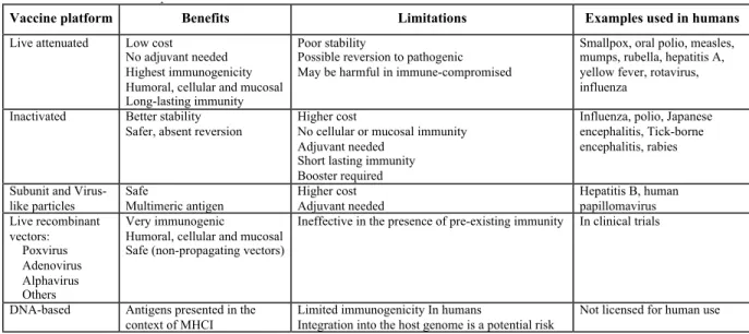
1.3. Current Vaccine Technologies
Most licensed vaccines fall into three categories: live attenuated (LAV), inactivated pathogen, and subunit/protein derived from infectious organisms (Table 1.1). Although some of them, like polio or smallpox and others, have been extremely efficacious, these vaccine
Table 1.1 - Current vaccine platforms
1.3.1. Live-attenuated vaccines
LAVs are essentially a weakened form of the pathogenic bacteria or viruses generated in the lab. Although the LAVs were originally designed through serial in vitro passage or random chemical mutagenesis without knowing its effects or risk of reversion, more recent designs includes removing one or more genes that encode virulence or metabolic factors (158). Their design reduces the risk of symptomatic disease, while mimicking a natural infection. Therefore, they elicit a long-lasting potent humoral, cellular, and mucosal immune response, often after one or two dose(s). Despite its advantages, LAVs have poor stability, ability to revert to the
pathogenic strain, and may cause complications in immune-compromised individuals. Some of the infectious diseases that have been successfully immunized against using LAVs are smallpox, oral poliomyelitis, mumps, rubella, influenza, etc.
TABLE 1.1. Current vaccine platforms
Vaccine platform Benefits Limitations Examples used in humans
Live attenuated Low cost
No adjuvant needed Highest immunogenicity Humoral, cellular and mucosal Long-lasting immunity
Poor stability
Possible reversion to pathogenic May be harmful in immune-compromised
Smallpox, oral polio, measles, mumps, rubella, hepatitis A, yellow fever, rotavirus, influenza
Inactivated Better stability Safer, absent reversion
Higher cost
No cellular or mucosal immunity Adjuvant needed
Short lasting immunity Booster required
Influenza, polio, Japanese encephalitis, Tick-borne encephalitis, rabies
Subunit and Virus-like particles
Safe
Multimeric antigen
Higher cost Adjuvant needed
Hepatitis B, human papillomavirus Live recombinant vectors: Poxvirus Adenovirus Alphavirus Others Very immunogenic Humoral, cellular and mucosal Safe (non-propagating vectors)
Ineffective in the presence of pre-existing immunity In clinical trials
DNA-based Antigens presented in the context of MHCI
Limited immunogenicity In humans
Integration into the host genome is a potential risk
1.3.2. Inactivated vaccines
Inactivated vaccines are produced after the pathogen is killed with chemicals (such as formalin), heat, or radiation. Because the pathogen is killed, there is no chance of reversion in inactivated vaccines. Additionally, these vaccines are very stable and can easily be stored. However, unlike LAVs, inactivated vaccines are not very immunogenic and require adjuvants to boost the immune response. Furthermore, immunization with inactivated vaccines may require multiple doses to maintain immunity. Inactivated vaccines that have been used in humans include influenza, polio, rabies, etc.
1.3.3. Subunit vaccines
Another class of vaccines is termed subunit vaccines, as they only contain antigens derived from a pathogen that best stimulate the immune system and not the entire pathogen. Therefore, subunit vaccines have even lower risk of adverse reaction. However, the process of determining the most immunogenic antigens and epitopes of a pathogen is a very time
consuming and expensive process. Furthermore, subunit vaccines require adjuvant for efficient priming and induction of immune response. Hepatitis B is an example of subunit vaccines that have been approved for human use. Human papillomavirus vaccines (Gardasil and Cervarix) can also be categorized with subunit vaccines as they only contain the surface glycoproteins that interact to form virus-like particles.
1.3.4. DNA vaccines
non-human primates (159, 160). Studies in humans have shown that DNA vaccines can induce a potent CTL response against malaria (161). DNA vaccines are also considered relatively stable and more cost-effective during manufacturing and storage (162). Other advantages that a DNA vaccine has includes the ability to immunize against multiple antigens using a single plasmid (162) and the stimulation of the innate immune system through the activation of TLR9 by unmethylated CpG motifs present on bacterial plasmid DNA (163). Finally, DNA vaccines can be advantageous in neonatal immunizations where passively transferred antibodies of maternal origin can interfere with vaccines, if the maternal antibodies recognize presented antigen (164). However, there are some limitations to DNA vaccines. DNA vaccines require a large dose to be effective in inducing immune response (162). Additionally, DNA vaccines are usually
administered intramuscularly, which are effective in small animal models; however, they have been relatively inefficient in the induction of antibody response in humans and non-human primates (162).
In addition to the current vaccine technologies described above, viral expression vector vaccines have been extensively studied. Below we will describe in detail the use and effect of these potential vaccine candidates.
1.3.5. Viral expression vectors
expressing viral proteins, and sometimes replicating to high titers. These viral vector vaccines are generated using molecular genetic approaches and use their efficient cell entry and
replication mechanism to express heterologous genes, which then act as the immunizing antigen [reviewed in (165-167)]. Vaccines that use bacterial genome as expression vectors have been reviewed elsewhere (168, 169). There are many examples of viral expression systems. Below we will focus on three of the most studied systems: poxviruses, adenoviruses, and alphaviruses.
1.3.5.1. Poxviruses
Poxviruses are linear, double-stranded DNA viruses that are relatively large with genome size of approximately 200,000 base pairs (170). As virus expression vectors, poxviruses have several advantages. First, poxvirus vaccine vectors are very stable and retain immunogenicity upon lyophilization, which overcomes an important hurdle in immunization, especially in the poor, developing world (171). Second, poxviruses can be easily genetically manipulated and can incorporate large amount of multiple foreign DNA, making them cost-effective (170, 172). And, thirdly, poxvirus vaccine vectors are quite versatile as they can induce effective immunity through multiple routes of immunization (170).
poxvirus immunization as well. There are several poxvirus vaccine vectors, but the two most well known are modified vaccinia Ankara (MVA) (175) and NYVAC (based on the Copenhagen strain) (176).
Poxvirus vaccine vectors have been shown to be effective inducer of humoral and cellular immunity, including upon mucosal delivery (177). In several studies, poxvirus vaccine vectors induced significant immunity to several infectious diseases, including influenza (178), measles (179), SHIV (180). The potential of poxvirus vaccine vectors should be further pursued.
1.3.5.2. Adenoviruses
The adenoviruses (Ad) are non-enveloped icosahedral virus with linear, double stranded DNA genome of 36 kb length encoding more than 30 proteins (171, 172, 181). Ad have a variety of attractive features that make them attractive candidates for vaccination: stability (because they are naked, non-enveloped), ease of manipulation, growth to very high titers (purified up to 1012 particles per ml), simple purification, broad tissue tropism, and they do not integrate into the host genome while existing extra-chromosomally (171, 181). Similar to poxviruses, Ad retain
immunogenicity after lyophilization and can be delivered through all routes of immunization (171). While replication-competent Ad vectors are useful where viral replication is desired for high expression of immunogen using low doses, replication-incompetent Ad vectors can also achieve robust immune response albeit with high dose vaccination (181).
Replication-incompetent Ad vectors are designed with either lack of structural genes in the genome or mutations in the structural components (182).
Because they are so prevalent, pre-existing immunity could be a concern for Ad vector
immunization. Additionally, Ad vectors, especially replication-competent, have limited capacity to carry heterologous antigens (165). Inflammation in the host due to induction of multiple signaling pathways can lead to dose-dependent toxicity (181). Also, at high multiplicity of infection replication-defective Ad vector can self-replicate, leading to further inflammation and tissue damage (183). Researchers have suggested use of different Ad strains to circumvent pre-existing immunity as well as oral inoculation to minimize the potential of a systemic hyper-inflammation (165, 171). Ad vectors have been demonstrated to be effective in rodent and NHP models against several pathogens, such as malaria (184), Ebola (185), SHIV (186), measles (187), and certain cancers (188). Due to the potential that Ad vector vaccines hold in the treatment of both infectious diseases and cancers, further studies should continue towards developing new candidate vaccines.
1.3.5.3. Alphaviruses
immunizing gene, is packaged into virions which can infect cells but not produce new virus particles (190).
Alphaviruses vectors are highly immunogenic as they express high levels of heterologous antigens and by targeting DCs (197, 198) induce potent humoral, cellular, and mucosal immunity (190-196). Additional advantages of Alphaviruse vectors are their ability to induce innate
immune signaling and relative absence of preexisting immunity in the general population (167, 199). Some of the protective immune response that have been demonstrated include against influenza virus (200-202), Ebola virus (203), and SIV (204, 205). A detailed discussion of one of the alphavirus replicon vector, VEE, is presented below in detail.
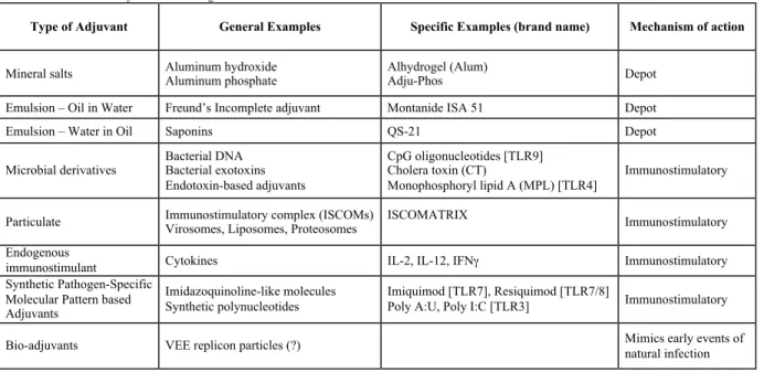
1.3.6. Adjuvants
The first instance of adjuvant use was done by Ramon in 1925, when it was demonstrated that it was possible to increase antigen-specific levels of diphtheria or tetanus anti-toxin levels by adding bread crumbs, tapioca, agar, starch oil, lecithin, or saponin [reviewed in (206)]. The few adjuvants that are currently in use, such as aluminum salts and oil-in-water emulsions, were developed empirically and there is still much to be discovered about their mechanism of action (207). However, effects of numerous adjuvant candidates have been described in humans:
introduce VEE replicon-based adjuvant as a new category of bio-adjuvant. Below, we will briefly introduce a few examples of some of the above listed adjuvant categories.
Table 1.2 - Current adjuvant technologies
1.3.6.1. Mineral salts (Alum)
Aluminum-based mineral salts, or alum, have been in use for almost a century and are the only adjuvants approved for use in licensed human vaccines by the FDA in the U.S. (206). The mechanism of action of alum is still in debate. However, it has been suggested that alum mediates its effect through “depot” effect, whereby antigens are retained in alum-induced (together with clotting agent fibrinogen) depot at the injection site and released slowly (209, 210). However, it has also been recently demonstrated that upon vaccination in fibrinogen-deficient mice, although alum depots were not formed, the antibody response was comparable to wild-type mice (210). This study shows that alum depots are dispensable for the adjuvant effect of alum, although it is still possible that alum can form “antigen depots” in the draining lymph nodes following transport from the site of immunization (211). The primary effect of alum is to regulate TH1/ TH2 environment (212) by promoting TH2 cytokines, in particular IL-4, and
TABLE 1.2. Current adjuvant technologies
Type of Adjuvant General Examples Specific Examples (brand name) Mechanism of action
Mineral salts Aluminum hydroxide Aluminum phosphate Alhydrogel (Alum) Adju-Phos Depot
Emulsion – Oil in Water Freund’s Incomplete adjuvant Montanide ISA 51 Depot
Emulsion – Water in Oil Saponins QS-21 Depot
Microbial derivatives Bacterial DNA Bacterial exotoxins Endotoxin-based adjuvants
CpG oligonucleotides [TLR9] Cholera toxin (CT)
Monophosphoryl lipid A (MPL) [TLR4]
Immunostimulatory
Particulate Immunostimulatory complex (ISCOMs) Virosomes, Liposomes, Proteosomes ISCOMATRIX Immunostimulatory
Endogenous
immunostimulant Cytokines IL-2, IL-12, IFNγ Immunostimulatory Synthetic Pathogen-Specific
Molecular Pattern based Adjuvants
Imidazoquinoline-like molecules
Synthetic polynucleotides Imiquimod [TLR7], Resiquimod [TLR7/8] Poly A:U, Poly I:C [TLR3] Immunostimulatory
improving TH2 IgG subtype, which is IgG1 in mice (213, 214). It has been recently shown that the mechanism of alum innate immune activation goes through NLRP3 inflammasome activation (215, 216). Further investigations demonstrated that the initial activation of the TH2 immune response by alum occurs through PI3 kinase activation and subsequent inhibition of IL12p70 secretion by DCs, a key cytokine that drives naïve TH response towards TH1 (217). Although there has been tremendous amount of discovery in alum adjuvanticity, further research is needed to understand the immunomodulatory effects of alum compounds and the protective antibody response it elicits.
1.3.6.2. Emulsion (Freund’s Adjuvants)
Freund’s adjuvant—incomplete (IFA) and complete (CFA)—were discovered over 50 years ago and have been experimentally used in various studies (218). However, much remains unknown about its mode of immune activation. While IFA is composed of a paraffin oil
surfactant mixture that forms a water-in-oil emulsion when mixed with an antigen, CFA additionally contains heat-killed Mycobacterium preparation, which further increases its immunogenicity (218). Both IFA and CFA induce potent humoral response, but only CFA induces cell-mediated immune response, suggesting that the Mycobacterium preparation drives the that response (218). Furthermore, the cytokine profile in CFA is more TH1 than IFA (219).
221, 222). Although potent adjuvants, safety concern should be further addressed before Freund’s adjuvants can be a candidate for human vaccination.
Saponins are triterpene glycosides obtained from the bark of Quillaja saponaria Molina tree, which are native to South America (223). Quil A is a partially purified saponin used as adjuvants in veterinary vaccines (224). Quil A has also been used as a component of immune-stimulating complexes (ISCOMs), which are small (40nm), cage-like particles additionally containing cholesterol and phospholipids (225). Although ISCOMs are very potent adjuvants, there are some safety concerns due to toxicity of Quil A in mice (225). Nonetheless less toxic saponins as part of ISCOMs are in development for human use (226).
1.3.6.3. Microbial derivative (CpG) and other TLR ligands
CpG motifs are considered pathogens associated molecular patterns (PAMPs) because they are ubiquitous in microbial genome but rare in vertebrates (227). They are very potent adjuvants of innate immune system inducing protection in both rodents and primates (163, 228). CpG are TLR agonists that activate TLR9. Just as any other TLR agonist (except for TLR3), TLR ligation initiates a complex signaling pathway involving MyD88, resulting in transcription factor NF-kB activation and the release of pro-inflammatory cytokines IL-6, IL-12, and TNF-α
(229, 230). Immune effect of CpG includes increased cellular migration towards draining lymph nodes (231), higher expression of DC co-stimulatory markers (232), higher cytokine production (233), production of IgG2a antibodies (234), and enhanced cell-mediated immune response (206). In summary, CpG are a promising candidate as adjuvants due to their potent immune response.
fact that vaccines containing bacterial cell walls, bacterial DNA, or viral RNA, can engage distinct TLRs and activate immune cells (235). Specific microbial molecules have been defined as ligands for these TLRs: lipopolysaccharides (LPS) (recognized by TLR4), flagellin (TLR5), and single (TLR7/8) or double-stranded RNA (TLR3) (236).
1.4. Venezuelan Equine Encephalitis Virus (VEE)
Venezuelan equine encephalitis virus (VEE) is a single stranded positive-sense RNA virus of the genus alphavirus and family Togoviridae. While alphaviruses are mosquito-borne infections, VEE is maintained in nature in an enzootic cycle between mostly Culex mosquitoes and small rodents (171). Alphaviruses are divided into two groups, Old World and New World viruses depending on their geographic origin. VEE is considered a member of New World alphavirus as it is endemic in Central America and the northern regions of South America and represents a significant burden of disease in these regions. The Old World alphaviruses, such as Sindbis virus (SIN), Semliki Forest virus (SFV), Ross River virus (RRV), Chikungunya virus (CHIK), and O’nyong-nyong virus (ONN) cause a somewhat different disease with arthritic and/or arthralgic symptoms, as well as fever and rash (237).
cells (240). TC-83 and its inactivated version C-84 have been used to vaccinate equines and humans with TC-83 proving to be extremely immunogenic (238). Due to the potential of
outbreaks and its use as an agent of biological weapon (238), VEE biology has been extensively studied.
1.4.1. VEE Genome and Replication
VEE is a single stranded, positive-sense RNA virus with a genome of approximately 11.5 kb length capped with a 5’ terminal 7-methylguanosine and 3’ polyadenylated tail (241). The genome is divided into two regions: 5’ two-third contains the viral non-structural proteins (nsp1-4) in a single open reading frame, while the 3’ one-third contains the structural components (capsid, E2, 6K, and E1 proteins) on a second open reading frame expressed from a subgenomic 26S promoter (242). There are four conserved sequence elements (CSE), important for
replication, spread throughout the genome: two near the 5’ end, one between non-structural and structural gene cassettes, and one immediately upstream of poly-A tail (243). Additionally, one short non-translated region (NTR) at each 5’ and 3’ terminal ends are also important for
replication (242). VEE genome is encapsidated in an icosahedral nucleocapsid surrounded by envelope containing spikes formed from trimers of glycoproteins, E1 and E2, heterodimers (242). While the molecular mechanism of VEE replication is relatively understudied, other alphaviruses, especially SIN and SFV, have been extensively studied. The review below summarizes the findings in other alphaviruses with VEE mentioned where appropriate.
receptor for SIN in both mammalian and insect cells (247). However, the authors also reported that RRV infection was NRAMP-independent, indicating that alphaviruses might use diverse set of receptors. Studies have indicated that E2 protein mediates attachment followed by entry through virus-cell membrane fusion in the early endosome. The fusion is mediated at low pH via a hydrophobic amino acid sequence in the E1-protein. Viral nucleocapsid release into the host cytoplasm occurs immediately after the receptor-mediated endocytosis (242). A viral fusion-independent entry mechanism has also been suggested for SIN, whereby following attachment the virus directly releases its genome into the plasma membrane without inducing membrane fusion or disassembling the viral protein shell (248).
Since alphavirus genomic RNA acts as a messenger RNA, immediately after release into the cytoplasm, the host translation machinery translates the viral genome into the precursor P123 or P1234 (249). The predominant species produced is P123, while 10-20% readthrough of the opaI codon at the nsp3-nsp4 junction produces P1234 polyprotein in SIN (242). The P1234 polyprotein is rapidly cleaved at the nsp3/4 junction by the cis-acting domains in nsp2, resulting in P123 and nsp4 production (242). Minus sense RNA strand is produced from a complex of P123 polyprotein and free nsp4, the viral RNA-dependent RNA polymerase, initiating at the 3’ CSE of the parental genome within 3-4 hours of infection (242). The production of minus strand is rather limited, due to the presence of few parental genomes and limited nsp4 quantity (250). Subsequently, P123 polyprotein is cleaved at nsp1/2 and nsp2/3 junctions yielding nsp1, nsp2, and nsp3 (251). Production of the nsp1-3, together with nsp4, irreversibly shifts the viral
gene cassette yields subgenomic RNA (242). The subgenomic RNA is produced in a 5-10 fold molar excess of the genomic RNA ensuring adequate production of structural components for assembly of new virions (242).
The translation of subgenomic RNA produces structural polyprotein capsid-PE2-6K-E1 followed by immediate cleavage of the capsid protein. The capsid protein binds the genomic RNA at the encapsidation signal (253). One molecule of genomic RNA interacts with 240 copies of the capsid protein in the cytoplasm to form the nucleocapsid (238).The rest of structural polyprotein (PE2-6K-E1) then translocates to the ER (254). After folding and conformational changes, the viral E1 and E2 are glycosylated (255). Subsequently, the viral glycoproteins egress from the ER into the Golgi network followed by passage into trans-Golgi network, where furin cleavage of PE2 yields mature E2 glycoproteins (256). Initiation of virus budding occurs when nucleocapsids, which diffuse throughout the cytoplasm, encounter and bind the E2-E1 complex at the plasma membrane (257). Overall, the alphavirus infection process leads to inhibition of host RNA production, protein synthesis, and eventual apoptosis (237, 258).
1.4.2. VEE pathogenesis
in convulsions, stupor, and coma. Death occurs in <1% of cases accompanied by diffuse congestion and edema with hemorrhage in the brain, lungs, and GI tract (260).
Infection of rodents with VEE has yielded many similarities between the observed pathogenesis to those observed in humans and equines (261, 262). Because of the ease of
handling mouse as well as the availability of molecularly cloned VEE and non-propagating VEE replicon particles (VRP), mice have been widely used to understand VEE pathogenesis (198, 263-266). VEE exhibits a biphasic disease in mice, similar to the pattern in equines, with an initial lymphotropic phase replication in the peripheral lymph nodes followed by infection of the CNS (neurotropic phase), which leads to lethal encephalitis by day 6-8 (261). Biphasic infection of VEE using both virulent and molecularly cloned mutants has been unequivocally
demonstrated in mice (263, 266, 267).
The early steps in the pathogenesis of VEE and induction of innate immune responses by VEE were discovered using VRPs, which are single-replication cycle, propagation-incompetent virus particles. Using VRP expressing GFP (GFP-VRP), initial target cells were identified as Langerhan cells, as they stained positively for DEC-205 and MHCII (198). Due to the
the DLN leads to viremia, which peaks at around 12 hpi with multiple organs infected by 18 hpi, including the spleen, heart, lung, and kidney (267). Peripheral titers peak between 24-48 hpi followed by CNS infection through the olfactory neuroepithelium and the trigeminal nerve (264, 266, 267).
Innate and adaptive immune responses have been important in host response to VEE
infection. Induction of robust systemic IFN-α/β occurs within 6 hpi, although its exact
mechanism is not known (269-271). The average survival time of IFN-α/β-receptor deficient
mice infected with VEE was reduced to 30 hours from 7.7 days in wild-type control, possibly due to an inability to control viral replication and dissemination into the periphery (271). The complement system also plays an important part in the clearance of VEE from the periphery and prevents CNS invasion in mice (272). Antibodies play an important protective role in anti-VEE immunity, with both VEE-specific IgM and IgG able to clear virus form the periphery (269). However, antibody response initiated during the primary immune response was not sufficient to prevent invasion of the CNS and lethal outcome in mice (237). Finally, the anti-VEE T-cell response has been shown to be crucial in the recovery from VEE-induced encephalomyelitis with IFNγ producing CD4+ T-cells more effective than CD8+ T-cells (273).
1.4.3. VEE-based vaccine vectors
Many insights about VEE-host interactions have been gained after the development of a full-length infectious cDNA from viral genomic RNA isolated from the Trinidad Donkey strain (201, 274, 275). This cDNA was cloned into a plasmid, pV3000, which can be in vitro
permissive cells. This easy-to-use, reverse genetics system can be further used to generate VEE vectors to deliver heterologous antigens, including two vectors that have been described. The first is termed double promoter vector and contains a second 26S subgenomic promoter downstream of the original 26S subgenomic promoter with structural genes (200). These
propagation-competent vectors allow for the expression of a foreign antigen from the second 26S promoter and induce strong humoral and cellular immune responses in the infected cell (200). A second VEE vector utilizes the reverse genetic system to replace the viral structural genes with a heterologous antigen and is called VRP. The VRP vectors are capable of infecting target cells, undergo a single round of replication, and do not propagate beyond the first infected cell as they can’t produce new virions due to the lack of structural genes in their RNA genome (201). It has been shown that VRP immunization induces protective immunity in rodent models of anthrax, botulism, Whipple’s disease, Lassa fever, Ebola, influenza, and human papillomavirus (202, 203, 276-281). Additionally, in non-human primate models, VRP immunization generated protective immunity against Marburg, Ebola, smallpox, dengue virus, and SIV (156, 204, 282-285).
1.4.4. VEE-based adjuvants
VEE-based adjuvants are related to the VRP vectors described above and are generated in a similar way. GVI3000 (previously known as nVRP) is a virus like particle that is generated by complementation of replicon RNA, which has the 26S subgenomic promoter and the structural genes deleted, together with the two helper RNAs (286). Just like the VRP vectors, GVI3000 particles do not propagate beyond the first infected cell and hence function as a first-in-class biological adjuvant. GVI3000 functions by inducing a strong natural and self-regulated innate immune response in the draining lymph node, which leads to an induction of immune response against the co-delivered killed virus antigen or the soluble protein antigen (287). The ensuing adaptive immune response towards the co-delivered antigen is comprehensive (humoral, cellular, and mucosal) and long lasting. Adjuvant studies using GVI3000 in mouse model induced robust immune response to co-delivered soluble antigens, (KLH, OVA, P. falciparum CelTOS, and soluble RSV), virus-like particles (norovirus), and inactivated viruses (dengue, influenza, and Sabin polio strains) [(286-290) and personal communication P. Jorquera, D. Tonkin, L. White, and R. Johnston]. More specifically, the immune responses observed using GVI3000 adjuvant included (1) enhanced antibody response, (2) balanced TH1/TH2 response, (3) antigen-specific T-cell response, (4) mucosal IgA, IgG, and T-T-cell response and (5) protection. While most of the studies looking at the adjuvanticity of GVI3000 were performed in mice, some studies have been successfully performed in rats, ferrets, and non-human primates (R. Johnston, personal
communication).
CHAPTER 2: A TETRAVALENT ALPHAVIRUS-VECTOR BASED DENGUE VACCINE PROVIDES EFFECTIVE IMMUNITY IN AN EARLY LIFE MOUSE
MODEL#
2.1. OVERVIEW
Dengue viruses (DENV1-4) cause 390 million clinical infections every year, several hundred thousand of which progress to severe hemorrhagic and shock syndromes. Preexisting immunity resulting from a previous DENV infection is the major risk factor for severe dengue during secondary heterologous infections. During primary infections in infants, maternal antibodies pose an analogous risk. At the same time, maternal antibodies are likely to prevent induction of endogenous anti-DENV antibodies in response to current live, attenuated virus (LAV) vaccine candidates. Any effective early life dengue vaccine has to overcome maternal antibody
interference (leading to ineffective vaccination) and poor induction of antibody responses (increasing the risk of severe dengue disease upon primary infection). In a previous study, we demonstrated that a non-propagating Venezuelan equine encephalitis virus replicon expression vector (VRP), expressing the ectodomain of DENV E protein (E85), overcomes maternal interference in a BALB/c mouse model. We report here that a single immunization with a
tetravalent VRP vaccine induced NAb and T-cell responses to each serotype at a level equivalent to the monovalent vaccine components, suggesting that this vaccine modality can overcome
#This chapter previously appeared as an article in the journal Vaccine. The original citation is as follows: Syed Muaz Khalil, Daniel R. Tonkin, Melissa D. Mattocks, Andrew T. Snead, Robert E. Johnston, Laura J. White. “A tetravalent alphavirus-vector based dengue vaccine provides
serotype interference. Furthermore, neonatal immunization was durable and could be boosted later in life to further increase NAb and T-cell responses. Although the neonatal immune response was lower in magnitude than responses in adult BALB/c mice, we demonstrate that VRP vaccines generated protective immunity from a lethal challenge after a single neonatal immunization. In summary, VRP vaccines expressing DENV antigens were immunogenic and protective in neonates, and hence are promising candidates for safe and effective vaccination in early life.
2.2. INTRODUCTION
The four serotypes of dengue virus (DENV) are the leading cause of the most important mosquito-borne viral disease worldwide, with annual estimates of approximately 390 million infections (151). The World Health Organization also estimates that up to half a million people are hospitalized with severe dengue disease (Dengue Hemorrhagic Fever/Dengue Shock Syndrome; DHF/DSS), and among them a large proportion are children (152). Children and adults are at increased risk of severe dengue upon a secondary infection with a different serotype. In addition, infants born to dengue immune mothers are at an increased risk of DHF/DSS during a primary infection, and account for more than 5% of all DHF cases (292, 293). This increased risk in infants seems to correlate with maternal antibody titers dropping to sub-neutralizing levels, and becoming potentially enhancing (292, 293). At present, there are no licensed dengue vaccines available, and the ones in development may not be effective in infants.
durable neutralizing antibodies (NAbs) against all 4 serotypes simultaneously, due to the theoretical enhanced risk of severe disease if incomplete immunity is induced. (B) Serotype interference has been described among the components of some TV LAV vaccines in development. The dominant serotype prevents other serotype(s) from inducing adequate
responses, resulting in incomplete immunity and the need for additional vaccinations over a one year period to achieve a tetravalent response (294). (C) In dengue endemic areas, most children are born with maternal antibodies (Abs) to DENV. These Abs protect in the first months, but also have the potential to interfere with and reduce the efficacy of LAV. Therefore, there is a need for early life vaccines that can induce balanced NAb responses after a single immunization given early in life, and that are not subject to maternal antibody interference.
Venezuelan equine encephalitis virus replicon particles (VRP) are non-propagating viral vectors that can express high levels of an antigen protein after a single round of replication. VRP-based vaccines expressing various antigens induced protective immunity in rodent models (202, 203, 276-281), and in non-human primates (NHP) (283, 285). A VRP-based dengue vaccine candidate is immunogenic and protective in adult mice and NHP (156, 295).
Furthermore, VRP expressing DENV2 prME was immunogenic in weanling mice even in the presence of maternal antibodies that prevented immunization with live virus (295).
Here we hypothesize that the VRP vectors are well suited as an effective early life vaccine platform for dengue. First, VRP are propagation incompetent, and therefore safe. Second, VRP immunization is not dependent on vector propagation and amplification.
Therefore, serotype interference is minimized as indicated by balanced responses to the 4 dengue serotypes in adult mice and non-human primates. Third, VRP induce strong Th1 immune
finally, since VRP contain no DENV antigens, maternal antibodies are less likely to interfere with the vaccine, as was demonstrated previously (295).
2.3. MATERIALS & METHODS
Cells and Viruses
Vero81, BHK-21, and C6/36 cells were obtained from the American Type Culture Collection (ATCC) and appropriately maintained as described previously (156). WHO reference DENV strains were used in the neutralization assays: DENV1 WP, DENV2 S-16803, DENV3
CH53489, and DENV4 TVP-360 (provided by R. Putnak from WRAIR). The viruses were propagated no more than three times in C6/36 cells, titrated on Vero cells, and stored at -80°C. The mouse-adapted, neurovirulent New Guinea C (NGC) strain of DENV2 used for challenge studies (provided by the late Robert Shope, UTMB, Galveston, TX) was amplified no more than
twice in C6/36 cells and stored at -80°C at a concentration of 1×10⁷PFU/ml.
DV1-4 VRP Vaccines
The construction and production of VRP vectors expressing a C-terminal truncated protein representing 85% of the E protein (E85) from DENV1-4 as well as their source have been described in detail elsewhere (156).
Mice and immunizations
and Use Committees. Pregnant mice were closely observed for date and approximate time of delivery. Adult mice (6-8 weeks old) or neonatal mice (7 days old) were anesthetized (only
adults) and injected in both rear footpads with vaccine or diluent (5µl in each rear footpad.
Neutralization assay
Neutralizing antibody titers specific for DENV1-4 were quantified using a flow cytometry-based neutralization assay on Vero cells, as previously described (156). MAb 2H2 (binds prM protein) was used for intracellular staining of dengue-infected cells.
Surface and intracellular staining (ICS)
Mousespleens were harvested, homogenized, and strained through 40µm cell strainers to
get single cell suspensions. The CD8/CD4 T cell responses were elicited after splenocytes were cultured in RPMI-10 media containing brefeldin A (GolgiPlug, BD Biosciences) with the
appropriate peptide pool (2 µg/ml) (see figure legends for detail) for 18 h at 37°C. Cells were
stained for CD8 and CD4 (eBioscience) in PBS at 4°C, fixed and permeabilized in
Cytofix/Cytoperm (BD). For ICS, fixed cells were stained with antibody specific for IFN-γ
(eBioscience). Cells were analyzed on an Accuri™ C6 Flow Cytometer (BD). The final results were obtained after normalizing the value in each sample by subtracting the value obtained in mock-stimulated cells. Spleens harvested from GFP-VRP immunized mice were also included as control.
IFN-γ enzyme-linked immunospot (ELISPOT) assay