INVESTIGATION OF HIV CURE STRATEGIES IN VIVO IN BLT HUMANIZED MICE
Perry Tsai
A dissertation submitted to the faculty at the University of North Carolina at Chapel Hill in partial fulfillment of the requirements for the degree of Doctor of Philosophy in the Department of
Microbiology and Immunology in the School of Medicine.
Chapel Hill 2016
Approved by:
J. Victor Garcia-Martinez Kristina De Paris
ii ©2016 Perry Tsai
iii ABSTRACT
Perry Tsai: Investigation of HIV cure strategies in vivo in BLT humanized mice (Under the direction of J. Victor Garcia-Martinez)
There is no cure yet for HIV. The development of HIV cure strategies will be accelerated by the use of animal models for HIV infection and treatment. Here, we investigated several HIV cure strategies in BLT humanized mice. First, we sought to explore the use of CCR5delta32 transplant to cure HIV-infected BLT mice. We found that CCR5delta32 stem cells engrafted in NSG mice and rendered them resistant to HIV infection. However, we were not able to successfully engraft CCR5delta32 cells in infected, suppressed mice.
Alternatively, a cure might be achieved by reversing HIV latency, so we tested a latency-reversing agent: histone deacetylase inhibitor panobinostat. Panobinostat treatment resulted in increased histone acetylation in BLT mice, but did not
significantly change levels of HIV cell-associated RNA, DNA, or latently infected cells in infected, suppressed BLT mice. Such an approach may require combination with other latency-reversing or cell-killing strategies.
HLA-iv
restricted, HIV-specific CD8+ T cells in the mice; and viremia increased rapidly in several mice after CD8-depletion. These studies suggest that CD8+ T cells are able to control infection in BLT mice only with nef-deficient virus.
A targeted immunological agent may be necessary for CD8+ T cells to kill infected cells. We tested a novel CD19xCD3 dual affinity retargeting (DART)
molecule for redirected lysis in BLT mice. DART molecule administration resulted in profound depletion of CD19+ cells in both peripheral blood and tissues, and this depletion was dependent on the presence of CD8+ T cells.
Overall, I present herein results from four approaches in HIV cure —
v
vi
ACKNOWLEDGEMENTS
I must express profound gratitude to my research mentor, Dr. J. Victor Garcia-Martinez. He has been my teacher, my role model, my coach, and my cheerleader over the past five years. I have had unprecedented opportunities and experiences learning and working in his laboratory; and his patience, his insight, and his enthusiasm have kept me going on my path toward becoming a
physician-scientist.
I would like to thank the other members of my thesis committee, Dr. Kristina De Paris, Dr. Joseph Eron, Dr. Edward Miao, and Dr. Jonathan Serody, for providing valuable feedback and always believing in me. I would also like to thank Dr. Nancie Archin, Dr. David Margolis, and Dr. Nilu Goonetilleke for numerous scientific
discussions and experimental input throughout my graduate career.
I would like to thank the members of the Garcia lab, past and present. We worked together, we laughed together, and we had cake together.
Finally, thank you to all of my family and friends for the advice, the
encouragement, the food, and the stories. And thank you to Shayla Birath, Michael Haas, Jenna Honeycutt, Lee Hong, Sian Lewis-Bevan, Matthew Moy, Erica
vii
TABLE OF CONTENTS
LIST OF TABLES ... xiii
LIST OF FIGURES ... xiv
LIST OF ABBREVIATIONS ... xvi
CHAPTER 1: INTRODUCTION ... 1
HIV EPIDEMIOLOGY ... 1
HIV ORIGIN ... 1
HIV ENTRY AND REPLICATION ... 1
HIV PATHOGENESIS ... 3
ANTIRETROVIRAL THERAPY ... 4
HIV PERSISTENCE ... 6
ACTIVE HIV RESERVOIRS... 7
LATENT HIV RESERVOIRS ... 8
A NEED FOR AN HIV CURE ... 10
THE BERLIN PATIENT ... 11
THE BOSTON PATIENTS ... 12
THE MISSISSIPPI BABY ... 13
THE VISCONTI COHORT ... 13
ELITE HIV CONTROLLERS ... 14
APPROACHES TO HIV CURE OR CONTROL ... 14
MECHANISMS OF HIV LATENCY ... 17
viii
TARGETED IMMUNOLOGIC AGENTS ... 21
IN VIVO PLATFORMS FOR HIV CURE RESEARCH ... 25
NON-HUMAN PRIMATES FOR HIV CURE RESEARCH ... 25
HUMANIZED MICE FOR HIV CURE RESEARCH ... 28
CHAPTER 2: INVESTIGATING THE EFFECT OF CCR5DELTA32 TRANSPLANTATION ON HIV INFECTION IN VIVO ... 31
SUMMARY... 31
INTRODUCTION ... 32
METHODS ... 36
ETHICS STATEMENT ... 36
GENERATION OF CCR5WT and CCR5DELTA32 NSG-HU MICE ... 36
CCR5WT AND CCR5DELTA32 GENOTYPING PCR ... 36
GENERATION OF BLT HUMANIZED MICE ... 37
HIV INFECTION AND ANTIRETROVIRAL TREATMENT OF BLT MICE ... 37
CONDITIONING AND ALLOGENEIC TRANSPLANT OF BLT MICE ... 37
METHYLCELLULOSE CULTURE ... 38
STATISTICAL TESTS ... 38
RESULTS ... 39
TRANSPLANTATION OF MICE WITH CCR5WT OR CCR5DELTA32 STEM CELLS ... 39
SUSCEPTIBILITY OF CCR5WT AND CCR5DELTA32 MICE TO HIV INFECTION ... 40
ANALYSIS OF CCR5WT AND CCR5DELTA32 GENOTYPE BY PCR/DIGEST ... 40
CONDITIONING OF BLT MICE FOR ALLOGENEIC TRANSPLANT ... 41
ix
CCR5 GENOTYPING OF BLT MICE AFTER ALLOGENEIC
TRANSPLANT ... 44
DISCUSSION ... 45
CONTRIBUTIONS ... 48
FIGURES ... 49
CHAPTER 3: IN VIVO ANALYSIS OF THE EFFECT OF PANOBINOSTAT ON CELL-ASSOCIATED HIV RNA AND DNA LEVELS AND LATENT HIV INFECTION ... 56
SUMMARY... 56
INTRODUCTION ... 57
METHODS ... 59
ETHICS STATEMENT ... 59
ISOLATION OF RESTING HUMAN CD4+ T CELLS FOR RNA INDUCTION AND QUANTITATIVE VIRAL OUTGROWTH ASSAY ... 59
MEASUREMENT OF RNA INDUCTION AND QUANTITATIVE VIRAL OUTGROWTH FROM RESTING CELLS ... 60
GENERATION OF BLT HUMANIZED MICE ... 61
ANALYSIS OF HISTONE ACETYLATION ... 62
HIV INFECTION AND TREATMENT OF BLT MICE ... 63
STATISTICAL TESTS ... 64
RESULTS ... 64
INDUCTION OF HIV EXPRESSION WITH PANOBINOSTAT FROM RESTING CD4+ T CELLS ISOLATED FROM HIV-INFECTED PATIENTS ON SUPPRESSIVE ANTIRETROVIRAL THERAPY ... 64
IN VIVO HISTONE ACETYLATION IN TISSUES AFTER TREATMENT WITH PANOBINOSTAT ... 65
ANALYSIS OF THE EFFECT OF PANOBINOSTAT TREATMENT IN HIV-INFECTED, ART-SUPPRESSED BLT MICE ... 66
x
DISCUSSION ... 68
CONTRIBUTIONS ... 74
FIGURES ... 75
CHAPTER 4: CD8-MEDIATED CONTROL OF INFECTION IN BLT MICE ... 82
SUMMARY... 82
INTRODUCTION ... 83
METHODS ... 85
ETHICS STATEMENT ... 85
GENERATION AND INFECTION OF BLT MICE ... 86
HLA HAPLOTYPING ... 87
PENTAMER STAINING ... 87
ELISPOT ASSAY ... 88
INTRACELLULAR CYTOKINE STAINING ... 89
STATISTICAL TESTS ... 89
RESULTS ... 90
IDENTIFICATION OF BLT COHORTS WITH HIV-PROTECTIVE HLA ALLELES ... 90
REPLICATION OF JRCSF AND JRCSFNefdd in BLT MICE ... 90
DETECTION OF FUNCTIONAL HIV-SPECIFIC CTL RESPONSES ... 91
EFFECT OF CD8+ T CELL DEPLETION ON HIV INFECTION IN BLT MICE ... 93
DISCUSSION ... 94
CONTRIBUTIONS ... 97
FIGURES ... 99
xi
SUMMARY... 106
INTRODUCTION ... 107
METHODS ... 110
ETHICS STATEMENT ... 110
GENERATION OF BLT HUMANIZED MICE ... 110
DART MOLECULES ... 110
TREATMENT OF BLT MICE ... 111
ANALYSIS OF BLT MICE ... 111
STATISTICAL ANALYSIS ... 112
RESULTS ... 112
DEPLETION OF HUMAN CD19+ B CELLS IN PERIPHERAL BLOOD AFTER ADMINISTRATION OF CD19xCD3 DART PROTEIN ... 112
DEPLETION OF HUMAN CD19+ B CELLS IN TISSUES AFTER ADMINISTRATION OF CD19xCD3 DART PROTEIN ... 115
REGENERATION OF HUMAN CD19+ B CELLS AFTER ADMINISTRATION OF CD19xCD3 DART PROTEIN ... 116
EFFECT OF CD19xCD3 DART PROTEIN ADMINISTRATION ON THE LEVELS OF HUMAN T CELLS IN VIVO ... 117
DEPENDENCE OF CD19xCD3 DART PROTEIN-MEDIATED DEPLETION ON THE PRESENCE OF HUMAN CD8+ T CELLS ... 118
DISCUSSION ... 120
CONTRIBUTIONS ... 123
FIGURES ... 124
CHAPTER 6: SUMMARY AND FUTURE DIRECTIONS ... 135
STUDY SUMMARY ... 135
SUMMARY OF STUDIES ON CCR5DELTA32 TRANSPLANTATION ... 136
xii
SUMMARY OF STUDIES ON CD8-MEDIATED CONTROL
OF HIV INFECTION ... 138
SUMMARY OF STUDIES ON CD19xCD3 DART MOLECULE ... 139
IMPLICATIONS OF CURRENT STUDIES ... 140
FUTURE DIRECTIONS ... 144
FINAL SUMMARY ... 147
xiii
LIST OF TABLES
xiv
LIST OF FIGURES
Figure 2.1: Reconstitution in CCR5WT and
CCR5delta32 NSG-Hu mice ... 49 Figure 2.2. Susceptibility of CCR5WT and
CCR5delta32 mice to HIV infection ... 50 Figure 2.3. CCR5 genotyping by PCR and restriction digest ... 51 Figure 2.4. Reconstitution of irradiated and non-irradiated BLT mice ... 52 Figure 2.5. Conditioning of BLT mice by
busulfan and antithymocyte globulin ... 53 Figure 2.6. CCR5WT or CCR5delta32 allogeneic transplant
in infected, ART-treated BLT mice ... 54 Figure 2.7. CCR5 genotyping of tissues from infected, ART-treated
BLT mouse transplanted with CCR5delta32 stem cells ... 55 Figure 3.1. Effect of panobinostat on histone acetylation, HIV RNA,
and viral outgrowth from patient cells ... 75 Figure 3.2. Panobinostat administration induces
systemic histone acetylation ... 76 Figure 3.3. Outline of panobinostat treatment of
HIV-infected, ART-suppressed BLT mice ... 77 Figure 3.4. Analysis of cell-associated HIV RNA levels in the tissues
of infected, suppressed, panobinostat-treated BLT mice ... 78 Figure 3.5. Analysis of HIV DNA levels in the tissues
of infected, suppressed, panobinostat-treated BLT mice ... 79 Figure 3.6. Analysis of panobinostat treatment on HIV latency
in infected, suppressed BLT mice ... 80 Figure 4.1. Flow cytometric analysis of HLA alleles ... 99 Figure 4.2. Viral loads of JRCSF and JRCSFNefdd
in BLT mice with protective HLA alleles ... 100 Figure 4.3. Analysis of peak viral loads and average viral loads in
xv
Figure 4.4. Pentamer staining of HIV-specific CD8+ T cells
from tissues of infected HLAB*2705 BLT mice ... 102 Figure 4.5. Functional assessment of HIV-specific CD8+ T cells
from tissues of infected HLAB*2705 BLT mice ... 103 Figure 4.6. Effect of CD8+ T cell depletion in infected BLT mice ... 104 Figure 5.1. CD19xCD3 DART protein administration depletes
human CD19+ B cells from the peripheral blood ... 124 Figure 5.2. Gating scheme for flow cytometry analysis ... 125 Figure 5.3. The percent human CD19+ cells out of human CD45+ cells
decreases as percent human CD3+ cells increases over time
in the peripheral blood of NSG/BLT humanized mice ... 126 Figure 5.4. 4420xCD3 DART protein administration does not deplete
human CD19+ B cells from the peripheral blood ... 127 Figure 5.5. CD19xCD3 DART protein administration depletes
human CD19+ B cells from the tissues ... 128 Figure 5.6. Immature human CD19+ B cells regenerate in NSG/BLT
mice after CD19xCD3 DART protein administration ... 129 Figure 5.7. CD19xCD3 DART protein administration results in transient
differences in the levels of human T cells in the peripheral blood of NSG/BLT mice, and over time there are no significant
differences in absolute numbers of human CD8+ T cells in the
peripheral blood or tissues as compared to vehicle-treated mice ... 131 Figure 5.8. Human CD8+ T cells are depleted after
administration of CD8-depleting antibody ... 133 Figure 5.9. Human CD19+ B cell depletion by CD19xCD3 DART
xvi
LIST OF ABBREVIATIONS
AIDS Acquired immune deficiency syndrome ANOVA Analysis of variance
AP-1 Activator protein 1
APOBEC3G Apolipoprotein B mRNA editing enzyme, catalytic polypeptide-like 3G
ART Antiretroviral therapy ATG Antithymocyte globulin BCL B cell line
BiTE Bispecific T cell engager BLT Bone marrow / liver / thymus Bp Base pair
Brd4 Bromodomain 4
BSA Bovine serum albumin CAR Chimeric antigen receptor CCR5 C-C chemokine receptor type 5 CCR5delta32 CCR5 delta32 mutant
CCR5WT CCR5 wildtype
CD Cluster of differentiation CD19 Cluster of differentiation 19 Cdk9 Cyclin-dependent kinase 9 cDNA Complementary DNA CTL Cytotoxic T lymphocyte
xvii CycT1 Cyclin T1
DART Dual affinity retargeting DMSO Dimethyl sulfoxide DNA Deoxyribonucleic acid
DSIF DRB sensitivity-inducing factor EBV Epstein-Barr virus
EC Elite controller
ELISA Enzyme-linked immunosorbent assay ELISPOT Enzyme-linked immunospot
EpCAM Epithelial cell adhesion molecule FTC Emtricitabine
Gag Group-specific antigen
G-CSF Granulocyte colony-stimulating factor HDAC Histone deacetylase
HDACi Histone deacetylase inhibitor
HEPES 4-(2-hydroxyethyl)-1-piperazineethanesulfonic acid HEXIM Hexamethylene bisacetamide-inducible protein HIV Human immunodeficiency virus
HLA Human leukocyte antigen HMBA Hexamethylene bisacetamide HSCT Hematopoietic stem cell transplant IFN-gamma Interferon-gamma
xviii
IMDM Iscove’s modified Dulbecco’s medium IUPB Infectious units per billion
IUPM Infectious units per million Liv Liver
LOQ Limit of quantitation LRA Latency-reversing agent LTR Long terminal repeat
MHC Major histocompatibility complex Nef Negative regulatory factor
Nefdd Nef-deleted
NELF Negative elongation factor
NFAT Nuclear factor of activated T cells
NF-kB Nuclear factor kappa-light-chain-enhancer of activated B cells NHP Non-human primates
NK Natural killer
NNRTI Non-nucleoside reverse transcriptase inhibitor NRG NOD-Rag2-/--gammachain-/-
NRTI Nucleoside/nucleotide reverse transcriptase inhibitor NSG NOD/SCID-gammachain-/-
PBMC Peripheral blood mononuclear cells PBS Phosphate-buffered saline
xix PHA Phytohemagglutinin
PI3K Phosphatidylinositol-4,5-bisphosphate 3-kinase PKC Protein kinase C
PMA Phorbol myristate acetate Pol Polymerase
P-TEFb Positive transcription elongation factor b QVOA Quantitative viral outgrowth assay R5-tropic CCR5-tropic
Rad Radiation absorbed dose
Rev Regulator of expression of the virion RLU Relative light units
RNA Ribonucleic acid
RPMI Roswell Park Memorial Institute medium SAHA Suberoylanilide hydroxamic acid
SAMHD1 Sterile alpha motif-domain and histidine aspartic-domain containing protein 1 SCID Severe combined immunodeficiency SD Standard deviation
SEM Standard error of the mean SFU Spot-forming units
xx Tat Transactivator of transcription TCR T cell receptor
TDF Tenofovir disoproxil fumarate
Thy Thymus
TNF Tumor necrosis factor UNC University of North Carolina Vif Viral infectivity factor
VISCONTI Virological and Immunological Studies in CONtrollers after Treatment Interruption
1
CHAPTER 1: INTRODUCTION
HIV EPIDEMIOLOGY
The global HIV/AIDS pandemic began over thirty years ago. The first reported cases of HIV/AIDS appeared in the Centers for Disease Control’s Morbidity and
Mortality Weekly Report on June 5, 1981 [1]. Five men in Los Angeles had been treated for Pneumocystis carinii pneumonia, an infection typically found only in patients that were severely immunocompromised. These unusual cases pointed to the possibility of a “cellular-immune dysfunction related to a common exposure that predisposes individuals to opportunistic infections” like Pneumocystis. Over the
following year, the number of cases of this immune deficiency rose, spanning the country – 158 in New York City, 10 elsewhere in New York State, 14 in New Jersey, and 71 in California [2] – and including occurrences of rare Kaposi’s sarcoma as well
[3]. The syndrome, initially referred to as gay-related immune deficiency because most of those affected were men who have sex with men, came to be known as acquired immune deficiency syndrome, or AIDS [4]. Soon after, two groups led by Robert Gallo [5] and by Luc Montagnier [6] discovered that the causative agent was a virus, first called human T-lymphotropic virus-III or lymphadenopathy-associated virus. Later, this pathogen would be renamed the human immunodeficiency virus, or HIV [7].
1
2014, the number of individuals infected with HIV was 36.9 million people, with 2 million people newly infected and 1.2 million having died from AIDS-related illnesses [9]. Sub-Saharan Africa continues to be the most severely affected area, where roughly 1 in 20 adults are infected, representing two thirds of all cases worldwide. Specific to the United States, about 1.2 million people are living with HIV, with approximately 50,000 new infections each year [10].
HIV ORIGIN
HIV originated from the simian immunodeficiency virus (SIV) found in non-human primates (NHP) [11]. The two types of HIV, HIV-1 and HIV-2, seem to have derived from species-crossover events, HIV-1 from SIV in chimpanzees and gorillas and HIV-2 from SIV in sooty mangabeys [11, 12]. HIV-1 can be further categorized into four groups, M, N, O, and P; and 95% of the viruses found worldwide fall within Group M and its nine subtypes (A-D, F-H, J, and K). Subtype C is found primarily in Africa; and subtype B, in Europe and North America. The other groups of HIV-1 (N, O, and P) are limited to Cameroon and thus classified as non-pandemic [12]. References to “HIV” that follow in this dissertation will refer to HIV-1.
HIV ENTRY AND REPLICATION
HIV enters target cells via binding to its primary receptor CD4, followed by binding to one of two potential co-receptors, CCR5 or CXCR4. CD4 is a glycoprotein found on the surface of immune cells, including T helper cells, monocytes/
macrophages, and dendritic cells. CCR5 and CXCR4 are both chemokine receptors from the superfamily of G-protein coupled receptors [13]. The HIV envelope
2
exposes the domains which bind to CCR5 or CXCR4. Binding to a co-receptor triggers another conformational change, allowing the HIV fusion peptide, gp41, to insert into the membrane of the host target cell and initiate fusion between the virus and host membranes, allowing the virus to enter the cell [14].
After HIV enters the host cell, the virion capsid is removed [15], and HIV reverse transcriptase synthesizes a complementary DNA (cDNA) strand from the single-stranded HIV RNA genome, followed by production of a second DNA strand to yield a double-stranded HIV DNA genome. HIV integrase then joins with the HIV DNA genome, forming the pre-integration complex, which is transported into the nucleus of the host cell by HIV Vpr (viral protein R) [16]. HIV integrase incorporates the HIV DNA genome into the genome of the host cell, and this integrated HIV DNA genome is referred to as a provirus. The proviral DNA is transcribed into RNA under a promoter present in the HIV long terminal repeat (LTR) region. This transcription is enhanced by HIV Tat (transactivator of transcription) which increases transcription processivity [17, 18].
3
the site of viral budding [25]. Finally, the HIV protease cleaves the Gag-Pol polyprotein into the functional forms of Gag and Pol, yielding mature infectious virions [26].
During the replication process, HIV is able to evade some of the host defense mechanisms, including APOBEC3G hypermutation and antigen presentation.
APOBEC3G is a host restriction factor that deaminates deoxycytidines in the HIV cDNA, resulting in hypermutation and generation of replication-incompetent provirus [27, 28]. To suppress this antiviral activity, HIV Vif (viral infectivity factor) binds to APOBEC3G and promotes its degradation [29, 30]. Also, HIV Nef downregulates major histocompatibility complex (MHC) class I molecules [31], thus preventing the recognition and killing of HIV-infected cells by CD8+ T cells (also known as cytotoxic T lymphocytes, CTLs) [32].
HIV PATHOGENESIS
4
an AIDS-defining condition, including candidiasis, cryptococcosis, Pneumocystic jirovecii pneumonia, Kaposi sarcoma, lymphoma, etc. [37].
ANTIRETROVIRAL THERAPY
The opportunistic infections and cancers associated with untreated HIV infection can be prevented if the host immune system and host CD4+ T cells are preserved through the use of antiretroviral therapy (ART) to suppress viral replication [38]. The first pharmaceutical approved for the treatment of HIV was zidovudine in 1987. Since then, the number of drugs approved to treat HIV infection has expanded to include thirty single antiretroviral agents and eight fixed-dose combination tablets [39]. These antiretroviral drugs fall into four major classes, categorized by their target enzyme in the HIV replication cycle: reverse transcriptase inhibitors, entry inhibitors, integrase inhibitors, and protease inhibitors.
The reverse transcriptase inhibitors include seven nucleoside/nucleotide analogs (NRTIs) and five non-nucleoside inhibitors (NNRTIs). NRTIs, such as emtricitabine (FTC) or tenofovir (TDF), are DNA nucleoside or nucleotide analogs. They compete with the naturally occurring nucleoside/nucleotide and become
incorporated into the cDNA strand, but then they terminate the strand elongation due to a chemical feature preventing 5’-3’ phosphodiester linkages [40]. NNRTIs, such
as nevirapine or efavirenz, bind reverse transcriptase at an allosteric site, inducing conformational changes that block substrate binding and polymerization [41].
The two approved entry inhibitors, enfuvirtide and maraviroc, prevent entry of HIV into the host cell. Enfuvirtide is a peptide inhibitor that binds to the fusion
5
is a CCR5 antagonist that binds to CCR5 and blocks its utilization as a co-receptor for entry [43].
Integrase inhibitors include raltegravir, elvitegravir, and dolutegravir. These drugs inhibit the integration of HIV DNA into the host genome by blocking the action of HIV integrase [44].
Protease inhibitors, like ritonavir or darunavir, block the action of HIV
protease to cleave precursor polyproteins into their active forms, which are needed for the maturation of virions into infectious particles. In the presence of protease inhibitors, only non-infectious and immature viral particles are produced [45, 46].
In the early days of the epidemic, options for antiretroviral therapy were limited, and the goals clinically were to improve the survival of already ill AIDS
6
The benefits of ART have been demonstrated in improvements in life expectancy and clinical outcomes in those receiving treatment [51-53]. Life expectancy has increased among treated HIV-positive individuals in the United States and Canada to 51.4 years after ART initiation, approaching that of the general population [54]. And while previous recommendations were to delay ART initiation in HIV patients until CD4+ T cell counts were less than 500 per microliter peripheral blood, these have been replaced with recommendations to initiate ART immediately for all HIV-diagnosed individuals, regardless of CD4+ T cell count [55, 56]. This shift arises from compelling evidence that early ART initiation reduces morbidity and mortality [57, 58] and prevents further transmission [49].
HIV PERSISTENCE
Despite the apparent efficacy and benefit of ART, it is not curative. Even though viral replication may be suppressed to the point of undetectable plasma viral load, the virus continues to persist in treated individuals. Because of this persistent infection, virtually all treated individuals experience a rapid rebound of viral
7 ACTIVE HIV RESERVOIRS
Patients on ART can maintain plasma viral loads below the limit of detection using standard clinical assays. However, steady-state levels of very low residual viremia (>1 HIV RNA copy per ml) have been observed using ultra-sensitive assays in patients who have been on treatment for years [62, 63], suggesting the presence of residual active HIV reservoirs that continue to release virus despite effective ART.
It is not yet clear if this residual viremia arises from the release of trapped virus, the ongoing production of virus in long-lived infected cells, and/or new cycles of infection/replication. The genetic stability of sequences obtained from residual viremia [64-66] and the futility of ART intensification (adding an additional drug to an ART regimen) to reduce residual viremia [67-69] suggest that new cycles of
infection/replication are not a major source of persistent viremia during ART.
However, the persistence of low-level viremia over the span of several years [63] implies that HIV is being continually produced from an active reservoir. Two randomized clinical trials showed that the addition of an integrase inhibitor
(raltegravir) resulted in a temporary increase of 2-LTR circles [70, 71]. These 2-LTR circles form from reverse-transcribed HIV DNA episomes before their integration into the host genome during the replication cycle, so the increase in 2-LTR circles means that the addition of raltegravir is blocking integration steps during ongoing
8
that drug concentrations are not fully suppressive in lymphoid tissues, thus allowing HIV replication to continue without development of resistance [73].
Persistent low-level viremia during ART could be explained by cell-to-cell spread, anatomical sanctuaries, or long-lived cellular reservoirs. In vitro experiments have shown that cell-to-cell transmission is less sensitive to antiretroviral drugs than infection by cell-free virus [74]. Cell-to-cell transmission has been observed between lymphocytes and astrocytes in culture [75], but it has not yet been demonstrated in vivo. Reports of pyroptotic death of CD4+ T cells after cell-to-cell transmission would predict progressive CD4+ T cell depletion in treated patients, but this is typically not the case. The virus may persist in anatomical sanctuaries in the lymphoid tissues or in the central nervous system due to poorer penetration of antiretroviral drugs into these anatomical locations [73, 76, 77]. Lower drug concentration was correlated with slower decay of follicular dendritic cell-associated virus and with detection of viral RNA in productively infected cells, though it is not clear why drug penetration is not as robust [76]. A recent study in elite controller rhesus macaques [78] suggests that HIV may persist in the B cell follicles of lymph nodes because of the exclusion of CD8+ T cells, which would typically control infection. Finally, tissue macrophages and microglia in the central nervous system are long-lived and highly resistant to viral cytopathic effects and apoptosis [79, 80], so infected macrophages could serve as long-lived cellular reservoirs [81, 82].
LATENT HIV RESERVOIRS
9
which HIV resides as transcriptionally silent provirus in the host genome [83]. The presence of an inducible latent HIV reservoir was first demonstrated in 1997 as viral outgrowth from peripheral blood mononuclear cells (PBMCs) of treated patients [84-86]. The most well-characterized latently infected cells are resting central memory CD4+ T cells. While some studies have suggested a potential for latent infection in other cell types (naïve CD4+ T cells, stem memory T cells, transitional memory CD4+ T cells, gamma-delta T cells, hematopoietic progenitor cells, and macrophages), there is so far limited evidence to support these claims [87]. The long-lived durability of these potential cellular reservoirs has yet to be demonstrated in patients or animal models as rigorously as has been shown in resting central memory CD4+ T cells according to the above criteria [61].
Latently infected cells could be infected directly while in the resting state [88] or while they are transitioning toward a resting memory state [89]. In this quiescent state, they are transcriptionally silent, producing very low levels of HIV RNA (<50 copies per 106 cells) [90]. Without HIV RNA for protein translation, latently infected cells are invisible to antiretroviral therapies and to the host immune responses that recognize HIV antigen. Longitudinal studies have shown that these latently infected cells are long-lived with an extremely slow decay rate (half-life of 44 months) which extrapolates to an estimate of >70 years of treatment needed to eradicate the latent reservoir with ART alone [91].
10
therapies are not able to clear these reservoirs, and so there is still a need for an HIV cure.
A NEED FOR AN HIV CURE
A cure for HIV would no doubt be beneficial to patients by eliminating the effects of chronic HIV infection. Even in patients adherent to ART, there is evidence of increased chronic immune activation and inflammation [92] in comparison to uninfected persons, and chronic inflammation has been associated with increased risk of cardiovascular disease, cancer, and osteoporosis [92-100].
A cure would eliminate the need for lifelong drug treatment and potential drug toxicities. Earlier drugs required a high pill burden and were associated with a long list of adverse side effects: nausea, diarrhea, rash, myopathy, pancreatitis,
lipodystrophy, hypercholesterolemia, mitochondrial toxicity, nephrotoxicity, and loss of bone mineral density [39]. Newer ART drugs avoid many but not all of the
toxicities of first-generation regimens. For example, long-term tenofovir disoproxil fumarate is associated with nephrotoxicity in patients who have pre-existing renal insufficiency or are taking other nephrotoxic medications [101], and ritonavir is
associated with hypertriglyceridemia [102, 103]. NRTIs are generally associated with mitochondrial toxicity due to the structural similarities between reverse transcriptase and mitochondrial DNA polymerase. Mitochondrial toxicity can manifest as
11
Curing a patient of HIV would also reduce the social and financial costs of infection. The status of being HIV-positive carries social stigma from
intrapersonal/interpersonal levels up to larger institutional/structural levels [108, 109], and the cost of lifelong ART has been estimated at $379,668 in 2010 [110]. The financial cost of lifelong ART not only affects individual access but also global access, particularly in low-income countries where the majority of HIV-infected people reside; therefore a cure would be particularly beneficial in these areas where ART can be cost-prohibitive.
A cure for HIV would allow a patient to discontinue ART and not experience viral rebound or progression to AIDS, and there are several potential strategies to achieve this state [111]. A sterilizing or eradicative cure means a complete removal of all infectious forms of HIV from a patient; no replication-competent provirus would be present. A functional cure describes a state of post-treatment control where infection is controlled without ART; this might be achieved through a reduction but not necessarily elimination of the reservoir, or through a modification of the immune system. Reductions in the HIV reservoir might also make it possible to achieve temporary ART-free remission or delay in rebound after ART interruption.
Several recent reports of HIV cure, delayed rebound, and control of infection have introduced the possibility of cure as well as potential approaches for cure [111]. THE BERLIN PATIENT
The only example of a possible eradicative HIV cure is the case of Timothy Ray Brown, also known as the “Berlin patient.” Brown received an allogeneic
12
acute myeloid leukemia. His donor was specifically chosen because the donor was homozygous for the CCR5delta32 mutation. People who are
CCR5delta32-homozygous do not express CCR5 protein on the surface of their cells; and, as CCR5 is a co-receptor for HIV entry, they are highly resistant to infection [112, 113]. By 61 days after his transplantation, Brown’s entire immune system had been
replaced by HIV-resistant CCR5delta32-homozygous donor cells; and, although he discontinued ART at the time of transplant, he did not experience any viral rebound [114, 115]. This case has been considered a possible sterilizing cure, as replication-competent HIV has still not been detected years later [116].
THE BOSTON PATIENTS
Following the Berlin patient, two more cases were described in 2012, the “Boston patients” [117]. Like the Berlin patient, the Boston patients received
allogeneic HSCTs, but with two major differences: their donors were CCR5-wildtype, and they continued ART during and after the transplant. This approach bypassed the need for a CCR5delta32 donor, and it tested the hypothesis that allogeneic HSCT alone could be curative for HIV as long as ART was present to protect the HIV-susceptible donor cells from becoming infected. The patients were followed for two to four years after transplant during which ART was continued; and neither of them had detectable HIV DNA or detectable replication-competent virus in their peripheral blood cells. This evidence suggested that their reservoirs might have been
13
be reduced after allogeneic HSCT, and that rebound can be delayed later than the typical rebound of 2 to 3 weeks [119].
THE MISSISSIPPI BABY
Another case of delayed rebound was reported in the “Mississippi baby” in
2013 [120]. A newborn that had been infected perinatally was started on ART immediately after birth. At 18 months old, the infant was lost from care and returned at 23 months, when it was discovered that the infant had not been taking ART since 15 months. Despite having not taken ART from 15 to 23 months, the infant had no detectable plasma viremia or HIV DNA in the peripheral blood. It was hypothesized that early treatment might have been able to prevent the establishment of a
reservoir. During follow-up, rebound was eventually detected at 26 months after ART discontinuation [121].
THE VISCONTI COHORT
Early treatment has also been implicated in cases of post-treatment control in fourteen patients from the VISCONTI cohort [122]. These patients began ART early during primary infection (a majority at Fiebig stage V) and continued treatment for a median of 36.5 months before ART interruption. At the time of the report, these patients had not been taking ART for a median of 89 months and were not
experiencing viral rebound. Their levels of plasma viremia were detectable but very low (median 5.0 copies per ml), and replication-competent virus was still present in their resting cells; so these cases were considered “post-treatment controllers,”
14 ELITE HIV CONTROLLERS
The possibility of immune control of infection is also implied in cases of “elite controllers” (ECs). These patients are able to control the virus on their own, usually
for at least 10 years, without ever initiating ART [124]; and they are very rare, representing only 0.15% of HIV-infected patients [125]. Genotypic and phenotypic analyses have suggested that viral isolates from ECs are fully virulent [126, 127], and there was even one case of HIV transmission from a patient who progressed to AIDS to a patient who remained an elite controller [128]. Therefore, EC status is likely due to features of the host. Indeed, certain MHC class I alleles are
overrepresented in ECs, including HLA-B27, -B57, -B14, and -B51 [129, 130]; and strong CTL responses to HIV Gag antigen have been reported in ECs, including characteristics of polyfunctional memory T cells [131], CD27 expression for long-term survival [132], and high functional avidity [133]. However, not all ECs exhibit intense CTL responses [134], so there are likely other factors contributing to control of the virus.
Though these reports are important for generating hypotheses toward strategies, only the Berlin patient has ever been possibly cured of infection. The challenge in curing HIV lies in the long-term persistence of HIV infection even with antiretroviral therapy, and several approaches are being investigated to clear this persistent infection.
APPROACHES TO HIV CURE OR CONTROL
15
1% of the Caucasian population carries the CCR5delta32 mutation [135]. Another way to replicate the CCR5delta32-homozygous HSCT is through gene therapy or gene-editing technology. Using zinc-finger nucleases, CD4+ T cells from twelve HIV patients were modified ex vivo to remove the CCR5 gene, thus mimicking
CCR5delta32-homozygous cells [136]. While the procedure was safe within the parameters of the study, the CCR5-deleted cells only represented a minority of the reinfused cells, and ART discontinuation resulted in viral rebound. Similar strategies are currently being investigated to delete CCR5 from hematopoietic stem cells in preclinical models as well [137].
Allogeneic HSCT itself might be effective in reducing the reservoir because (1) pre-transplant conditioning regimens designed to reduce tumor burden and to prevent graft rejection may also reduce the number of HIV-infected host cells, and (2) graft-versus-host effects may result in post-transplant clearance of HIV-infected cells. However, allogeneic transplant alone was not sufficient for cure in the Boston patients. Also, allogeneic HSCT is not generalizable due to the need for
HLA-matched donors and the risks inherent to the procedure, so it would be inappropriate for healthy, HIV patients without an oncological indication for allogeneic HSCT.
16
days after exposure [141]. Also, early treatment requires a diagnosis during acute or primary HIV infection. This can be challenging because the symptoms of acute infection are vague and nonspecific [142], and intensive resources are needed to detect HIV RNA in blood before seroconversion. The development and use of new technologies to detect HIV RNA or p24 antigen should increase diagnoses of acute HIV infection and identification of candidates for early treatment [143].
The evidence for immune control in elite controllers suggests the possibility of recapitulating this control in other patients through immunization. Passive
immunization could be accomplished by injection of broadly neutralizing antibodies that would block new infections and target infected cells for destruction by antibody-dependent cellular cytotoxicity [144-147]. Vaccination may also confer the ability to either resist or control infection. The bivalent RV144 vaccine was designed to induce humoral and cellular immune responses, and it demonstrated a 31.2% reduction in transmission in a phase III clinical trial in Thailand [148]. Hansen et al. vaccinated rhesus macaques with a cytomegalovirus vector containing SIV genes, before challenging them with SIV. This vaccine induced the generation of SIV-specific effector memory CD8+ T cells, and 13 out of 24 animals were able to control and clear infection [149, 150]. These results suggest that preexisting robust CTL responses may be able to clear the latent reservoir or prevent the formation of a latent reservoir.
17
produced by isolating CTLs from a patient, then selecting and expanding the clones with robust anti-HIV responses for reinfusion. This approach was safe in a phase 1 clinical trial, but its effect on viremia was transient and not statistically significant [151]. High-affinity TCRs are also being investigated [152], but this approach would be restricted to HLA-matched patients.
To bypass HLA restriction, chimeric antigen receptor (CAR) gene therapy is being developed. CARs are engineered by joining an extracellular targeting domain derived from CD4 and single-chain variable fragments specific for HIV gp120, with an intracellular activating domain to trigger CTL killing activity. Clinical trials using this technology have thus far shown safety and long-term persistence of CAR-transduced T cells, but no significant change in reservoir size [153-155].
Finally, the “shock-and-kill” or “kick-and-kill” approach seeks to target the
latent HIV reservoir by reversing latency and inducing HIV gene expression, thereby allowing the clearance of infected cells through viral cytopathic effects, immune responses, or targeted cytotoxic agents [156]. Strategies to disrupt the latent reservoir are currently being developed to target the mechanisms of HIV latency. MECHANISMS OF HIV LATENCY
There are three broad mechanisms identified so far by which HIV establishes latent infection and maintains latency: (1) transcriptional interference, (2) epigenetic silencing, and (3) unavailability of transcription factors.
Transcriptional interference occurs when HIV integrates within an actively transcribed host gene in the sense orientation, 5’LTR-to-3’LTR. As RNA polymerase
18
elongation at the polyA site in the HIV 5’LTR, thus preventing transcription of HIV
RNA [157-159]. If transcription of the upstream host gene were to be silenced, then transcriptional interference would no longer occur, and HIV could be reactivated. Alternatively, transcriptional interference could be overcome by cellular activation, elevated NF-kB levels, and binding of NF-kB to the 5’LTR [160].
Integrated proviruses can also be silenced by epigenetic changes which promote the formation of condensed heterochromatin that is inaccessible to
transcription factors or transcription machinery. After histone deacetylases (HDACs) are recruited to the HIV LTR by host factors, the HDACs remove acetyl groups from lysine residues on histone proteins [161, 162]. This removal of histone acetyl groups increases the ionic interactions between positively charged histones and negatively charged DNA, yielding a more compact chromatin structure and inducing
transcriptional silencing of HIV [163, 164]. Several studies also report the
association of histone methylation with chromatin condensation and silenced proviral DNA, specifically methylation of histone H3 lysines at positions 9 and 27 [165-170].
Certain transcription factors are needed for HIV expression, such as HIV Tat protein, as well as host NF-kB and P-TEFb (positive transcription elongation factor b). TEFb consists of cyclin CycT1 with cyclin-dependent kinase Cdk9 [171]. P-TEFb is recruited by Tat to the trans-activation response (TAR) element [172], where Cdk9 phosphorylates the C-terminal domain of RNA polymerase II and promotes elongation of the viral transcript [173]. P-TEFb also phosphorylates SPT5
19
phosphorylates the RD (named for Arg-Asp dipeptide repeat sequence) subunit of the NELF (negative elongation factor) complex, causing it to disengage [174]. P-TEFb levels are regulated through microRNA-mediated inhibition of translation of the CycT1 subunit, and CycT1 is present at very low levels in resting cells [175]. This inhibition can be counteracted by cellular activation which raises P-TEFb levels [176], but P-TEFb is also sequestered and inactivated by HEXIM (hexamethylene bisacetamide-inducible protein) within the 7SK snRNP (small nuclear
ribonucleoprotein) complex [177, 178]. P-TEFb is released and activated in response to cellular stress or to molecules that change chromatin or DNA methylation [177-179].
These mechanisms of HIV latency — transcriptional interference, epigenetic silencing, and unavailability of host transcription factors — represent potential
targets for interventions to reverse HIV latency and induce HIV expression. LATENCY-REVERSING AGENTS
Multiple classes of latency-reversing agents (LRAs) have been proposed and are currently being investigated for their ability to induce HIV expression from
latently infected cells [180]. Latency reversal could be accomplished by (1)
disrupting epigenetic silencing, (2) activating T cells, or (3) increasing availability of host transcription factors [181].
Histone deacetylase (HDAC) inhibitors are a class of latency-reversing agent designed to disrupt latency by inhibiting epigenetic silencing mechanisms. As
20
thereby preventing transcription [182]. HDAC inhibitors block the activity of HDACs, thus allowing histone acetyltransferases to re-acetylate histone proteins, neutralizing the lysine positive charge and relaxing the histone-DNA binding, making proviral DNA accessible to transcription factors and RNA polymerase II. HDAC inhibitors such as vorinostat, panobinostat, and romidepsin are already being tested for their ability to induce HIV expression from latently infected cells [183-185]. Methylation of histones, specifically methylation of histone 3 at lysine 9 and 27, has also been associated with condensed heterochromatin at the HIV LTR DNA [167]. Several histone methyltransferase inhibitors (BIX01294, chaetocin, DZNep) have been shown to reactivate latent HIV from cell lines transfected with LTR-driven luciferase reporter and from resting CD4+ cells of ART-treated patients [186, 187], with a notable synergistic effect in combination with HDAC inhibitors.
Protein kinase C (PKC) enzymes have been identified as a central factor in the signaling cascade of activated T cells [188]. After T cell activation, PKC localizes to the immunological synapse and is activated by diacylglycerol. Alternatively, PKC can also be activated by the administration of phorbol ester compounds [189-191]. PKC then initiates multiple signaling cascades that result in the activation of
21
treatment induced reporter expression in latently infected monocytic and lymphocytic cell lines [195].
Transcription factor P-TEFb can contribute to reversal of HIV latency by promoting the elongation of HIV transcription, therefore agents that release P-TEFb are of interest in latency reversal. HMBA is a molecule which activates the PI3K/Akt pathway, leading to the phosphorylation of HEXIM and release of P-TEFb from 7SK snRNP complex [196]. HMBA has been found to have a weak effect in a primary cell latency model [197], possibly due to the negative feedback loop by which HMBA increases the expression of new HEXIM [198]. Bromodomain inhibitors are another set of candidate agents that can disrupt the binding of bromodomain Brd4 with P-TEFb and allow Tat-mediated recruitment of P-P-TEFb. The molecule JQ1(S) inhibits Brd4 and has been shown to reactivate HIV in cell line models and to induce HIV outgrowth in resting cells from one out of three ART-treated patients [199].
TARGETED IMMUNOLOGIC AGENTS
The next step in the “kick-and-kill” strategy is clearance of latently infected
cells that have been reactivated to induce HIV expression. Immunologic agents that effect the clearance of cells expressing HIV proteins could enhance the “kill” step in “kick-and-kill” of the latent reservoir. On their own, they could also serve to reduce
active HIV reservoirs.
22
killing of infected cells in vitro through antibody-dependent cellular cytotoxicity and antibody-dependent cellular phagocytosis [206] and to accelerate the clearance of infected cells in vivo [144].
The targeting ability of anti-HIV antibodies can be linked to a cytotoxic agent in the form of recombinant immunotoxins. Recombinant immunotoxins are fusion proteins with an antibody-derived targeting arm that binds to a target protein and a cytotoxic arm that mediates cell-killing [207]. One example is 3B3-PE38, a fusion protein formed from the 3B3 single-chain variable fragment specific for HIV gp120 and Pseudomonas aeruginosa exotoxin A [208]. The 3B3 targeting arm recognizes HIV gp120 expressed on the surface of actively infected cells; and exotoxin A
cell-23
associated HIV RNA in tissues, thus demonstrating a depletion of productively infected cells in vivo.
There is evidence that HIV-specific CTLs develop in response to HIV infection in patients and are associated with initial control of viremia [212-214]. This control may be mediated in part by the ability of CTLs to recognize and kill HIV-infected cells [215]. However, in most patients, the presence of HIV-specific CTLs is not sufficient to control or clear infection completely, possibly due to the emergence of escape mutants [216] or due to CTL exhaustion [217, 218]. Therefore, strategies are being investigated to enhance the ability of CTLs to recognize HIV-infected cells for killing.
24
acute lymphoblastic leukemia, and carcinoembryonic antigen for gastrointestinal tumors [227].
An anti-HIV BiTE, VRC07-antiCD3, has been developed by inserting the variable region from the VRC07 broadly neutralizing antibody into the targeting arm, in order to target the CD4 binding site of gp120. Addition of VRC07-antiCD3 BiTE resulted in cell lysis of latently infected cell lines co-cultured with purified human T cells, and it was shown to be well-tolerated in infected rhesus macaques [228].
A newer class of bispecific antibody-derived molecules for redirected lysis are dual affinity retargeting (DART) molecules. The mechanism of action is the same for DART molecules as for BiTEs, but DART molecules differ from BiTEs in two ways. There is no intervening linker sequence between the V regions of the DART
25
Anti-HIV DART molecules have been developed using variable regions that target gp120: PGT121, PGT145, VRC01, and 10E8 from broadly neutralizing antibodies, and A32 and 7B2 from non-neutralizing antibodies. Two studies of anti-HIV DART molecules demonstrated clearance of anti-HIV-infected cells in vitro [232, 233], but efficacy still has yet to be determined in vivo.
IN VIVO PLATFORMS FOR HIV CURE RESEARCH
Although an HIV cure will ultimately need to be proven in humans, testing in human subjects carries ethical considerations of creating risk for HIV patients who are otherwise healthy while taking ART, as well as practical difficulties, including variable host/infection parameters, medication compliance, and limited access to tissue material [234, 235]. Animal models that faithfully replicate key aspects of HIV/AIDS are therefore critical for the preclinical investigation of candidate HIV cure strategies for safety and efficacy. The two most commonly used animal models for HIV cure research are non-human primates (NHPs) and humanized mice [236-240]. NON-HUMAN PRIMATES FOR HIV CURE RESEARCH
26
outcomes depending on the specific strain and host [243, 244]. NHPs such as African green monkeys, sooty mangabeys, and mandrills naturally control SIV infection and do not progress to AIDS; comparative studies are useful in these particular species to identify mechanisms that prevent SIV-mediated disease [245-248].
SIV infection in macaques shares some important features with HIV that are key to cure research. Like HIV, SIV DNA is integrated in the target cell genome [249, 250], and latently infected cells can be induced to express SIV through costimulatory signals [251]. SIV-infected cells are distributed similarly in the peripheral blood, lymph nodes, and mucosal sites, as are HIV-infected cells in humans [252, 253]. Also, as with HIV, CTLs can lose the ability to clear SIV-infected cells due to the emergence of CTL-escape mutations [254, 255].
There are some limitations to the use of SIV and non-human primates as an animal model for HIV research. First, there are differences between SIV and HIV with respect to their genome. SIV contains the gene Vpx, not found in HIV, which encodes the Vpx protein that counteracts the activity of SAMHD1 (Sterile Alpha Motif-domain and Histidine Aspartic-domain containing protein 1) in macrophages thus enhancing infectivity in macrophages [256-258]. Conversely, HIV has the gene
27
Third, some SIV isolates are able to utilize alternative co-receptors (GPR1, GPR15, STRL33, CXCR6) in addition to CCR5 [263-265], but they rarely utilize CXCR4 [266, 267].
As mentioned above, a recent study in rhesus macaques was key in characterizing the timeline for establishment of the latent reservoir. Rhesus macaques were infected intrarectally with SIVmac251, and ART was initiated at days 3, 7, 10, and 14 after infection [141]. Even though the macaques were treated for 24 weeks, virus rebounded in all animals after ART interruption, demonstrating that the latent reservoir was seeded by day 3.
Rhesus macaques have also been used to test several cure strategies. The HDAC inhibitor SAHA has been shown to increase histone acetylation in SIV-infected ART-suppressed rhesus macaques and to induce a small amount of virus reactivation [268, 269]. However, SAHA has also been shown to inhibit ex vivo
proliferation of effector CD8+ T cells isolated from rhesus macaques [270],
suggesting that HDAC inhibitors may negatively affect the ability of CD8+ T cells to kill infected cells. There are no published studies using protein kinase C activators in SIV-infected NHP, but a narrow therapeutic window with substantial toxicity in NHPs has been described [240].
28
the conditioning and autologous HSCT were insufficient to eliminate the reservoir [271].
Non-human primates may be useful as preclinical models for testing
candidate HIV cure strategies because of the similar features between SIV and HIV infection (CD4+ depletion, viral rebound) and because of the ability to sample large numbers of cells at successive time points. However, suppressive antiretroviral therapies will need to be optimized in this model, and differences between SIV and HIV will need to be considered in the interpretation of NHP experiments.
HUMANIZED MICE FOR HIV CURE RESEARCH
Humanized mice are another animal model for HIV cure research in which HIV and human immune cells can be studied in vivo. Humanized mice are
bioengineered by implantation of human thymus/liver tissue and/or transplantation of human hematopoietic stem cells [272, 273]. The mice then become repopulated with human immune cells and are susceptible to infection with HIV; and they have been utilized for in vivo studies of HIV replication, transmission, and prevention, as well as evaluation of therapeutic interventions [211, 274-281].
Humanized mice are useful for cure research because they provide an in vivo
system with access to tissue analysis, they serve as a platform for the study of HIV clones or isolates with human immune cells, they generate HLA-restricted T cell responses if human thymus is implanted, and they are able to suppress viremia with the same antiretroviral drugs used in humans. However, humanized mice are
29
that must be considered in the interpretation of results. In addition, the cell numbers and sample volumes that can be collected from a single animal are limited.
Initial HIV cure studies in humanized mice were conducted in SCID-hu mice. SCID-hu mice are SCID (severe combined immunodeficiency) mice with a
thymus/liver implant; they generate human T cells only in the implant and can be infected with HIV by direct injection into the implant. This model was used to
demonstrate HIV latency [282], reactivation of latently infected cells using prostratin or interleukin-7 [283, 284], and clearance with an immunotoxin [285].
NRG (NOD-Rag2-/--gammachain-/-) mice can support human cell engraftment after sublethal irradiation and intrahepatic injection of newborn pups with human hematopoietic stem cells [286-288], to generate NRG-Hu mice. These mice engraft with T cells, B cells, macrophages, and dendritic cells systemically for about 6 months. NRG-Hu mice have been shown to generate latent infection [289]; and a combination of LRAs (vorinostat, I-BET151, and anti-CTLA4) has been used with broadly neutralizing antibodies to prevent rebound in ART-suppressed NRG-Hu mice [145].
Bone marrow/liver/thymus (BLT) humanized mice combine a human
thymus/liver transplant with an autologous human hematopoietic stem cell transplant for reconstitution in NSG (NOD/SCID-gammachain-/-) mice [290]. They reconstitute with T cells, B cells, monocyte/macrophages, NK cells, and dendritic cells
30
utilized to demonstrate infection, ART suppression, and rebound, as well as
generation of latency [292, 293]. As mentioned above, 3B3-PE38 immunotoxin [211] has been tested in the BLT model, with evidence of an enhanced reduction of cell-associated HIV RNA in tissues when immunotoxin was added to ART.
Humanized mice are an excellent platform for evaluation of HIV cure strategies and acceleration of progress towards an HIV cure. Toward this goal, I have evaluated several HIV cure interventions in BLT humanized mice. First, to reproduce the conditions of the Berlin patient, I tested the effect of a CCR5delta32 allogeneic transplant in infected, suppressed BLT mice (Chapter 2). Next, as a “kick”
candidate, I evaluated the effects of a latency-reversing agent, HDAC inhibitor panobinostat, on HIV infection in vivo (Chapter 3). For the “kill” strategy, I studied
31
CHAPTER 2: INVESTIGATING THE EFFECT OF CCR5DELTA32
TRANSPLANTATION ON HIV INFECTION IN VIVO
SUMMARY
32
CCR5delta32 stem cells intravenously. ART was discontinued five and a half weeks later, and all mice rebounded with viremia. Using a CCR5 genotyping PCR/digest assay, we determined that the CCR5delta32 stem cells did not engraft in the transplanted BLT mice. In these studies, we have demonstrated that CCR5delta32 cells are resistant to HIV infection in a humanized mouse model, and we have identified opportunities for improving conditioning/transplant procedures for allogeneic transplant in BLT humanized mice.
INTRODUCTION
The human immunodeficiency virus (HIV) remains a major source of morbidity, mortality, and healthcare cost worldwide. While antiretroviral therapy (ART) can drastically reduce viremia in HIV patients, ART must be continued throughout life, or the infection rebounds. Without a cure, the necessity of lifelong adherence to ART presents challenges of cost, long-term toxicity, adverse
interactions, and potential resistance.
The primary target cell for HIV infection is the CD4+ T cell. The envelope glycoprotein of HIV first binds CD4 as a primary receptor, then binds CCR5 or CXCR4 as a co-receptor before membrane fusion and viral entry. Viruses that use CCR5 as a co-receptor are termed “R5-tropic” or “R5,” and those that use CXCR4, “X4-tropic” or “X4.” R5 viruses are the dominant type detected during HIV
33
expressed on the cell surface for HIV binding/entry [112]. Without CCR5 available on the surface to serve as a co-receptor for HIV entry, individuals who are homozygous for the CCR5delta32 allele are highly resistant to HIV infection [112, 113].
Heterozygous individuals may have a reduced susceptibility to infection and delayed disease progression [113]. The allele frequency is 0.092 in Caucasian populations, but it is rare or absent in Western/Central Africa and Japanese populations [113].
In 2009, Hutter et al. [114] first reported the case of “the Berlin patient,” a
40-year-old man with HIV who had undergone an allogeneic hematopoietic stem cell transplant (HSCT) using cells from a CCR5delta32-homozygous donor. After having been diagnosed with HIV more than ten years earlier and taking ART for four years, the patient was newly diagnosed with acute myeloid leukemia. For initial treatment of the leukemia, he underwent two courses of induction chemotherapy and one course of consolidation chemotherapy. During this treatment, ART was discontinued due to liver and kidney toxicity, resulting in viral rebound, after which ART was resumed and viremia returned to below detection.
Seven months after the initial treatment, the leukemia relapsed, and so the patient underwent allogeneic HSCT using CD34+ peripheral blood stem cells from an HLA-matched, unrelated donor who was homozygous for the CCR5delta32 allele. As conditioning for the transplant, the patient was treated with antithymocyte globulin, cyclosporine, and mycophenolate mofetil; then he received a graft
34
leukemia relapsed again 332 days after the first transplant; and so on day 391, he received a single dose of whole-body irradiation (200cGy) and a second graft from the same donor. The second transplant resulted in complete remission of the leukemia.
Eight years after these transplants and discontinuation of ART, the patient has maintained undetectable viremia [115, 116, 298]. HIV-1 DNA could not be detected in a rectal biopsy 159 days after transplant, and HIV-specific CD8+ T cell responses became undetectable [114]. At follow-up, his CD4+ T cells reconstituted in the peripheral blood to normal ranges after 2 years, and in the gut after 29 months [115]. No CCR5-expressing T cells or macrophages were detected in liver, brain, and colon biopsies, taken at 12, 17, and 24 months post transplant, respectively [115]. In a collaborative report among several laboratories, HIV RNA, HIV DNA, and replication-competent viral outgrowth could not be detected in peripheral blood cells collected three years after the transplant [116].
35
be resistant to viral entry and unable to sustain infection. It is not yet clear to what extent each of these factors contributed to the cure of the Berlin patient.
Previous studies have reported the persistence of HIV after conditioning and autologous HSCT in humans [302-304] and persistence of SHIV after conditioning and autologous HSCT in macaques [271] despite continued ART, demonstrating that pre-transplant conditioning for autologous BMT is not sufficient for a cure. The two “Boston patients” described by Henrich et al. received CCR5-wildtype allogeneic
HSCTs while continuing ART, thus providing an opportunity to examine the allogeneic graft-versus-host effect on the HIV reservoir. Over two to four years following their transplants, the HIV reservoir in their peripheral blood cells had been reduced below detection, as measured by HIV DNA and viral outgrowth [117]. However, after the patients discontinued ART, they experienced viral rebound three and eight months later respectively [118]. In this case, allogeneic transplant may have reduced the HIV reservoir, at least in the peripheral blood compartment, and it may have delayed rebound; but the procedure was not sufficient for long-term remission or cure of HIV.
In order to test the hypothesis that a CCR5delta32 allogeneic transplant is sufficient for HIV cure, we have acquired adult hematopoietic stem cells from a CCR5WT donor and a CCR5delta32 donor. In the following studies, we investigated susceptibility of CCR5WT and CCR5delta32 NSG-Hu mice to HIV infection,
36 METHODS
ETHICS STATEMENT
All animal experiments were conducted following guidelines for housing and care of laboratory animals in accordance with University of North Carolina at Chapel Hill (UNC Chapel Hill) regulations after review and approval by the UNC Chapel Hill Institutional Animal Care and Use Committee.
GENERATION OF CCR5WT and CCR5DELTA32 NSG-HU MICE
Hematopoietic stem cells were mobilized in a CCR5WT donor (M001) and a CCR5delta32 donor (M004) after five daily doses with G-CSF (Neupogen, Amgen, Thousand Oaks, CA), enriched from leukapheresis product using CliniMACS CD34 Reagent System (Miltenyi Biotec, Bergisch Gladbach, Germany). The cells were live frozen and stored in cryovials in liquid nitrogen. Prior to transplant, cells were
thawed and counted by trypan blue exclusion, then 1-2x10⁶ CCR5WT (M001) cells or 4x10⁶ CCR5delta32 (M004) cells were injected intrahepatically into neonatal NSG pups that had been sublethally irradiated with 200 rad.
CCR5WT AND CCR5DELTA32 GENOTYPING PCR
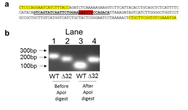
Genotyping PCRs were prepared with 5’ CTCCCAGGAATCATCTTTACC 3’ forward primer and 5’ TCATTTCGACACCGAAGCAG 3’ reverse primer [114]
37
(R0566, New England Biolabs) at 50°C for one hour. PCR products and digests were separated by gel electrophoresis on a 2% agarose gel.
GENERATION OF BLT HUMANIZED MICE
BLT mice were generated as described previously [305]. NOD/SCID-gamma chain-/- mice (NSG, Stock #5557, The Jackson Laboratories, Harbor, ME) were implanted with human thymus and liver tissue, then transplanted with autologous human liver CD34+ cells (Advanced Bioscience Resources, Alameda, CA); then they were monitored for human reconstitution in peripheral blood by flow cytometry as previously described [275, 305, 306].
HIV INFECTION AND ANTIRETROVIRAL TREATMENT OF BLT MICE
Mice were infected by intravenous exposure to 30,000 tissue culture infectious units of HIV-1JRCSF, HIV-1LAI, or HIV-189.6. Infection was monitored in peripheral blood by measuring plasma levels of vRNA as described (limit of
detection = 750 copies per ml plasma from 40µl plasma sample volume) using one-step reverse transcriptase real-time PCR (Applied Biosystems custom TaqMan Assays-by-Design, Thermo Fisher, Waltham, MA) [277, 278, 292]. Antiretroviral therapy in infected BLT mice was administered by daily intraperitoneal injection (tenofovir disoproxil fumarate 208mg/kg, emtricitabine 240mg/kg, raltegravir 56mg/kg) as previously described [292]. Tissues were harvested, and cells were isolated for DNA extraction as previously described [46, 47].
CONDITIONING AND ALLOGENEIC TRANSPLANT OF BLT MICE
38
administration. Mice were injected intraperitoneally with two doses of 25mg/kg, 24 hours apart. Lyophilized antithymocyte globulin (Genzyme, Cambridge, MA) was reconstituted in sterile water, filtered through a 0.22 micron filter, then injected
intraperitoneally with two doses of 0.3mg per mouse, four days apart. Hematopoietic stem cells were isolated and frozen as described above from a CCR5WT patient (M002) and a CCR5delta32 patient (M004). Prior to transplant, cells were thawed and counted by trypan blue exclusion, and then 3.5x10⁶ cells were injected
intravenously into conditioned BLT mice. METHYLCELLULOSE CULTURE
Cells were isolated from the bone marrow of mice as previously described [275, 306]. Bone marrow cells (2x10⁶) were incubated overnight in IMDM with 1% bovine serum albumin and penicillin/streptomycin for adherence depletion. The following day, non-adherent cells were harvested and counted by trypan blue exclusion. 100,000 cells were plated with 3ml Methocult H4434 Classic (04444, Stemcell Technologies, Vancouver, Canada) and incubated at 37°C for three weeks. Colonies were aspirated, pelleted, and frozen at -80°C before DNA extraction by digest with Proteinase K (03115887001, Roche, Basel, Switzerland).
STATISTICAL TESTS
39 RESULTS
TRANSPLANTATION OF MICE WITH CCR5WT OR CCR5DELTA32 STEM
CELLS
First, we confirmed the ability of the CCR5WT and CCR5delta32 stem cells to engraft in NSG mice. Neonatal NSG mice were irradiated and injected
intrahepatically with either CCR5WT or CCR5delta32 stem cells to generate NSG-Hu mice, and then they were monitored for human immune reconstitution in the peripheral blood by flow cytometry. Specifically, peripheral blood cells were stained for surface markers human CD45 (hematopoietic cells), CD3 (T cells), and CD4 (CD4+ T cells). At week 23 post transplant, the cohort of mice that received
CCR5delta32 cells had an average 51.6% human (CD45+) cells in peripheral blood. The CCR5WT cohort had an average of 10.6% human cells in peripheral blood (Fig. 2.1a). This difference was statistically significant, and the percentage of human cells in the peripheral blood remained higher in the CCR5delta32 cohort through week 39. The percentage CD3+ T cells was also higher in the CCR5delta32 cohort at week 23, but the differences were not significant at other time points (Fig. 2.1b). By week 31, the percentage of CD3+ T cells was greater than 90% in both cohorts. The percentage of CD4+ T cells was significantly higher in the CCR5delta32 mice (80%) than in the CCR5WT mice (60%) at weeks 31 and 35 (Fig. 2.1c).
40
with the CCR5WT donor. Therefore, it is unlikely that any difference in susceptibility of the CCR5delta32 mice to HIV infection could be explained by lower levels of human CD4+ T cells, which are the target cells for infection.
SUSCEPTIBILITY OF CCR5WT AND CCR5DELTA32 MICE TO HIV INFECTION
To determine their susceptibility to R5-tropic HIV infection, CCR5WT and CCR5delta32 NSG-Hu mice were challenged by intravenous exposure to R5-tropic HIVJRCSF. After exposure, all three CCR5WT mice had detectable viral loads by week 3, while none of the four CCR5delta32 mice had detectable viral loads over 8 weeks (Fig. 2.2a). To determine their susceptibility to X4-tropic HIV infection, mice from both cohorts were exposed to X4-tropic HIVLAI. CCR5WT mice (3/3) were viremic by week 2 after exposure, and CCR5delta32 mice (2/2) were viremic as early as week 1 after exposure (Fig. 2.2b). To determine the susceptibility of CCR5delta32 mice to dual-tropic virus, three CCR5delta32 mice were exposed to HIV89.6, and 2 out of 3 mice were viremic by week 1, with the third mouse becoming viremic at week 2 (Fig. 2.2c). These results show that both R5-tropic and X4-tropic HIV replicated in vivo in the CCR5WT mice, and that only X4- or dual-tropic HIV replicated in vivo in the CCR5delta32 mice, while R5-tropic virus did not.
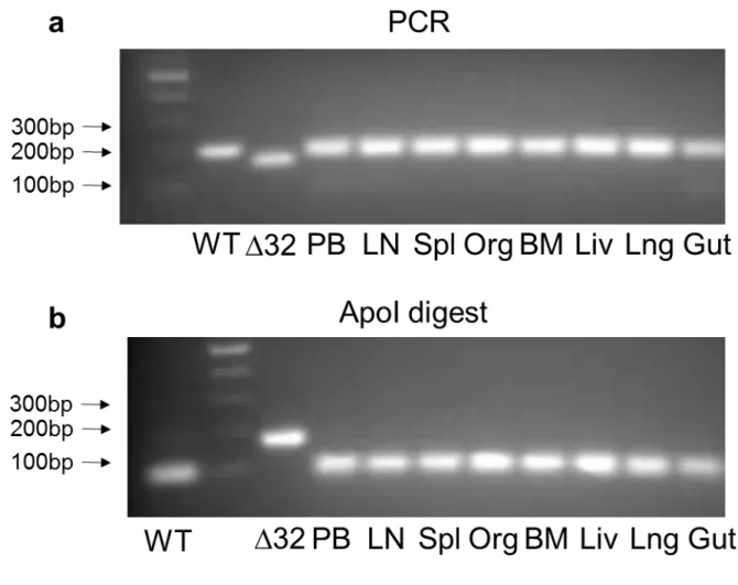
ANALYSIS OF CCR5WT AND CCR5DELTA32 GENOTYPE BY PCR/DIGEST