Francisella tularensis interactions with the lung
Joshua David Hall
A dissertation submitted to the faculty of the University of North Carolina at Chapel Hill in partial fulfillment of the requirements for the degree of Doctor of Philosophy
in the Department of Microbiology and Immunology .
Chapel Hill 2008
Approved by: Advisor: Thomas Kawula Reader: Miriam Braunstein
Reader: Janne Cannon Reader: Raymond Pickles
ABSTRACT Joshua David Hall
Francisella tularensis interactions with the lung (Under the direction of Thomas H. Kawula)
Francisella tularensis, a gram-negative bacterium, is the etiological agent of the human disease, tularemia. Inhalation of as few as 10 bacteria can result in a rapidly progressing, disseminating, and often lethal infection. The biology and pathogenesis of Francisella within the lung following inhalation is not well understood. In vitro, the bacterium replicates within macrophages, but the cellular targets and intracellular niche within an animal host are unknown.
We determined the suite of cells infected by Francisella during inhalation-acquired tularemia and assessed how pulmonary innate immune cell populations are impacted during infection. Initially, alveolar macrophages were the predominant host cell for replicating Francisella. Later, monocytes and neutrophils were recruited to the lung and neutrophils, normally potent microbial killers, became a major host cell for bacterial replication. Francisella was also found within interstitial macrophages, monocytes, dendritic cells, and alveolar type II epithelial (ATII) cells.
recruitment into the lung over wild-type mice, though the infected cell profile and bacterial burden in lungs, liver, or spleen throughout the disease course were similar, indicating that this receptor did not play a critical role during pulmonary tularemia.
Of all the identified infected cell types in the lung, only ATII cells are stationary structural cells. ATII cells possess many properties important to respiratory function including the production of pulmonary surfactant which maintains lung surface tension and contributes to protection against infectious agents. Francisella invaded ATII cells via host-cell dependent processes, and the bacterium replicated equally well within ATII host-cells as within macrophages. We isolated an insertion mutation disrupting the Francisella gene, ripA, that ablated the bacterium’s ability to replicate within ATII cells but not macrophages. This strain was significantly attenuated in a mouse model of tularemia suggesting that
TABLE OF CONTENTS
List of Tables ... vi
List of Figures ... vii
List of Abbreviations and Symbols... ix
Chapter 1 Introduction and Background... 1
Chapter 2 Francisella tularensis replicates within alveolar type II epithelial cells in vitro and in vivo following inhalation ... 28
Chapter 3 Infected host cell repertoire and cellular response following inhalation of Francisella tularensis Schu S4, LVS, or subspecies novicida U112... 41
Chapter 4 Chemokine receptor CX3CR1 deficiency leads to increased cell recruitment in the lung following inhalation of Francisella tularensis but does not impact disease outcome ... 66
Chapter 5 Francisella tularensis invasion of lung epithelial cells ... 82
Chapter 6 Use of transposon-transposase complexes to create stable insertion mutant strains of Francisella tularensis LVS... 114
Chapter 7 RipA, a cytoplasmic membrane protein unique to Francisella species is required for intracellular survival ... 130
Chapter 8 Discussion and Conclusions... 162
LIST OF TABLES Table
3.1 Comparison of Francisella tularensis strains ... 56
3.2 Identification of specific cell-types from lung homogenates... 57
6.1 Transposon insertion frequencies ... 123
LIST OF FIGURES Figure
2.1 F. tularensis LVS invades and replicates within
ATII cell lines in vitro... 35
2.2 Following inhalation F. tularensis localizes to the alveolus... 36
2.3 Following inhalation, F. tularensis LVS expressing GFP co-localized with proSP-B and proSP-C; proteins produced by ATII epithelial cells ... 37
2.4 F. tularensis LVS expressing GFP co-localized with cells expressing the macrophage marker F4/80, the dendritic cell marker CD11c, and the ATII cell markers proSP-B and proSP-C ... 38
3.1 Identifying Francisella-infected lung cells... 58
3.2 Identifying cell types in the lung by flow cytometry... 59
3.3 Lung cell types infected by Francisella following inhalation ... 60
3.4 Number of Francisella-infected cells following inhalation... 61
3.5 Effects of Francisella infection on cell number within lung populations... 62
4.1 Francisella recovery from CX3CR1-deficient mice following intranasal inoculation ... 77
4.2 Impact of Francisella infection on pulmonary cell populations from CX3CR1-deficient mice ... 78
4.3 Abundance of Francisella-infected cells in CX3CR1-deficient mice during pulmonary infection... 79
5.1 F. tularensis LVS association with and internalization by TC-1 lung epithelial cells... 103
5.2 The effect of actin and microtubule polymerization on F. tularensis LVS invasion of lung epithelial cells... 104
5.4 Representative fluorescence microscopy images demonstrating LVSgfp localization within EEA1 or LAMP-1containing
endosomes in TC-1 cells... 106
5.5 Transmission electron micrographs of TC-1 infected with F. tularensis LVS... 107
5.6 LVSgfp was analyzed by flow cytometry for escape from vacuoles into the cytoplasm of TC-1 cells……….108
6.1 Graphic representation of the chromosomal positions of the 27 identified insertion sites... 125
6.2 Southern blot of five insertion mutants and wild-type F. tularensis LVS probed with labeled Tn5………..……….126
7.1 Gentamicin protection assays of LVS and LVS ripA::tn5... 149
7.2 Graphical representation of the genomic organization of the LVS ripA locus ... 150
7.3 Gentamicin protection assays of LVS and LVS ∆ripA... 151
7.4 LVS and ∆ripA escape from vacuoles to cytoplasm ... 152
7.5 Bacterial organ burdens in infected mice... 153
7.6 Graphical representation of the ripA region and ripA expression analysis ... 154
LIST OF ABBREVIATIONS AND SYMBOLS µg microgram
µm micrometer
ATCC American Type Culture Collection ATII alveolar type II epithelial cells BEI Biodefense and Emerging Infections
bp base pair
BSA bovine serum albumin
CDC Centers for Disease Control and Prevention CFU colony forming unit
DAPI 4',6-diamidino-2-phenylindole DC dendritic cell
DNA deoxyribonucleic acid DNase deoxyribonuclease
E. Escherichia
F. Francisella
FPI Francisella pathogenicity island g gram
GFP green fluorescent protein
IACUC Institutional Animal Care and Use Committee kb kilobase
L liter
LPS lipopolysaccharide LVS Live Vaccine Strain
mL milliliter
MOI multiplicity of infection
oC degree Celsius
OD optical density ORF open reading frame PBS phosphate buffered saline
PE phycoerythrin
PerCP Peridinin-chlorophyll-protein complex RNA ribonucleic acid
ROS reactive oxygen species rRNA ribosomal RNA
SP surfactant protein T4P type 4 pili
TEM transmission electron microscopy TLR toll-like receptor
U unit ∆ deletion
CHAPTER 1
Introduction and Background
Discovery of Francisella tularensis
During the spring of 1911 in Tulare County, California, a plague-like disease
afflicting hundreds of ground squirrels was documented by Dr. George McCoy (65). Within a year, McCoy and his associate isolated and identified the causative agent as a
gram-negative bacterium which they named Bacterium tularense (66). Edward Francis, a
prominent scientist of the day, named the disease caused by Bacterium tularense, “tularemia” and demonstrated that this disease of small mammals could be transmitted to man through various routes including handling of infected carcasses and biting insects (34). Dr. Francis determined that similar diseases observed in Europe and Japan were, in fact, tularemia and were caused by the same bacterial pathogen. As methods of bacterial taxonomy became more sophisticated, it was evident that the agent of tularemia did not fit into previously described bacterial phylogenetic groups (88). Thus, in 1947 the bacterial genus Francisella was named in honor of Dr Francis, and the causative agent of tularemia was renamed, Francisella tularensis.
Francisella in humans
severe disease in guinea pigs, rabbits, rats, mice, ground squirrels, gophers, and monkeys (67). Infections in monkeys led the researchers to believe that the organism could also cause disease in humans. Soon afterwards, infections of meat-packers and animal hunters were reported with the causative agent identified as B. tularense (98). As scientific interest in studying this newfound pathogen grew, so did the incidence of laboratory-acquired infections. Francisella infections in the laboratory have been common since the very
beginning of tularemia research. In the initial papers describing Bacterium tularense, McCoy reported that his serum and the serum from a fellow lab worker agglutinated the bacteria (66), indicating prior exposure during the processing of hundreds of diseased rodents. There are reports of McCoy taking extended leaves of absence from his research due to acute illness which included high fever (36). By 1959, there were 55 reported laboratory-acquired infections although other accounts estimate this number at as much as 200 (47).
Seven clinical types of human tularemia were recognized by 1947 including,
Worldwide Incidence of Francisella infections
Incidence of human tularemia is widespread among the northern hemisphere, though the degree of incidence varies greatly from region to region. Almost 60% of the 778 reported cases of tularemia in the United States between 2000 and 2005 occurred in 5 states:
Missouri, Arkansas, Oklahoma, Massachusetts, and South Dakota (49). Tularemia is likely to be highly underreported due to general symptoms, such as fever, which often results in antibiotic treatment which could allow recovery from infection in the absence of
identification. Reported incidence of tularemia in European countries is higher than in the United States with 2,697 reports of tularemia in Finland and 1,828 in Sweden from 2000-2005 (49). Severity of disease varies widely among Francisella subspecies, and various subspecies are localized within different geographic regions.
Taxonomy
In 1959 it was first proposed that Francisella tularensis from North America and Europe divided into two groups which were indistinguishable serologically, but displayed extreme differences with regard to virulence in small mammals and humans (75). Today, Francisella tularensis is divided into four subspecies based on biochemical determinants and virulence in rabbits; tularensis, holarctica, novicida, and mediasiatica. Found predominantly in North America (49), subspecies, tularensis is the most virulent in humans, causing
are isolated and rare. Subspecies tularensis, holarctica, and novicida all cause lethal
infections in mice which has resulted in the mouse being the most common animal model for the study of tularemia.
Natural reservoirs
The environmental reservoir allowing F. tularensis to persist in nature is unclear. Tularemia is a zoonosis, as the bacterium establishes infection within a wide range of mammals, however all of these species develop acute infections making it unlikely that any mammal species is a true environmental reservoir of Francisella. Infections of
water-dwelling animals such as beavers and lemmings (8) have been observed indicating a potential water reservoir. F. tularensis has been detected in water samples taken from sources near human tularemia outbreaks (8, 50), though it is unclear if bacteria are free-living or
associated with a unicellular host. Evidence for the latter is that Francisella has the ability to survive and replicate within amoebae species, though once infected, these cells are eventually killed by Francisella in vitro (2, 9). Insect vectors could provide an environmental reservoir for Francisella. The organism has been isolated from tick populations (44, 45, 63) and early work on Francisella identified disease acquired from biting flies (33). In Martha’s Vineyard, as many as 5% of dog ticks in certain areas contained F. tularensis, though the most common primary form of disease is pneumonic for unknown reasons (63). A likely conclusion from these varied isolation sources is that Francisella persists in many different environmental niches among different geographical locations.
Francisella as a biological weapon
confirmed cases of anthrax including 5 deaths (6). The estimated cost of dealing with this event was greater than US $1 billion (52).
In response to this act of bioterrorism, the U.S. government greatly increased the amount of funding for organisms that pose a threat for purposeful release. Those given highest priority were defined as “Category A Diseases/Agents”(1). Organisms in this group pose a risk to national security due to their ease of dissemination or transmission, high mortality rate, and potential for causing panic and social disruption. Members of this
category are Bacillus anthracis (anthrax), toxin produced by Clostridium botulinum, Yersinia pestis (plague), variola major (smallpox), viral hemorrhagic fevers (ebola, lassa), and
Francisella tularensis (tularemia).
Characteristics of Francisella tularensis have made it attractive for development as a biological weapon. The organism is extremely infectious, with a dose of less than 10
bacteria necessary to cause disease in humans (85). The organism causes the most acute and severe disease when inhaled (26), and F. tularensis is easily aerosolized (18, 19). In the environment, Francisella can survive for weeks to months in water (31), and potentially as long in dead animal carcasses, or grasses and hay which would result in prolonged and repeated exposure long after the initial release event. Since Francisella is a natural host to a wide variety of animal species, bacterial populations could persist in numerous and varied animal reservoirs which would lead to continued exposure for quite some time.
executive order terminated U.S. biological weapons programs and all stocks of these
weapons were destroyed by 1973 (14). Parallel programs existed in the former Soviet Union until the 1990s which allegedly led to the production of Francisella strains resistant to
antibiotics and vaccines (3). There are claims that a massive tularemia outbreak that affected more than 10,000 German and Soviet soldiers during World War II was the result of an intentional Soviet release (3), though this assertion is challenged and the real result of such an outbreak could be due to increased proximity to infected rodents during times of war (20).
In 1969, the World Health Organization developed a committee to assess the medical, social, and financial impacts of biological attacks. The committee estimated that an aerosol dispersal of 50 kg of live Francisella tularensis over a metropolitan area would result in 250,000 incapacitated individuals including 19,000 deaths (102). The appearance of acute, non-specific, respiratory illness would appear in the exposed population 3-5 days following release. In the early stages, it would be difficult for public health authorities to diagnose a tularemia outbreak due to the initial similarities with more common community-acquired outbreaks such as influenza (discussed in 23). Illness would persist for weeks to months with multiple relapses being probable. The CDC estimates that the financial burden of dealing with a tularemia outbreak would be approximately $5.4 billion per 100,000 exposed people (48).
Pathogenic factors of Francisella tularensis
Despite the potential risks of a purposeful Francisella release, very little is
to light concurrently with the studies presented here. In Chapter 8, I will attempt to frame this body of work within the current (April 2008) literature and discuss how these data filled a knowledge gap within the field, as well as will influence future studies.
Replication within macrophages
The most commonly studied Francisella phenotype related to pathogenesis is the ability to replicate within macrophages. Intra-macrophage replication has been observed within cell lines and primary cells from mice, humans, and other mammalian hosts (5, 39). Francisella entry into macrophages may occur via a novel mechanism involving spacious pseudopod loops (15). Immediately following phagocytosis, Francisella is contained within a membrane-enclosed compartment (39) that contains the early endosomal marker, EEA-1 (13). This vacuole becomes associated with lysosome-associated membraneglycoproteins (LAMPs), but acidification does not take place (13, 16). Instead, by an unknown
mechanism, the phagosomal membrane degrades within 1-4 hours and Francisella replicates within the host cell cytoplasm (13, 16, 39). There is some evidence that intracellular
Francisella re-enters a LAMP-positive compartment by 24 hours following phagocytosis (13) prior to death of the host cell.
The ability of F. tularensis to cause disease may be directly tied to the organism’s ability to escape the endocytic pathway and avoid degradation by the lysosome. Francisella mutants that are unable to replicate within macrophages are attenuated for virulence in animal hosts (69, 72, 84, 94). Three of these intracellular replication-deficient mutants,
Dissemination in an animal host
Independent of inoculation route, Francisella disseminates from the initial site of infection to distal tissues. Within a day following infection, bacteria are found in the
draining lymph nodes in close proximity of the infection site, followed by bacterial presence in the lungs, liver, and spleen (32). Once bacteria arrive in these tissues, they begin
replicating logarithmically. Due to the extreme bacterial burden in various tissues, multiple organ failure is a probable cause of death in fatal cases of tularemia.
Pathogenicity island
The sequencing of Francisella genomes, as well as genetic studies within F.
tularensis novicida, have facilitated the discovery of a Francisella pathogenicity island (FPI). The FPI has been classified as such due to it’s deviation in G+C content from the rest of the chromosome as well as the fact it contains a cluster of genes required for full virulence (71). The FPI is made up of 17-19 genes and is duplicated within holarctica and tularensis strains. Because of this, much of the preliminary work on the FPI has been performed in the novicida strain U112, which has only one copy, making genetic manipulations of the FPI more
are required for intracellular replication within macrophages and for full virulence within an animal host (21, 72, 83, 92). The FPI genes are clearly critical for pathogenesis of
Francisella, however their function remains a mystery. Sequence homology of several FPI ORFs has led to the proposal that these genes function as a type VI secretion system (71), though further work is needed to substantiate this claim.
LPS
A major component of the gram-negative outer membrane is lipopolysaccharide (LPS). Taking advantage of this, many organisms produce receptors and pattern-recognition molecules that detect the presence of LPS which results in stimulation of an immune
response aimed at clearing the bacterial invader. LPS recognition occurs primarily through toll-like receptor 4 (TLR4) which is present on macrophages and dendritic cells among other cell types (68). Activation of the TLR4 complex leads to translocation of NFκB to the nucleus where transcription of genes important in the inflammatory response are initiated (24).
Francisella immune evasion appears to partly be due to a lack of host-cell
recognition. In the presence of increasing amounts of Francisella LPS, human and mouse monocytic cells produce very little, if any, inflammatory cytokines such as TNF-α, IL-6, and IL- 8 (17, 25, 42). In contrast, Salmonella and E. coli LPS induce high levels of these cytokines (25, 42). The lack of host immune stimulation is most likely because Francisella LPS does not interact with TLR4 (17, 41).
Pilus
mainly homopolymeric structures that extend from the bacteria and can mediate attachment to a host cell (79). The F. tularensis holarctica Live Vaccine Strain (LVS), and the highly virulent F. tularensis tularensis Schu S4 strain displayed long thin fibers on their surface that were reminiscent of type 4 pili (T4P) produced by other gram-negative pathogens (38, 62, 79). Analysis of Francisella genome sequences revealed the presence of gene clusters with homology to T4P genes found in Neisseria spp.and Pseudomonas aeruginosa (38). The Schu S4 genome contained a pilAEV locus predicted to encode three full length genes. In holarctica isolates, pilE and pilV are truncated due to nonsense mutations that introduce a stop codon. In a subset of holarctica isolates, including LVS, the majority of pilA is deleted due to homologous recombination of two 120bp direct repeats flanking the gene (30).
Novicida ssp. are similar to tularensis ssp. with the exception of differences in the C-terminal portion of pilA. In other gram-negative pathogens, PilA is the major pilin subunit.
Functional studies demonstrate the pilA of Francisella is expressed and PilA protein is exported to the cell surface where it forms multimeric complexes (30). Schu S4 and F. tularensis novicida U112 pilA cloned into a heterologous Neisseria T4P expression system resulted in expression of a functional pilus (29). A pilA deletion strain retained the ability to replicate in macrophages, though to a lesser degree than wildtype, but was attenuated for dissemination to the spleen following sub-cutaneous infection of mice (30). Though LVS lacks full length pilA, TEM studies have identified T4P-like structures on the bacterial surface, whose expression has been linked to pilB (29). These results indicate that
major pilin subunits between subspecies; particularly in strains that lack full length pilA, such as LVS.
Capsule
Evading the host immune system is an important criteria for a successful pathogen. One strategy employed by several bacterial human pathogens is the presence of a
polysaccharide capsule which surrounds the bacterial outer membrane and prevents host recognition of bacterial components. Often, the presence of a capsule is required for full virulence of an organism, as is the case with Staphylococcus aureus and Streptococcus pneumoniae (76, 80). The capsule often confers the ability of bacteria to avoid phagocytosis, however intracellular replication of Francisella may be an important component of the bacterium’s patho-biology within an animal host.
Francisella strains lacking an electron dense zone, proposed to be a capsule, have been isolated (82, 89). These strains are more sensitive to serum killing and are attenuated in mice (82). However, these strains also contain significant differences in their outer
Inhalation tularemia
From a biological weapon perspective, the most serious tularemia transmission route is via inhalation (77). An inhalation exposure is predicted to incapacitate the largest number of civilians and carry the largest financial burden to an affected community (102). To understand how Francisella instigates a lethal infection following inhalation, it is important to understand the host mechanisms in place to deal with inhaled foreign particles. This will enable deciphering of how Francisella subverts these pulmonary immune functions in order to establish disease.
Pulmonary innate immunity
The lung is the largest epithelial surface in the body, and is important for efficient gas exchange from the air to the blood stream. Respiration moves 15,000 liters of air over 150 m2 of epithelial surface area (64). A byproduct of this interaction is constant exposure of external particles and potentially harmful pathogens to the lung during respiration. To protect the host, a complex defense system has evolved that combines physical barriers, pathogen-ingesting cells, microbicidal molecules, and molecular signals that enable
communication among all of the components. These systems must work in concert to control potential infections without interfering with respiration, a primary function necessary for the life of the organism.
Physical barrier
the lung, sending foreign matter back from whence it came, up the airway where it can be coughed out or swallowed.
Alveolar macrophages
The alveolus is a sterile environment, and alveolar macrophages are key scavengers and sentinels that play a critical role in maintaining alveolar sterility. These cells are efficient at phagocytosing organisms that invade the alveolar space (37). Following phagocytosis, alveolar macrophages often single-handedly eliminate intruding
microorganisms through production of microbicidal molecules such as reactive oxygen radicals and nitric oxides (37). If inhaled pathogenic microorganisms cannot be quickly and efficiently contained and eliminated, alveolar macrophages become potent producers of cytokines, such as TNF-α and IL-6 which recruit neutrophils and monocytes from the blood stream into the lung to aid in clearing the infection (70). In this way, the alveolar
macrophage is central to initiating an innate immune response within the lung in response to respiratory pathogens.
Dendritic cells
attracted to chemokines produced by T-cells which are found in large numbers within lymph nodes. This leads to DC migration from the initial infection site to the draining lymph node where the antigen is presented to lymphocytes, leading to proliferation of antigen-specific T-cells which can travel to the site of infection and aid in controlling the disease (96). In this way, the DC acts as a bridge joining the innate and adaptive immune systems together.
Monocytes
Monocytes are cells that circulate in the bloodstream and can be recruited to infected tissues by cytokine signals. Monocytes are precursors to macrophages (95) and dendritic cells (81) and upon entry into tissue, differentiate into mature phagocytes that can aid in the clearance of invading pathogens (10). During infection, endothelial cells respond to
inflammatory signals produced by macrophages and DCs which leads to upregulation of chemokines on their cell surface (7). Through interaction with cognate molecules on the surface of recruited cells, such as monocytes and neutrophils, extravasation of these cells into infected tissues is facilitated (46).
Neutrophils
Perhaps the cell with the most potent antimicrobial potential is the neutrophil. These cells are highly phagocytic and have several pathways available to eliminate invading microorganisms (73). Neutrophils are crucial to the protection of the host from microbial infections. People with inherited disorders affecting neutrophil number or function suffer from frequent and severe microbial diseases (74).
pathogens, but also to host cells. Therefore regulation of their production is tightly
controlled. When a neutrophil is in an inactivated state, NADPH subunits are segregated into different cellular compartments which prevents the enzyme from being active. Two subunits, gp91phox and p22phox are targeted to the membranes of phagosomes whereas the remaining
NADPH subunits, p47phox, p67phox, and rac2, are distributed within the cytoplasm of resting
neutrophils (73). Within 30 seconds after particle binding on the neutrophil cell surface, oxidase subunits localize to the invaginating membrane that will become the phagosome (4, 22). Assembled and active NADPH oxidase is limited to the particle-containing phagosome which is critical in effectively killing potentially pathogenic organisms while simultaneously protecting host cells and tissues from oxidative damage (22, 73).
Neutrophils also possess non-oxidative means to kill microorganisms. Anaerobic killing in neutrophils can occur via phagosome maturation and fusion with a lysosome, similar to macrophages. However, this process occurs more rapidly in neutrophils and is augmented through the fusion of the phagosome with granules containing a suite of cationic anti-microbial peptides (27, 51). Activated neutrophils also possess the ability to expel the contents of the anti-microbial containing vesicles into the extracellular environment as a means of controlling an infection, however this often occurs at the expense of damage to host tissues (27). Neutrophils are often critical to controlling bacterial infections with a
ATII cells
The primary function of the lung is to facilitate gas exchange from the air into the bloodstream. To mediate efficient gas transfer, the lung epithelial cell surface is quite large. Mucociliary clearance is an important host defense mechanism utilized in the airways, but it requires an air-liquid interface. Surface tension at this interface creates collapsing forces that would make respiration difficult (100). The system that has evolved to circumvent this problem is the production of pulmonary surfactant, which creates a lipid-rich monolayer separating liquid and gas at the epithelial cell surface, thus reducing the surface tension and allowing respiration to occur easily. A lack of sufficient surfactant leads to respiratory failure (99).
The primary producer of pulmonary surfactant is the alveolar type II (ATII) epithelial cell. Surfactant is made up of 90% phospholipids and 10% protein, which includes two hydrophilic proteins, surfactant protein A (SP-A) and surfactant protein D (SP-D), and two hydrophobic proteins, surfactant protein B (SP-B) and surfactant protein C (SP-C) (28, 64). SP-A and SP-D have homology to proteins labeled “collectins” found in serum. These proteins have the ability to recognize common patterns found on the surface of
The hydrophobic surfactant proteins, SP-B and SP-C do not have identified roles in host defense, but are necessary for maintaining proper surface tension at the liquid-air interface within the lung (100). Both proteins are produced primarily by ATII cells and are stored in lamellar bodies before being secreted into the alveolus. Once secreted, interactions with phospholipids occur which leads to the formation of stable monolayers and bilayers, thus reducing surface tension in the lung.
The ATII cell has been referred to as the “stem cell of the alveolar epithelium” (28). In response to alveolar injury, ATII cells proliferate and become new ATII cells or
differentiate into ATI cells. In this way, ATII cells are critical for repair of damaged alveolar epithelium. In addition, ATII cells aid in the innate immune response to invading pathogens by producing pro-inflammatory cytokines as well as responding to signals produced by macrophages and DCs (28). This leads to increased epithelial layer permeability which facilitates the extravasation of neutrophils and monocytes from the blood stream into the alveolus to aid in battling an infection.
Pulmonary immune response to Francisella
Within one hour following inhalation of Francisella, bacteria are found within
resulting in no production of TNF-α (91). This suppression may be a result of TGF-β
production, which is activated during Francisella infection (11). Francisella infection also suppresses T-cell activation by macrophages via production of prostaglandin-E2 (101). In this way, Francisella pathogenesis in the lung is most likely achieved through a balance of avoiding immune recognition and actively subverting the host response.
Key questions
Understanding pathogenic mechanisms utilized during pulmonary tularemia is important to deciphering more effective vaccines or therapeutics to combat the disease. Also, understanding Francisella’s manipulation of the host immune response can provide more specific insight into how lung pathogens interact with and exploit the host immune response to cause disease. Francisella replicates within macrophages in vitro and the bacterium can avoid phagosomal killing and suppresses pro-inflammatory signaling, clearly aiding the organism’s survival. However the suite of cells infected by Francisella following inhalation is unknown. Also, it is unclear how infection by Francisella affects cell
populations in the lung as disease progresses. These are fundamental pieces of information that must be in place to most efficiently make directed hypotheses concerning how
Francisella causes disease when inhaled. A major goal of this work was to fill in these gaps and determine which cells become infected by Francisella in the lung and how those
populations are affected over time.
identified; the ATII cell. This work follows a top-down approach, beginning from the understanding that inhaling F. tularensis causes a rapidly progressing lethal disease. Knowing that Francisella survives in host cells in vitro, this led us to determine which cells were infected within the lung following inhalation. Subsequently, we could ask more specific questions about how the bacterium gains entry into those cells and what is the intracellular biology of the organism. To discover the bacterial factors important, or even necessary, for aspects of Francisella’s lifestyle within the host, we needed the ability to make mutants in specific genes and decipher how disruption of those genes changed specific components of the bacterium’s pathogenesis. Lastly, understanding how Francisella causes disease in a host gives us a baseline to study how host factors are important during inhalation tularemia through the use of mice deficient for specific immune components. In this body of work, we move from gross pathology progressively towards the molecular details of disease.
REFERENCES 1. http://www.bt.cdc.gov/agent/agentlist-category.asp
2. Abd, H., T. Johansson, I. Golovliov, G. Sandstrom, and M. Forsman. 2003. Survival and growth of Francisella tularensis in Acanthamoeba castellanii. Appl Environ Microbiol 69:600-6.
3. Alibek, K., and S. Handelman. 1999. Biohazard : the chilling true story of the largest covert biological weapons program in the world, told from the inside by the man who ran it, 1st ed. Random House, New York.
4. Allen, L. A., F. R. DeLeo, A. Gallois, S. Toyoshima, K. Suzuki, and W. M. Nauseef. 1999. Transient association of the nicotinamide adenine dinucleotide phosphate oxidase subunits p47phox and p67phox with phagosomes in neutrophils from patients with X-linked chronic granulomatous disease. Blood 93:3521-30. 5. Anthony, L. D., R. D. Burke, and F. E. Nano. 1991. Growth of Francisella spp. in
6. Atlas, R. M. 2002. Bioterriorism: from threat to reality. Annu Rev Microbiol 56:167-85.
7. Bazan, J. F., K. B. Bacon, G. Hardiman, W. Wang, K. Soo, D. Rossi, D. R. Greaves, A. Zlotnik, and T. J. Schall. 1997. A new class of membrane-bound chemokine with a CX3C motif. Nature 385:640-4.
8. Berdal, B. P., R. Mehl, H. Haaheim, M. Loksa, R. Grunow, J. Burans, C. Morgan, and H. Meyer. 2000. Field detection of Francisella tularensis. Scand J Infect Dis 32:287-91.
9. Berdal, B. P., R. Mehl, N. K. Meidell, A. M. Lorentzen-Styr, and O. Scheel. 1996. Field investigations of tularemia in Norway. FEMS Immunol Med Microbiol 13:191-5.
10. Blusse van Oud Alblas, A., B. van der Linden-Schrever, and R. van Furth. 1981. Origin and kinetics of pulmonary macrophages during an inflammatory reaction induced by intravenous administration of heat-killed bacillus Calmette-Guerin. J Exp Med 154:235-52.
11. Bosio, C. M., H. Bielefeldt-Ohmann, and J. T. Belisle. 2007. Active suppression of the pulmonary immune response by Francisella tularensis Schu4. J Immunol
178:4538-47.
12. Bosio, C. M., and S. W. Dow. 2005. Francisella tularensis induces aberrant activation of pulmonary dendritic cells. J Immunol 175:6792-801.
13. Checroun, C., T. D. Wehrly, E. R. Fischer, S. F. Hayes, and J. Celli. 2006.
Autophagy-mediated reentry of Francisella tularensis into the endocytic compartment after cytoplasmic replication. Proc Natl Acad Sci U S A 103:14578-83.
14. Christopher, G. W., T. J. Cieslak, J. A. Pavlin, and E. M. Eitzen, Jr. 1997. Biological warfare. A historical perspective. Jama 278:412-7.
15. Clemens, D. L., B. Y. Lee, and M. A. Horwitz. 2005. Francisella tularensis enters macrophages via a novel process involving pseudopod loops. Infect Immun 73:5892-902.
16. Clemens, D. L., B. Y. Lee, and M. A. Horwitz. 2004. Virulent and avirulent strains of Francisella tularensis prevent acidification and maturation of their phagosomes and escape into the cytoplasm in human macrophages. Infect Immun 72:3204-17.
18. Cox, C. S. 1971. Aerosol survival of Pasteurella tularensis disseminated from the wet and dry states. Appl Microbiol 21:482-6.
19. Cox, C. S., J. S. Derr, Jr., E. G. Flurie, and R. C. Roderick. 1970. Experimental technique for studying aerosols of lyophilized bacteria. Appl Microbiol 20:927-34. 20. Croddy, E., and S. Krcalova. 2001. Tularemia, biological warfare, and the battle for
Stalingrad (1942-1943). Mil Med 166:837-8.
21. de Bruin, O. M., J. S. Ludu, and F. E. Nano. 2007. The Francisella pathogenicity island protein IglA localizes to the bacterial cytoplasm and is needed for intracellular growth. BMC Microbiol 7:1.
22. DeLeo, F. R., L. A. Allen, M. Apicella, and W. M. Nauseef. 1999. NADPH oxidase activation and assembly during phagocytosis. J Immunol 163:6732-40.
23. Dennis, D. T., T. V. Inglesby, D. A. Henderson, J. G. Bartlett, M. S. Ascher, E. Eitzen, A. D. Fine, A. M. Friedlander, J. Hauer, M. Layton, S. R. Lillibridge, J. E. McDade, M. T. Osterholm, T. O'Toole, G. Parker, T. M. Perl, P. K. Russell, and K. Tonat. 2001. Tularemia as a biological weapon: medical and public health management. Jama 285:2763-73.
24. Doyle, S. L., and L. A. O'Neill. 2006. Toll-like receptors: from the discovery of NFkappaB to new insights into transcriptional regulations in innate immunity. Biochem Pharmacol 72:1102-13.
25. Duenas, A. I., M. Aceves, A. Orduna, R. Diaz, M. Sanchez Crespo, and C. Garcia-Rodriguez. 2006. Francisella tularensis LPS induces the production of cytokines in human monocytes and signals via Toll-like receptor 4 with much lower potency than E. coli LPS. Int Immunol 18:785-95.
26. Ellis, J., P. C. Oyston, M. Green, and R. W. Titball. 2002. Tularemia. Clin Microbiol Rev 15:631-46.
27. Faurschou, M., and N. Borregaard. 2003. Neutrophil granules and secretory vesicles in inflammation. Microbes Infect 5:1317-27.
28. Fehrenbach, H. 2001. Alveolar epithelial type II cell: defender of the alveolus revisited. Respir Res 2:33-46.
30. Forslund, A. L., K. Kuoppa, K. Svensson, E. Salomonsson, A. Johansson, M. Bystrom, P. C. Oyston, S. L. Michell, R. W. Titball, L. Noppa, E.
Frithz-Lindsten, M. Forsman, and A. Forsberg. 2006. Direct repeat-mediated deletion of a type IV pilin gene results in major virulence attenuation of Francisella tularensis. Mol Microbiol 59:1818-30.
31. Forsman, M., E. W. Henningson, E. Larsson, T. Johansson, and G. Sandstrom. 2000. Francisella tularensis does not manifest virulence in viable but non-culturable state. FEMS Microbiol Ecol 31:217-224.
32. Fortier, A. H., M. V. Slayter, R. Ziemba, M. S. Meltzer, and C. A. Nacy. 1991. Live vaccine strain of Francisella tularensis: infection and immunity in mice. Infect Immun 59:2922-8.
33. Francis, E. 1919. Deerfly fever or Pahvant Valley plague. A disease of man of hitherto unknown etiology. Public Health Reports 34:2061-2062.
34. Francis, E. 1921. The occurence of tularemia in nature as a disease of man. Public Health Reports 36:1731-1738.
35. Francis, E. 1947. Streptomycin in treatment of tularemia. Transactions of the Association of American Physicians 60:181-187.
36. Francis, E. 1925. Tularemia. JAMA 84:1243-1250.
37. Franke-Ullmann, G., C. Pfortner, P. Walter, C. Steinmuller, M. L. Lohmann-Matthes, and L. Kobzik. 1996. Characterization of murine lung interstitial macrophages in comparison with alveolar macrophages in vitro. J Immunol 157:3097-104.
38. Gil, H., J. L. Benach, and D. G. Thanassi. 2004. Presence of pili on the surface of Francisella tularensis. Infect Immun 72:3042-7.
39. Golovliov, I., V. Baranov, Z. Krocova, H. Kovarova, and A. Sjostedt. 2003. An attenuated strain of the facultative intracellular bacterium Francisella tularensis can escape the phagosome of monocytic cells. Infect Immun 71:5940-50.
40. Gray, C. G., S. C. Cowley, K. K. Cheung, and F. E. Nano. 2002. The identification of five genetic loci of Francisella novicida associated with intracellular growth. FEMS Microbiol Lett 215:53-6.
42. Hajjar, A. M., M. D. Harvey, S. A. Shaffer, D. R. Goodlett, A. Sjostedt, H. Edebro, M. Forsman, M. Bystrom, M. Pelletier, C. B. Wilson, S. I. Miller, S. J. Skerrett, and R. K. Ernst. 2006. Lack of in vitro and in vivo recognition of Francisella tularensis subspecies lipopolysaccharide by Toll-like receptors. Infect Immun 74:6730-8.
43. Harris, S. 1992. Japanese biological warfare research on humans: a case study of microbiology and ethics. Ann N Y Acad Sci 666:21-52.
44. Hubalek, Z., and J. Halouzka. 1997. Mosquitoes (Diptera: Culicidae), in contrast to ticks (Acari: Ixodidae), do not carry Francisella tularensis in a natural focus of
tularemia in the Czech Republic. J Med Entomol 34:660-3.
45. Hubalek, Z., F. Treml, J. Halouzka, Z. Juricova, M. Hunady, and V. Janik. 1996. Frequent isolation of Francisella tularensis from Dermacentor reticulatus ticks in an enzootic focus of tularaemia. Med Vet Entomol 10:241-6.
46. Imai, T., K. Hieshima, C. Haskell, M. Baba, M. Nagira, M. Nishimura, M. Kakizaki, S. Takagi, H. Nomiyama, T. J. Schall, and O. Yoshie. 1997.
Identification and molecular characterization of fractalkine receptor CX3CR1, which mediates both leukocyte migration and adhesion. Cell 91:521-30.
47. Jellison, W. L. 1974. Tularemia in North America, 1930-1974. University of Montana, University of Montana Foundation, Missoula.
48. Kaufmann, A. F., M. I. Meltzer, and G. P. Schmid. 1997. The economic impact of a bioterrorist attack: are prevention and postattack intervention programs justifiable? Emerg Infect Dis 3:83-94.
49. Keim, P., A. Johansson, and D. M. Wagner. 2007. Molecular epidemiology, evolution, and ecology of Francisella. Ann N Y Acad Sci 1105:30-66.
50. Leblebicioglu, H., S. Esen, D. Turan, Y. Tanyeri, A. Karadenizli, F. Ziyagil, and G. Goral. 2007. Outbreak of tularemia: a case-control study and environmental investigation in Turkey. Int J Infect Dis.
51. Lee, W. L., R. E. Harrison, and S. Grinstein. 2003. Phagocytosis by neutrophils. Microbes Infect 5:1299-306.
52. Lengel, A. 2005. Little Progress In FBI Probe of Anthrax Attacks, Washington Post, Washington D.C.
54. LeVine, A. M., J. Elliott, J. A. Whitsett, A. Srikiatkhachorn, E. Crouch, N. DeSilva, and T. Korfhagen. 2004. Surfactant protein-d enhances phagocytosis and pulmonary clearance of respiratory syncytial virus. Am J Respir Cell Mol Biol 31:193-9.
55. LeVine, A. M., K. Hartshorn, J. Elliott, J. Whitsett, and T. Korfhagen. 2002. Absence of SP-A modulates innate and adaptive defense responses to pulmonary influenza infection. Am J Physiol Lung Cell Mol Physiol 282:L563-72.
56. LeVine, A. M., K. E. Kurak, M. D. Bruno, J. M. Stark, J. A. Whitsett, and T. R. Korfhagen. 1998. Surfactant protein-A-deficient mice are susceptible to
Pseudomonas aeruginosa infection. Am J Respir Cell Mol Biol 19:700-8.
57. LeVine, A. M., J. A. Whitsett, J. A. Gwozdz, T. R. Richardson, J. H. Fisher, M. S. Burhans, and T. R. Korfhagen. 2000. Distinct effects of surfactant protein A or D deficiency during bacterial infection on the lung. J Immunol 165:3934-40.
58. LeVine, A. M., J. A. Whitsett, K. L. Hartshorn, E. C. Crouch, and T. R.
Korfhagen. 2001. Surfactant protein D enhances clearance of influenza A virus from the lung in vivo. J Immunol 167:5868-73.
59. Lindgren, H., I. Golovliov, V. Baranov, R. K. Ernst, M. Telepnev, and A. Sjostedt. 2004. Factors affecting the escape of Francisella tularensis from the phagolysosome. J Med Microbiol 53:953-8.
60. Lippmann, M., D. B. Yeates, and R. E. Albert. 1980. Deposition, retention, and clearance of inhaled particles. Br J Ind Med 37:337-62.
61. Mandic-Mulec, I., J. Weiss, and A. Zychlinsky. 1997. Shigella flexneri is trapped in polymorphonuclear leukocyte vacuoles and efficiently killed. Infect Immun 65:110-5.
62. Mattick, J. S., C. B. Whitchurch, and R. A. Alm. 1996. The molecular genetics of type-4 fimbriae in Pseudomonas aeruginosa--a review. Gene 179:147-55.
63. Matyas, B. T., H. S. Nieder, and S. R. Telford, 3rd. 2007. Pneumonic tularemia on Martha's Vineyard: clinical, epidemiologic, and ecological characteristics. Ann N Y Acad Sci 1105:351-77.
64. McCormack, F. X., and J. A. Whitsett. 2002. The pulmonary collectins, SP-A and SP-D, orchestrate innate immunity in the lung. J Clin Invest 109:707-12.
43:53-66. McCoy, G. W., and C. W. Chapin. 1912. Bacterium tularense the cause of a plague-like disease of rodents. Public Health Bulletin 53.
67. McCoy, G. W., and C. W. Chapin. 1912. Further observations on a plague-like disease of rodents with a preliminary note on the causative agent, Bacterium tularense. J Infect Dis 10:61-72.
68. Miller, S. I., R. K. Ernst, and M. W. Bader. 2005. LPS, TLR4 and infectious disease diversity. Nat Rev Microbiol 3:36-46.
69. Mohapatra, N. P., A. Balagopal, S. Soni, L. S. Schlesinger, and J. S. Gunn. 2007. AcpA is a Francisella acid phosphatase that affects intramacrophage survival and virulence. Infect Immun 75:390-6.
70. Monton, C., and A. Torres. 1998. Lung inflammatory response in pneumonia. Monaldi Arch Chest Dis 53:56-63.
71. Nano, F. E., and C. Schmerk. 2007. The Francisella pathogenicity island. Ann N Y Acad Sci 1105:122-37.
72. Nano, F. E., N. Zhang, S. C. Cowley, K. E. Klose, K. K. Cheung, M. J. Roberts, J. S. Ludu, G. W. Letendre, A. I. Meierovics, G. Stephens, and K. L. Elkins. 2004. A Francisella tularensis pathogenicity island required for intramacrophage growth. J Bacteriol 186:6430-6.
73. Nauseef, W. M. 2007. How human neutrophils kill and degrade microbes: an integrated view. Immunol Rev 219:88-102.
74. Newburger, P. E. 2006. Disorders of neutrophil number and function. Hematology Am Soc Hematol Educ Program:104-10.
75. Olsufiev, N. G., O. S. Emelyanova, and T. N. Dunayeva. 1959. Comparative study of strains of B. tularense in the old and new world and their taxonomy. J Hyg
Epidemiol Microbiol Immunol 3:138-49.
76. O'Riordan, K., and J. C. Lee. 2004. Staphylococcus aureus capsular polysaccharides. Clin Microbiol Rev 17:218-34.
77. Oyston, P. C., A. Sjostedt, and R. W. Titball. 2004. Tularaemia: bioterrorism defence renews interest in Francisella tularensis. Nat Rev Microbiol 2:967-78. 78. Pammit, M. A., E. K. Raulie, C. M. Lauriano, K. E. Klose, and B. P.
79. Pizarro-Cerda, J., and P. Cossart. 2006. Bacterial adhesion and entry into host cells. Cell 124:715-27.
80. Preston, J. A., and D. H. Dockrell. 2008. Virulence factors in pneumococcal respiratory pathogenesis. Future Microbiol 3:205-21.
81. Randolph, G. J., K. Inaba, D. F. Robbiani, R. M. Steinman, and W. A. Muller. 1999. Differentiation of phagocytic monocytes into lymph node dendritic cells in vivo. Immunity 11:753-61.
82. Sandstrom, G., S. Lofgren, and A. Tarnvik. 1988. A capsule-deficient mutant of Francisella tularensis LVS exhibits enhanced sensitivity to killing by serum but diminished sensitivity to killing by polymorphonuclear leukocytes. Infect Immun 56:1194-202.
83. Santic, M., M. Molmeret, J. R. Barker, K. E. Klose, A. Dekanic, M. Doric, and Y. Abu Kwaik. 2007. A Francisella tularensis pathogenicity island protein essential for bacterial proliferation within the host cell cytosol. Cell Microbiol 9:2391-403. 84. Santic, M., M. Molmeret, K. E. Klose, S. Jones, and Y. A. Kwaik. 2005. The
Francisella tularensis pathogenicity island protein IglC and its regulator MglA are essential for modulating phagosome biogenesis and subsequent bacterial escape into the cytoplasm. Cell Microbiol 7:969-79.
85. Saslaw, S., H. T. Eigelsbach, J. A. Prior, H. E. Wilson, and S. Carhart. 1961. Tularemia vaccine study. II. Respiratory challenge. Arch Intern Med 107:702-14. 86. Sertl, K., T. Takemura, E. Tschachler, V. J. Ferrans, M. A. Kaliner, and E. M.
Shevach. 1986. Dendritic cells with antigen-presenting capability reside in airway epithelium, lung parenchyma, and visceral pleura. J Exp Med 163:436-51.
87. Simpson, W. M. 1929. Tularemia: History, Pathology, Diagnosis and Treatment. Paul B. Hoeber, Inc, New York.
88. Sjostedt, A. 2007. Tularemia: history, epidemiology, pathogen physiology, and clinical manifestations. Ann N Y Acad Sci 1105:1-29.
89. Sorokin, V. M., N. V. Pavlovich, and L. A. Prozorova. 1996. Francisella tularensis resistance to bactericidal action of normal human serum. FEMS Immunol Med Microbiol 13:249-52.
91. Telepnev, M., I. Golovliov, T. Grundstrom, A. Tarnvik, and A. Sjostedt. 2003. Francisella tularensis inhibits Toll-like receptor-mediated activation of intracellular signalling and secretion of TNF-alpha and IL-1 from murine macrophages. Cell Microbiol 5:41-51.
92. Tempel, R., X. H. Lai, L. Crosa, B. Kozlowicz, and F. Heffron. 2006. Attenuated Francisella novicida transposon mutants protect mice against wild-type challenge. Infect Immun 74:5095-105.
93. Thomas, E. L., R. I. Lehrer, and R. F. Rest. 1988. Human neutrophil antimicrobial activity. Rev Infect Dis 10 Suppl 2:S450-6.
94. Twine, S., M. Bystrom, W. Chen, M. Forsman, I. Golovliov, A. Johansson, J. Kelly, H. Lindgren, K. Svensson, C. Zingmark, W. Conlan, and A. Sjostedt. 2005. A mutant of Francisella tularensis strain SCHU S4 lacking the ability to express a 58-kilodalton protein is attenuated for virulence and is an effective live vaccine. Infect Immun 73:8345-52.
95. van oud Alblas, A. B., and R. van Furth. 1979. Origin, Kinetics, and characteristics of pulmonary macrophages in the normal steady state. J Exp Med 149:1504-18. 96. Vermaelen, K., and R. Pauwels. 2005. Pulmonary dendritic cells. Am J Respir Crit
Care Med 172:530-51.
97. Weinrauch, Y., D. Drujan, S. D. Shapiro, J. Weiss, and A. Zychlinsky. 2002. Neutrophil elastase targets virulence factors of enterobacteria. Nature 417:91-4. 98. Wherry, W. B., and B. H. Lamb. 2004. Infection of man with Bacterium tularense.
1914. J Infect Dis 189:1321-9.
99. Whitsett, J. A. 2006. Genetic disorders of surfactant homeostasis. Paediatr Respir Rev 7 Suppl 1:S240-2.
100. Whitsett, J. A., and T. E. Weaver. 2002. Hydrophobic surfactant proteins in lung function and disease. N Engl J Med 347:2141-8.
101. Woolard, M. D., J. E. Wilson, L. L. Hensley, L. A. Jania, T. H. Kawula, J. R. Drake, and J. A. Frelinger. 2007. Francisella tularensis-infected macrophages release prostaglandin E2 that blocks T cell proliferation and promotes a Th2-like response. J Immunol 178:2065-74.
CHAPTER 2
Francisella tularensis replicates within alveolar type II epithelial cells in vitro and in vivo following inhalation
Joshua D. Hall, Robin R. Craven, James R. Fuller, Raymond J. Pickles, Thomas H. Kawula Department of Microbiology and Immunology, School of Medicine, University of North
Carolina at Chapel Hill, Chapel Hill, NC
ABSTRACT
Francisella tularensis replicates in macrophages and dendritic cells, but interactions with other cell types have not been well described. F. tularensis LVS invaded and replicated within alveolar epithelial cell lines. Following intranasal inoculation of C57BL/6 mice, Francisella localized to the alveolus and replicated within alveolar type II epithelial cells.
INTRODUCTION
this organism as a biological weapon (9), yet little is known about the basis of its
pathogenesis or virulence. It has been reported that macrophages and potentially dendritic cells serve as the primary host cells for Francisella growth in humans and other animals (6, 18). This conclusion is supported by the observation that F. tularensis and other closely related Francisella species survive and replicate in human and rodent macrophages (2, 5), and that many of the identified attenuating mutations impair intra-macrophage growth (3, 11, 12, 14, 15).
Clearly intra-macrophage survival is an important Francisella virulence property. However, apart from one report of hepatocyte association in infected liver (8), the potential contribution of other host cell interactions to F. tularensis virulence and pathogenesis remains untested. Given the severity of inhalation-acquired tularemia, we sought to address this possibility by using in vitro and in vivo models to examine whether Francisella invades and replicates within epithelial cells of the lung.
RESULTS
100:1. Four hours following inoculation, the cells were washed with PBS and media containing gentamicin (25 µg/mL) was added to kill extracellular bacteria.
Intracellular organisms were recovered from all cell lines 6 and 24 hours inoculation (Figure 1A). The number of intracellular organisms recovered 6 hours post-inoculation as well as the percentage of infected cells varied among the lung epithelial cell lines with the fewest intracellular Francisella recovered from the human ATII cell line, A549. The percentage of infected epithelial cells ranged from 0.2% infected A549 cells infected to 17% infected Tc-1 cells (Figure 1A). The number of intracellular bacteria recovered from each lung epithelial cell line at 6 hours was less than the recovery from J774A.1 cells as was the percentage of infected cells, indicating that entry into epithelial cells may be a less efficient process than uptake by macrophages.
The number of intracellular bacteria increased by an average of 100 fold during the following 18 hours in each cell line representing an intracellular doubling time of roughly 3 hours (Figure 1A). Intracellular localization and proliferation was confirmed using
fluorescence microscopy and digital image deconvolution (Figure 1B-E). From 6 to 24 hours post-inoculation, the relative increase of intracellular bacteria within lung epithelial cells was similar to that observed within J774A.1 cells, indicating that F. tularensis LVS replicated to the same degree within lung epithelial cells as in macrophages.
There are many reports of Francisella replicating within macrophages in vitro and here we report Francisella’s ability to replicate within cultured lung epithelial cells.
bronchioalveolar lavage fluid of mice one hour following inhalation (6). However, the localization of bacteria in lung tissue throughout the course of infection has not been addressed.
Following inhalation, Francisella localizes to the alveolus.
To determine the localization of Francisella in the airway following inhalation, anesthetized 6-8 week old female C57BL/6 mice were inoculated intranasally with 105 CFU of F. tularensis LVS expressing GFP suspended in 50 µl of PBS (All mouse work was performed under IACUC-approved protocol.). Immunofluorescence analysis was performed on formalin fixed and paraffin embedded tissue sections obtained from nasal turbinates, trachea, and lungs harvested 1, 3 and 7 days post-inoculation. No bacteria were observed in turbinates or trachea. In the lung, few bacteria associated with the apical surface of bronchial epithelial cells while the majority localized with alveolar cells (Figure 2). Fluorescence imaging of sequential vertical planes revealed that most infected alveolar cells contained multiple bacteria throughout the cytoplasm, indicative of intracellular replication (data not shown). Bacterial replication within ciliated or non-ciliated (Clara) bronchial epithelial cells was not observed. The absolute number of bacteria (Figure 2D) and the number of infected alveolar cells increased by day 3 (Figure 2B). By day 7, F. tularensis within the lung was widespread but remained exclusively in the alveolus (Figure 2C). Throughout the course of infection, the amount of extracellular space observed in the network of alveolar cells
decreased leading to the alveolar cells appearing more condensed (Figure 2).
Inhaled Francisella co-localizes to and replicates within ATII cells in vivo.
precursors proSP-B and proSB-C (Chemicon) to determine if Francisella localized to and replicated within ATII cells in vivo following inhalation. proSP-B is produced by ATII cells and non-ciliated bronchiolar Clara cells (13) whereas proSP-C is produced by ATII cells exclusively (4). Three days following inoculation, the majority of alveolar cells associated with F. tularensis also bound antibodies to non-secreted surfactant protein precursors, proSP-B and proSproSP-B-C (Figure 3A-D). Some cells associated with bacteria were not positive for proSP-B or proSP-C indicating that Francisella interacts with other cell types in the lung following inhalation.
In addition to the co-localization with proSP proteins, microscopy of sequential vertical planes confirmed that bacteria were inside infected ATII cells (data not shown). Rabbit antibody specific for β-tubulin (Abcam) did not co-localize with F. tularensis (Fig 3E) indicating that the co-localization of Francisella with rabbit proSP antibodies was not due to bacteria non-specifically binding rabbit antibodies, nor did the proSP antibodies cross-react with cultured Francisella (data not shown). These collective data demonstrate that F. tularensis replicates within ATII cells following inhalation, and the proportion of ATII cells harboring bacteria increases during the course of pneumonic tularemia.
Bacteria were also observed in probed sections that did not co-localize with proSP-B and proSP-C (Figure 3) indicating that ATII cells are not the only alveolar cell type
Three days following inoculation with LVS expressing GFP, lungs were infused and incubated with the neutral protease, dispase (BD Biosciences). Digested tissue was washed in PBS and subsequently filtered through 40 µm and 20 µm mesh. Staining with
fluorescently-labeled antibody specific for the surface markers F4/80 (eBioscience) and CD11c (eBioscience) was performed prior to fixation with 4% paraformaldehyde. Staining for the intracellular markers proSP-B and proSP-C occurred following fixation and required the use of Cytoperm (BD Biosciences) to permeabilize the cells. F. tularensis LVS was observed within cells expressing F4/80, CD11c, proSP-B, and proSP-C (Figure 4) indicating that following inhalation, bacteria survive and replicate within macrophages, dendritic cells, and ATII cells.
Here we report that in addition to replicating within macrophages and dendritic cells, F. tularensis also invades and replicates within alveolar type II epithelial cells, indicating that interaction with these cells following inhalation may be an important component of
pneumonic tularemia. Future work will analyze the proportion of various cell types that are infected in the lung as well as attempt to identify the contribution of ATII cell invasion and replication to the disease progression of pneumonic tularemia.
ACKNOWLEDGEMENTS
Southeast Regional Center of Excellence in Biodefense and Emerging Infections grant (NIH/NIAID U54-AI057157) and by the National Institutes of Health (R21-AI053399).
ATTRIBUTIONS
FIGURES
Figure 2.2. Following inhalation F. tularensis localizes to the alveolus. Mice were intranasally inoculated with 105 CFU of F. tularensis LVS expressing GFP. One, three, and seven days post-inoculation, lungs were harvested and prepared for immunofluorescence analysis. Bacterial localization was determined by probing lung sections with a
fluorescently-labeled antibody to GFP (green). Nuclei were stained with DAPI (blue) to visualize lung cells. Representative images of the alveolus of infected mice (A) 1, (B) 3, and (C) 7 days post-inoculation. (D) Bacterial recovery from lungs 1, 3, and 7 days following intranasal inoculation with 105 CFU LVS. Each bar represents mean recovery from 3 mice with error bars representing standard deviations of the means.
1.E+00 1.E+01 1.E+02 1.E+03 1.E+04 1.E+05 1.E+06 1.E+07 1.E+08 1.E+09 1.E+10
Day 1 Day 3 Day 7
CF
U /
l
u
n
REFERENCES
1. 2005. Tularemia transmitted by insect bites--Wyoming, 2001-2003. MMWR Morb Mortal Wkly Rep 54:170-3.
2. Anthony, L. D., R. D. Burke, and F. E. Nano. 1991. Growth of Francisella spp. in rodent macrophages. Infect Immun 59:3291-6.
3. Baron, G. S., and F. E. Nano. 1998. MglA and MglB are required for the intramacrophage growth of Francisella novicida. Mol Microbiol 29:247-59. 4. Beers, M. F., and S. Mulugeta. 2005. Surfactant protein C biosynthesis and its
emerging role in conformational lung disease. Annu Rev Physiol 67:663-96.
5. Bolger, C. E., C. A. Forestal, J. K. Italo, J. L. Benach, and M. B. Furie. 2005. The live vaccine strain of Francisella tularensis replicates in human and murine
macrophages but induces only the human cells to secrete proinflammatory cytokines. J Leukoc Biol 77:893-7.
6. Bosio, C. M., and S. W. Dow. 2005. Francisella tularensis induces aberrant activation of pulmonary dendritic cells. J Immunol 175:6792-801.
7. Conlan, J. W., W. Chen, H. Shen, A. Webb, and R. KuoLee. 2003. Experimental tularemia in mice challenged by aerosol or intradermally with virulent strains of Francisella tularensis: bacteriologic and histopathologic studies. Microb Pathog 34:239-48.
8. Conlan, J. W., and R. J. North. 1992. Early pathogenesis of infection in the liver with the facultative intracellular bacteria Listeria monocytogenes, Francisella tularensis, and Salmonella typhimurium involves lysis of infected hepatocytes by leukocytes. Infect Immun 60:5164-71.
9. Dennis, D. T., T. V. Inglesby, D. A. Henderson, J. G. Bartlett, M. S. Ascher, E. Eitzen, A. D. Fine, A. M. Friedlander, J. Hauer, M. Layton, S. R. Lillibridge, J. E. McDade, M. T. Osterholm, T. O'Toole, G. Parker, T. M. Perl, P. K. Russell, and K. Tonat. 2001. Tularemia as a biological weapon: medical and public health management. Jama 285:2763-73.
10. Ellis, J., P. C. Oyston, M. Green, and R. W. Titball. 2002. Tularemia. Clin Microbiol Rev 15:631-46.
12. Gray, C. G., S. C. Cowley, K. K. Cheung, and F. E. Nano. 2002. The identification of five genetic loci of Francisella novicida associated with intracellular growth. FEMS Microbiol Lett 215:53-6.
13. Guttentag, S. H., M. F. Beers, B. M. Bieler, and P. L. Ballard. 1998. Surfactant protein B processing in human fetal lung. Am J Physiol 275:L559-66.
14. Lauriano, C. M., J. R. Barker, F. E. Nano, B. P. Arulanandam, and K. E. Klose. 2003. Allelic exchange in Francisella tularensis using PCR products. FEMS
Microbiol Lett 229:195-202.
15. Lauriano, C. M., J. R. Barker, S. S. Yoon, F. E. Nano, B. P. Arulanandam, D. J. Hassett, and K. E. Klose. 2004. MglA regulates transcription of virulence factors necessary for Francisella tularensis intraamoebae and intramacrophage survival. Proc Natl Acad Sci U S A 101:4246-9.
16. Saslaw, S., H. T. Eigelsbach, J. A. Prior, H. E. Wilson, and S. Carhart. 1961. Tularemia vaccine study. II. Respiratory challenge. Arch Intern Med 107:702-714. 17. Saslaw, S., H. T. Eigelsbach, H. E. Wilson, J. A. Prior, and S. Carhart. 1961.
Tularemia vaccine study. I. Intracutaneous challenge. Arch Intern Med 107:689-701. 18. Titball, R. W., A. Johansson, and M. Forsman. 2003. Will the enigma of
CHAPTER 3
Infected host cell repertoire and cellular response following inhalation of Francisella tularensis Schu S4, LVS, or subspecies novicida U112
Joshua D. Hall, Matthew D. Woolard, Bronwyn M. Gunn, Robin R. Craven, Sharon Taft-Benz, Jeffrey A. Frelinger, and Thomas H. Kawula
Department of Microbiology and Immunology, School of Medicine, University of North Carolina at Chapel Hill, Chapel Hill, NC
ABSTRACT
Background: Francisella tularensis causes systemic disease in humans and other mammals with high morbidity and mortality associated with inhalation-acquired infection. F.
tularensis is a facultative intracellular pathogen, but the scope and significance of cell-types infected during disease is unknown. Methods: Using flow cytometry, we identified and quantified infected cell-types and assessed the impact of infection on cell populations following inhalation of F. tularensis U112, LVS, and Schu S4. Results: Initially alveolar macrophages comprised over 75% of Schu S4- and LVS-infected cells whereas
approximately 51% and 27% of U112-infected cells were alveolar macrophages and neutrophils, respectively. After three days roughly half the Schu S4- and LVS- and nearly 80% of U112-infected cells were neutrophils. All strains infected interstitial macrophages, dendritic cells, monocytes and ATII cells throughout infection. Macrophage, monocyte, and dendritic cell populations were reduced during U112, but not Schu S4, infection.
promiscuous intracellular pathogen that invades and replicates within cell-types ranging from migratory immune cells to structural tissue cells. However, the proportion of cell-types infected, and the cellular immune response evoked by the human pathogenic strain Schu S4 differs from that of the human avirulent U112.
INTRODUCTION
Since 2001, there has been increased interest in understanding pathogens with virulence characteristics making them dangerous for purposeful release. One such organism is the gram-negative bacterium, Francisella tularensis, the etiological agent of tularemia. Virulent strains of Francisella can cause incapacitating or lethal disease in humans, mice, and other mammals (24). Tularemia can be acquired via insect-bites (13), handling infected animal carcasses (30) , contaminated water (11), or inhalation (15). Inhalation exposure results in the most acute, rapidly progressing, manifestation of disease (6). When inhaled, as few as 10 organisms can cause a debilitating, and potentially fatal, infection in humans (22).
Growth within host cells is recognized as an important aspect of Francisella
pathogenesis, however the range and scope of cells infected throughout disease have not been determined. In order to understand mechanisms pivotal to the virulence of this organism we identified and quantified the repertoire of infected lung cells and characterized the cellular immune response following inhalation of three different F. tularensis subspecies.
RESULTS Identifying Francisella-Infected Lung Cells
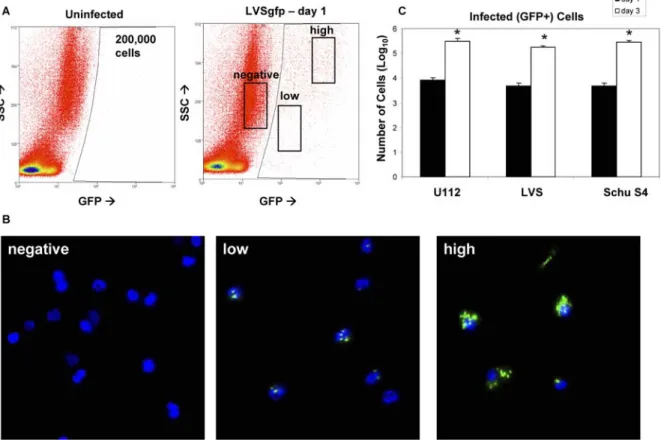
U112, Live Vaccine Strain (LVS), and Schu S4 are commonly studied closely related F. tularensis strains that exhibit differences in host range, virulence, and host response (Table 1). Single cell suspensions prepared from whole lung digests of mice inoculated intranasally with Francisella strains expressing green fluorescent protein (GFP) were analyzed by flow cytometry and fluorescence microscopy to identify the host-cell niche occupied by these organisms. GFP+ lung cells, indicating association with Francisella, were readily detectable by flow cytometry (Figure 1A). Microscopic examination of cells sorted from the high GFP+ population presented significant punctate GFP+ fluorescence, indicative of highly infected cells (Figure 1B), whereas low GFP+ cells had as few as 1 GFP+ bacterium
indicating that cells containing a single bacterium could be detected by flow cytometry. No GFP+ bacteria were observed in the negative population. Projection images compiled from multiple planes throughout the Z-axis of membrane-stained cells revealed that bacteria were intracellular (data not shown).