Original Research
Cervical Cancer in Women Aged 35 Years and Younger
Elizabeth Pelkofski, MD
1,*
; Jessica Stine, MD
2,†; Nolan A. Wages, PhD
3;
Paola A. Gehrig, MD
2; Kenneth H. Kim, MD
2; and Leigh A. Cantrell, MD, MSPH
1 1Division of Gynecologic Oncology, Department of Obstetrics and Gynecology, University of Virginia,
Charlottesville, Virginia;
2Division of Gynecology Oncology, Department of Obstetrics and Gynecology,
University of North Carolina, Chapel Hill, North Carolina; and
3Division of Translational Research and
Applied Statistics, Department of Public Health Sciences, University of Virginia, Charlottesville, Virginia
ABSTRACT
Purpose: Age has been evaluated as a prognostic
factor in cervical cancer in both hospital- and population-based studies. Results regarding the rela-tion of age and cervical cancer prognosis are confl ict-ing. This study pursued a contemporary assessment of the association of extreme young age at the time of a cervical cancer diagnosis on survival.
Methods: Institutional review board approval was
obtained, and retrospective data collection at 2 aca-demic institutions was performed. Inclusion criteria
involved women ≤35 years diagnosed with cervical
cancer between 1990 and 2012. Data included dem-ographic and prognostic information pertinent to survival and progression. Characteristics of very
young (r25 years) and young (425–35 years)
women were compared. Kaplan-Meier estimates, the log-rank test, and Cox proportional hazards modeling were used to assess the association of age, tumor histology, grade, stage, and parametrial involvement with progression-free survival (PFS) and overall survival (OS).
Findings: Incident cases (n ¼ 126) of cervical
cancer in patients ≤35 years of age were identified of which complete clinical information was available for 114 women. Fifteen percent (17 of 114) werer25
years, with the remaining 85% (97 of 114) being 26 to 35 years of age. Race, smoking status, and marital status were comparable between the 2 groups. Squ-amous histology dominated overall (77 of 114; 68%)
with adenocarcinoma contributing 25% (30 of 114;
26%) of cases. The majority (96 of 114, 84%) had either stage 1A (31 of 114, 27%) or 1B (65 of 114, 57%) disease. A log-rank test revealed no evidence to infer a difference in either PFS or OS among the age
groups (P ¼ 0.511 and P ¼ 0.340). In a univariate
analysis, grade and stage significantly affected OS
(P o 0.0001, P ¼ 0.045), and stage significantly
affected PFS (Po0.0001). In multivariate modeling, presence of parametrial involvement and histologic cancer type significantly affected both PFS (P¼0.002,
P ¼0.001) and OS (P ¼0.001,P ¼0.001).
Implications: Tumor histology, parametrial
in-volvement, and stage continue to be strong prognos-ticators for PFS and OS. Progression and survival outcomes are age independent in women with cervical cancer r35 years of age. Further study of a larger young cohort may potentially yield different out-comes. (Clin Ther.2016;38:459–466)&2016 Elsevier HS Journals, Inc. All rights reserved.
Key words:age, cervical cancer, progression, survival, young women.
Scan the QR Code with your phone to obtain FREE ACCESS to the articles featured in the Clinical Therapeutics topical updates or text GS2C65 to 64842. To scan QR Codes your phone must have a QR Code reader installed.
Accepted for publication January 7, 2016.
http://dx.doi.org/10.1016/j.clinthera.2016.01.024 0149-2918/$ - see front matter
&2016 Elsevier HS Journals, Inc. All rights reserved.
*Current affiliation: Department of Gynecologic Oncology, Baptist Health Lexington, Lexington, Kentucky.
INTRODUCTION
Cervical cancer, although largely preventable, is the most common site of gynecologic malignancy in
women o35 years of age in the United States.
Worldwide, cervical cancer is second only to breast cancer in cancers that affect women.1 Young patient age has been posited as a risk factor for more aggressive cervical cancers. Alternatively, although no genetic predisposition for cervical cancer has been accepted, researchers have proposed that there is a heritable inability to clear human papillomavirus (HPV) infection because population studies have found an increased incidence of cervical cancer in some families. In a Swedish study of49000 siblings or half-siblings with cervical cancer or dysplasia, 64% of cases were attributed to genetics and only 36% to environmental exposures.2 It seems improbable for young women to develop advanced disease, given the classic teaching that the risk of progression from mild dysplastic changes of the cervix to severe dysplasia, let alone cancer, is only 1% per year.3Therefore, the development of cancer in young women, especially the very young, has led to the theory that cervical cancer in the very young must be more aggressive.4 Others blame changes in sexual behavior with an earlier age of first intercourse, greater frequency of multiple partners and HPV infection, and tobacco use for the observations.5–8 Current estimates put the prevalence of HPV (all types) at 59% in 20- to 24-year-old
women and 50% in 25- to 29-year-old women.9
Several investigators have examined the relation between age at diagnosis and prognosis with conflicting results. In a study by Rutledge et al,10250 womenr35 years were matched by stage and treatment to older women. Younger women with advanced stage disease were noted to have worse overall survival (OS), yet they survived longer when diagnosed with early-stage dis-ease. Conversely, Clark et al11 concluded that cervical cancer behaved more aggressively in their comparison of 41 womenr35 years old with 96 women agedZ36 years in that there was a higher incidence of nodal metastases observed in the younger patients despite less-advanced clinical stage of disease. Paradoxically, they simultaneously observed that youth conferred better survival outcomes overall. In other studies, clinical behavior was age independent, but these studies com-pared womeno35 years with older women.12–14Our hypothesis is that cervical cancer in the very young (womeno25 years) is a more aggressive disease.
We sought to evaluate the relation of very young age to aggressiveness of cancer by comparing the young with the very young. This is a contemporary investigation after changes to practice that followed the 1999 National Cancer Institute alert that all patients with cervical cancer treated with radiation should also receive sensitizing cisplatin.
The primary objective of this study was to assess the effect of age on progression-free survival (PFS) and OS in women with cervical cancer r35 years of age. Secondarily, we sought to evaluate the impact of tumor histology, grade, stage, and parametrial in-volvement on PFS and OS in this cohort.
METHODS
Retrospective data collection was performed after approval from the institutional review boards at 2 tertiary academic medical centers (University of Vir-ginia Health System, Charlottesville, VA; University of North Carolina, Chapel Hill, NC). Data for patients with cervical cancer aged r35 years treated between 1990 and 2012 were abstracted.
Chart review included abstraction of demographic information (age, race, smoking status, and marital status), disease characteristics (histology, grade, stage, parametrial involvement), treatment history (surgery, radiation, chemotherapy, combination), and outcome data (OS and time to recurrence).
To assess the primary outcome of the effect of age on PFS and OS, patients were classified according to age at diagnosis. These age groups were defined as very young (r25 years old) and young (425–35 years old)
women. PFS was defined as the time from date of
diagnosis to disease progression or death from any
cause. OS was defined as the time from date of
diagnosis to death from any cause. Secondary outcomes included assessing the effect of tumor histology, grade, stage, and parametrial involvement on PFS and OS.
Differences in OS and PFS among the age groups were evaluated with Kaplan-Meier survival estimates and the log-rank test. Multivariate Cox proportional hazards modeling was used to perform time-to-event analysis of OS and PFS, including the predictors tumor histology, parametrial involvement, and age.
Tumor grade was not stated for 420% of women,
determined with a Wald test, and differences were considered significant for P values (2-sided) r0.05. Assumptions of the Cox proportional hazards model were assessed with graphical methods.
RESULTS
A total of 126 women with cervical cancer were identified from the 2 institutions. Complete clinical information was available for 114 women, and they comprise the study group. Approximately 15% (17 of 114) were very young, with the remaining 85% (97 of 114) belonging to the young group. Despite a 23-year searchable study time frame, the oldest case in the
final cohort dated back to 1998. Demographic and
clinical characteristics are summarized inTable I. The mean age at diagnosis was 29.7 years (median,
30 years; range, 17–35 years). The majority of
patients were Caucasian (75%), 13% were black,
and 11% were Hispanic. Nearly half were married. Most patients were never-smokers. Of the 114 with data available, 22% of women were nulliparous, and parity wasZ2 in 63% of the cohort.
Squamous histology dominated overall (68%)
with adenocarcinoma contributing 25% of cases.
Squamous histology was present in 10 of the 17 very young women (58%) versus 67 of the 97 young women (69%) with adenocarcinoma being the next most prevalent (4 of 17, 24% and 26 of 97, 27%, respectively). There were 5 cases of small cell carci-noma with 2 occurring in womeno25 years of age.
More than 80% of women in both groups had early-stage disease (stage 1A or 1B). Of the 109 women for whom a description of parametrial disease extension was available, only 15 had involvement, and this was unrelated to patient age. Of the 88 patients for whom tumor grade was stated, cancers were more often grade 2 (35 of 88, 40%) or grade 3 (35 of 88, 40%), with only 20% (18 of 88) being grade 1.
Associations of age with PFS and OS, as assessed by Kaplan-Meier estimation, are presented inFigures 1A
and 1B. Neither PFS (log-rank P ¼ 0.511) nor OS
(log-rank P ¼ 0.340) were significantly different by age group (r25 vs 425 years old).
The entire cohort had a 5-year OS rate of 42.1% (48 of 114). When analyzed according to disease distribution, this equated to a rate of 46% for patients
Table I. Patient demographic and clinical chara-cteristics (N¼114).
Characteristic Value
Mean age at diagnosis (range), years
29.7 (17–35)
Race, n (%)
Caucasian 86 (75.4) Black 15 (13.2) Hispanic 13 (11.4) Marital status, n (%)
Single 39 (34.2) Married 55 (48.2) Divorced 13 (11.4) Widowed 1 (0.88) Unknown 6 (5.3) Smoking status, n (%)
Current 35 (30.7) Former 13 (11.4) Never 62 (54.4) Unknown 4 (3.5) Stage at diagnosis, n (%)
1A1 25 (21.9)
1A2 6 (5.3)
1B1 37 (32.5) 1B2 28 (24.5)
IIA1 0
IIA2 2 (1.7) IIB 10 (8.7)
IIIA 0
IIIB 4 (3.5)
IV 2 (1.7)
Histology, n (%)
Squamous 77 (67.5) Adenocarcinoma 30 (26.3) Small cell 5 (4.4%) Adenosquamous 2 (1.7) Grade, n (%)
1 18 (15.8)
2 35 (30.7)
3 35 (30.7)
Unknown 26 (22.8) Parametrial involvement, n (%)
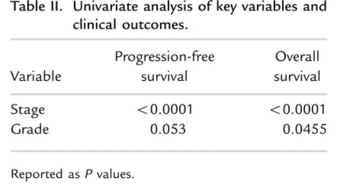
with localized disease, 25% for patients with region-alized disease, and 0% for patients with distant disease. In univariate analyses (Table II), PFS and
OS were evaluated across 5 categories of stage (1A1, 1A2, 1B1, 1B2, and stage II to IV) and 3 levels of tumor grade (1–3). Higher tumor grade conferred an
Product-Limit Survival Estimates
With Number of Subjets at Risk
0.0 2.5 5.0 7.5 10.0 12.5
Progression-free survival time (years)
+ Censored
0.0 0.2 0.4 0.6 0.8 1.0
group 25 or younger 26 or older
25 or younger 26 or older
0 3 1
6 2
16 2
25 10
49 17
97
Sur
vival Pr
obability
0.0 2.5 5.0 7.5 10.0 12.5
Product-Limit Survival Estimates
With Number of Subjects at Risk
+ Censored 1.0
0.8
0.6
0.4
0.0 0.2
Sur
viv
al Probability
Overall survival time (years)
25 or younger 26 or older
17 97
10 64
3 45
3 36
1 20
0 5 group 25 or younger 26 or older
A
B
increased risk of recurrence or progression. As stage
increased, recurrence increased, and survival
decreased. Both stage and grade significantly affected OS (Po 0.0001,P¼ 0.045), and stage significantly affected PFS (Po0.0001). A trend was found toward
grade significantly affecting PFS; however, this
relation did not reach statistical significance (P ¼ 0.053).
Cox proportional hazard modeling (Table III) of variables associated with PFS or OS indicated that there was no association between age and PFS (hazard ratio [HR]¼1.07;P¼0.315) or OS (HR¼1.05;P¼
0.468). Absence of parametrial involvement was
associated both with improved PFS (HR ¼ 8.71; P ¼
0.002) and OS (HR¼7.39;P¼0.001). Approximately 40% of patients with parametrial involvement recurred or progressed compared with only 10% of patients without parametrial involvement. Results from the multivariate model in Table III indicate that both squamous and adenocarcinoma tumor histology were associated with
improved PFS and OS. Women with adenocarcinoma were the least likely to recur or progress (3%) compared with 14% of women with squamous histology and 57% of women with other histologic types.
CONCLUSIONS
According to estimates, cervical cancer is the most
common gynecologic malignancy in women o35
years old with an incidence of 16.5 per 100,000.15,16
An abnormal Pap smear or cervical biopsy was the presenting complaint in nearly 60% of women in our cohort. This finding provides additional support for the need for continued screening in this young pop-ulation to the extent set forth by the American College of Obstetricians and Gynecologists.17 In addition to
screening, efforts at primary prevention via HPV vaccination should continue. Large clinical trials have found HPV vaccination to be effective in preventing cervical disease associated with HPV-16 and HPV-18;
however, immunization is most beneficial in
HPV-naive women.18 Screening remains important because
25% of cancers will not be prevented by earlier
vaccine types that did not immunize against all HPV types.
For women found to have cervical cancer, our
findings indicate age at diagnosis in and of itself in
women r35 years of age is not associated with a
worse prognosis (Table III). Because there were only
17 patients o25 years of age in the very young
cohort, statistical difference may not have been noted, but that group did have worse survival rates, as displayed inFigures 1A and 1B. In addition both
Table II. Univariate analysis of key variables and clinical outcomes.
Variable
Progression-free survival
Overall survival
Stage o0.0001 o0.0001 Grade 0.053 0.0455
Reported asPvalues.
Table III. Multivariate analysis of key variables and clinical outcomes.
PFS OS
Variable Hazard ratio P Hazard ratio P
Histology* 0.001† 0.001†
Squamous 0.066 0.001 0.069 0.0009 Adenocarcinoma 0.035 0.004 0.022 0.002 Parametrial involvement 8.710 0.002 7.395 0.001
Age 1.071 0.315 1.05 0.468
*The reference group is‘Other’tumor histology. †Cumulative
groups fared worse than survival in older women. The percentage of women survivingZ5 years in this study according to local and regional disease was 46% and 25%, respectively, compared with historic 5-year relative survival by stage at diagnosis for women Z50 years of age of 87% for localized disease and 54% for regional disease.19 This could point to a different disease pathway in younger women, leading to a more aggressive disease due to host or other unknown factors.
Similar to their older counterparts, tumor grade and histology affected prognosis and specifically PFS in our cohort. Analogous to patients with cervical cancer of all ages, squamous histology predominated in nearly 68% of cases in this study, and 26% of
patients had adenocarcinoma.20 Compared with
historic trends, this rising proportion of invasive adenocarcinoma especially in younger white women is consistent with thefindings of other researchers.21,22
This may be due to greater recognition and more awareness of adenocarcinoma and improved detection of its precursor lesions through better endocervical sampling techniques.23
It has been reported that adenocarcinoma confers poorer survival rates than squamous cell carcinoma, particularly for patients with regional disease.21,24 All patients in our study with adenocarcinoma had localized disease, and cases were fairly evenly distrib-uted among the 2 age groups. Despite the absence of adenocarcinoma histology in women having regional and distant disease, we found that histology still substantially affected OS and that the aggregate
histologic proportions were similar to larger
population-based studies.
More than 2% of patients in this study had small cell carcinoma. Historically, this rare and aggressive histology has been observed to occur at a mean incidence of only 0.06 per 100,000 women; thus, its substantially higher rate in this series may affect our
findings.25 Nevertheless, although the recent work of Castanon et al26in a series that examined 1800 cases of cervical cancer in women aged 20 to 29 years between 2007 and 2012 found a low non-squamous, non-adenocarcinoma, and non-adenosquamous rate of 2%, the rare histologic subtypes were most com-monly found in the youngest women in the series, aged 20 to 24 years, at a rate of nearly 6%. Furthermore, 20% of the time, these younger women were diagnosed at a higher stage of disease (stageZ2)
compared with frequencies of 4% and 7% for their
older counterparts (age 25 years and age 26–29 years, respectively). This trend was similar in our study with 17% of patients aged 20 to 29 years having been diagnosed at stageZ2.
Nearly 30% of cases in the youngest women occurred in those r20 years old. This is in contrast to an analysis of national surveillance data by Bernard
et al27 in which 1% of cervical carcinoma was
diagnosed in this young age range. This discrepancy may indicate a regional variation in our patient cohort or may be due to referral bias of younger, higher risk women to academic medical centers and thus may limit the generalizability of our findings.
Recent evidence from Japan epidemiologic data found a higher risk of death from cervical cancer in a younger cohort. This population-based cancer regis-try study covered 9 million people in a single geo-graphic area in Japan.28 If the aggressiveness and
presence of cervical cancer in a young woman is due to a genetic alteration, we would expect that would most likely be seen in regional studies such as the Japanese study compared with the genetically diverse population in the United States. There does appear to be an association of cervical cancer with a large variety of polymorphisms in a wide variety of genes,
including genes that regulate immunity and
susceptibility,29 cytokine production,30,31 angiogene-sis, tumor suppressor pathways,32,33 and signal
trans-ducer and activator of transcription pathways.34
Investigations are ongoing to identify genetic
alterations that may make women less likely to clear persistent HPV infections and more susceptible to the development of cervical cancer.
This study did not include a comparison group because it was our primary intention to determine whether degree of youth was clinically affected. Lymphovascular space invasion (LVSI) status was
unknown for 50% of the young women and for
425% of the very young women. Of the results
chemoradiation and analysis not according to treat-ment modality are other weaknesses. All 3 cases eligible for treatment with chemosensitization diag-nosed before the 1999 National Cancer Institute alert had received cisplatin. In addition, we recognize that this study is limited by the small numbers of cases, and additional multi-institutional studies may clarify these
findings. Nonetheless, our findings offer further
hypothesis-generating data and serve to emphasize the importance of considering disease distribution, tumor histology, and grade when determining prog-nosis in young patients.
ACKNOWLEDGMENTS
Supported in part by the Biostatistics Shared Re-source, University of Virginia Cancer Center, Univer-sity of Virginia (P30 CA044579). Elizabeth Pelkofski, Jessica Stine, and Leigh Cantrell were involved in study design, data collection, data interpretation, and manuscript preparation. Paola Gehrig and Kenneth Kim contributed to study design, data interpretation, and manuscript preparation. Nolan Wages provided all statistical support. All authors materially partici-pated in this research and its preparation and approve this work in itsfinal form.
CONFLICTS OF INTEREST
The authors have indicated that they have no conflicts of interest regarding the content of this article. No
financial support or funding was provided for this project.
REFERENCES
1. Boyle B, Levin B. World Cancer Report 2008. Lyon, France: International Agency for Research on Cancer; 2008. 2. Hemminki K, Chen B. Familial risks for cervical tumors in
full and half siblings: etiologic apportioning. Cancer Epidemiol Biomarkers Prev. 2006;15:1413–1414.
3. Holowaty P, Miller A, Rohan T. To T. Natural history of dysplasia of the uterine cervix. J Natl Cancer Inst. 1999;91:252–258.
4. Hildesheim A, Hadjimachael O, Schwartz P, et al. Risk factors for rapid-onset cervical cancer.Am J Obstet Gynecol. 1999;180:571–577.
5. Cuzick J, Sasieni P, Singer A. Risk factors for invasive cervix cancer in young women.Eur J Cancer. 1996;32A:836–841. 6. Parazzini F, Chatenoud L, La Vecchia C, et al. Determi-nants of risk of invasive cervical cancer in young women. Br J Cancer. 1998;77:838–841.
7. Walboomers J, Jacobs M, Manos M, et al. Human papillomavirus is a necessary cause of invasive cervical cancer worldwide.J Pathol. 1999;189:12–19.
8. Plummer M, Herrero R, Franceschi S, et al. Smoking and cervical cancer: pooled analysis of the IARC multi-centric case–control study.Cancer Causes Control. 2003;14:805–814. 9. Markowitz L, Hariri S, Lin C, et al. Reduction in human papillomavirus (HPV) prevalence among young women following HPV vaccine introduction in the United States, National Health and Nutrition Examination Surveys, 2003-2010.J Infection. 2013;208:385–393.
10. Rutledge F, Mitchell MF, Munsell M, et al. Youth as a prognostic factor in carcinoma of the cervix: a matched analysis.Gynecol Oncol. 1992;44:123–130.
11. Clark M, Naahas W, Markert R, Dodson M. Cervical cancer: women aged 35 and younger compared to women aged 36 and older.Am J Clin Oncol. 1991;14:352–356. 12. Carmichael J, Clarke D, Moher D, et al. Cervical
carci-noma in women aged 34 and younger.Am J Obstet Gynecol. 1986;154:264–269.
13. Smales E, Perry CM, Ashby MA, Baker JW. The influence of age on prognosis in carcinoma of the cervix.Br J Obstet Gynaecol. 1987;94:784–787.
14. Poka R, Juhasz B, Lampe L. Cervical cancer in young women: a poorer prognosis?Int J Gynaecol Obstet. 1994;46:33–37. 15. Surveillance, Epidemiology, and End Results (SEER)
program. SEER Cancer Statistics Review 1975-2012 data-base: Cancer of the Cervix Uteri (Invasive), Table 5.7; Cancer of the Corpus and Uterus (NOS), Table 7.7; Cancer of the Ovary (Invasive), Table 21.7. National Cancer Institute.http://seer.cancer.gov. Accessed Septem-ber 27, 2015.
16. Surveillance, Epidemiology, and End Results (SEER) pro-gram. SEER Cancer Statistics Review 1975-2012 database: Cancer of the Cervix Uteri (Invasive), Table 5.7; Cancer of the Ovary (Invasive), Table 21.7. National Cancer Institute. http://seer.cancer.gov. Accessed September 27, 2015. 17. American College of Obstetricians and Gynecologists.
ACOG Practice Bulletin no. 109: cervical cytology screen-ing.Obstet Gynecol. 2009;114:1409–1420.
18. FUTURE II Study Group. Quadrivalent vaccine against human papillomavirus to prevent high-grade cervical lesions.N Engl J Med. 2007;356:1915–1927.
19. Surveillance, Epidemiology, and End Results (SEER) pro-gram. SEER Cancer Statistics Review 1975-2012 database: Cancer of the Cervix Uteri (Invasive), Table 5.8: 5-Year Relative and Period Survival by Race, Diagnosis year, Age and Stage at Diagnosis. National Cancer Institute.http:// seer.cancer.gov. Accessed September 27, 2015.
Cases, 2008-2012 Females by Race. National Cancer Institute. http:// seer.cancer.gov. Accessed September 27, 2015.
21. Smith HO, Tiffany MF, Qualls CR, Key CR. The rising incidence of adenocarcinoma relative to squa-mous cell carcinoma of the uterine cervix in the United States–a 24-year population-based study.Gynecol On-col. 2000;78:97–105.
22. Sherman ME, Wang SS, Carreon J, Devesa SS. Mortality trends for cervical squamous and ad-enocarcinoma in the United States. Cancer. 2005;103:1258– 1264.
23. Wang SS, Sherman ME, Hilde-sheim A, et al. Cervical adenocar-cinoma and squamous cell carcinoma incidence trends among white women and black women in the United States for 1976-2000. Cancer. 2004;100:1035–1044. 24. Lau HY, Juang CM, Chen YJ, et al.
Aggressive characteristics of cervi-cal cancer in young women in Taiwan.Int J Gynaecol Obstet. 2009; 107:220–223.
25. Chen J, Macdonald OK, Gaffney DK. Incidence, mortality, and prognostic factors of small cell carcinoma of the cervix. Obstet Gynecol. 2008;111:1394–1402. 26. Castanon A, Leung VM, Landy R,
et al. Characteristics and screening history of women diagnosed with cervical cancer aged 20-29. Br J Cancer. 2013;109:35–41.
27. Benard VB, Watson M, Castle PE, Saraiya M. Cervical carcinoma rates among young females in the United States.Obstet Gynecol. 2012; 120:1117–1123.
28. Motoki Y, Mizushima S, Taguri M, et al. Increasing trends in cervi-cal cancer mortality among young Japanese women below the age of 50 years: an analysis using the Kanagawa population-based Cancer Registry, 1975-2012.Cancer Epidemiol. 2015;39: 700–706.
29. Liu L, Yang X, Chen X, et al. Association between TNF-α poly-morphisms and cervical cancer risk: a meta-analysis. Mol Biol Rep. 2012;39:2683–2688.
30. Grimm C, Watrowski R, Baumühl-ner K, et al. Genetic variations of interleukin-1 and -6 genes and risk of cervical intraepithelial neopla-sia. Gynecol Oncol. 2011;121:537– 541.
31. Wang Q, Zhang C, Walayat S, et al. Association between cytokine gene polymorphisms and cervical cancer in a Chinese population. Eur J Obstet Gynecol Reprod Biol. 2011;158:330–333.
32. Craveiro R, Bravo I, Catarino R, et al. The role of p73 G4C14-to-A4T14 polymorphism in the sus-ceptibility to cervical cancer.DNA Cell Biol. 2012;31:224–229. 33. Wang L, Gao R, Yu L.
Com-bined analysis of the association between p73 G4C14-to-A4T14 polymorphisms and cancer risk. Mol Biol Rep. 2012;39:1731–1738. 34. Wang K, Zhou B, Zhang J, et al.
Association of signal trans-ducer and activator of transcrip-tion 3 gene polymorphisms with cervical cancer in Chinese women.DNA Cell Biol. 2011;30: 931–936.
Address correspondence to: Elizabeth Pelkofski, MD, Baptist Health