
Original Article Application value of ultrasound contrast in gastric cancer examinations and its correlation with microvessel density
Full text
Figure

Related documents
Results: Tissue microarrays showed that 72.2% of gastric cancer tissues exhibited down-regulated expression of TOB1, and the expression level was significantly lower in cases
In conclusion, our study shown that NEDD9 was high expressed in gastric cancer, but weakly expressed in adjacent non-tu- mor tissues, besides, the NEDD- 9 expression was
could be detected using real-time PCR and western blot in gastric cancer patients com- pared with health subjects or adjacent normal tissues in gastric cancer patients which is in
The data was not in agreement with our findings, where the expression of IL-33 was up- regulated in cervical cancer tissues compared with that in paired adjacent non-cancerous
There was no correlation of 15-PGDH expression between non-cancerous gastric tis- sues and GC tissues (P=0.519), while 15-PGDH expression level in patients with NACT group was
The relative expression levels of Tgif2 in gastric cancer tis- sues and the adjacent non-tumor tissues were shown in Figure 2 , which were obtained by real- time PCR and western
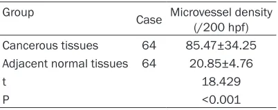
Positive rates of integrin αvβ3 and VEGF were higher, MVD counting was larger, and T-cadherin positive rate was lower in gastric cancer tissues than those in the adjacent
This study used microvessel-slice spiral com- puted tomography (MSCT) perfusion imaging to quantitatively evaluate tumor angiogenesis, with the aim of analyzing differences between
