Jiangsu, China; 4Prenatal Diagnosis Center, Departments of Obstetrics and Gynecology, Nanjing Drum Tower Hos-pital, The Affiliated Hospital of Nanjing University School of Medicine, Nanjing, Jiangsu, China. *Equal contributors and co-first authors.
Received November 3, 2015; Accepted February 10, 2016; Epub April 15, 2016; Published April 30, 2016
Abstract: Background: Von Hippel-Lindau (VHL) disease is an autosomal dominant familial cancer syndrome that occurs as a consequence of an inactivation of the VHL gene. Point mutations in VHL are a major cause for VHL disease, and fragment deletions contribute to ~30% of VHL patients. Array Comparative Genomic Hybridization (Array-CGH) is a powerful tool for the identification of genomic deletions, and often reveals unexpected information as a non-targeted method. Method: We extracted the DNA from the patients’ blood andscreened for point mutations in VHL by PCR and large deletions by Array Comparative Genomic Hybridization (Array-CGH). Real-time quantitative
PCR was used to confirm the deletion of VHL gene and expression of VHL in tumor tissue. Results: We present a family case of VHL syndrome, showing hemangioblastomas in the central nervous system and retina, multiple pan-creatic cysts and clear-cell renal cell carcinoma. After excluding point mutations in VHL gene exons and conjunctive regions, we identified a ~9 kb deletion of chromosome 3p25.3 with Array-CGH analysis. Real-time PCR confirmed a heterozygous deletion in exon 3 of the VHL gene. Additional 90 DNA deletions were detected ranging from 5 kb to 160 kb in size. Moreover, 70.3% of the breakpoints were located in Alu elements. Conclusions: This study confirmed that DNA deletions contribute to familial VHL disease. It also highlights that coding deletions appear to be more frequent than expected, and that Alu-mediated recombination is the major mechanism.
Keywords: Von Hippel-Lindau, array comparative genomic hybridization, coding deletion, alu element
Introduction
Von Hippel-Lindau (VHL) disease is a hereditary cancer syndrome that is characterized by mul-tiple benign and malignant neoplasias in many organs, especially hemangioblastomas in the central nervous system (CNS) and retina at a young age [1]. It also presents with clear-cell renal cell carcinoma (ccRCC) and cysts, pheo-chromocytomas, multiple pancreatic cysts or tumors, epididymal/ovarian cystadenomas and endolymphatic sac tumors [2]. The inactivation of the VHL gene (OMIM 608537), a well-known tumor-suppressor gene, directly leads to famil-ial VHL diseases, as well sporadic cases. The VHL gene is located on chromosome 3p26-25, spans a 10 kb region consisting of three exons [3]. VHL mRNA encodes a 213 amino acid
pro-tein (pVHL) with a molecular weight of ~24 to 30 kDa (VHL30) [4], as well a second pVHL isoform of approximately 19 kDa (VHL19) [5]. Both iso-forms appear to retain tumor suppressor activ-ity [6]. Inactivation of VHL leads to an accumu-lation of hypoxia-inducible factor (HIF), the tar-get of pVHL, which contributes to the overpro-duction of vascular endothelial growth factor (VEGF) [7] and erythropoietin (Epo) [8], and, ultimately, vascular proliferation and tumori- genesis.
pancreatic tumors and epididymal/ovarian cystadennoma and endolymphatic sac tumor [10]. Generally, germline mutations in the VHL gene account for approximately 100% classical non-mosaic VHL disease and somatic from 17.9% to 71% of ccRCC [11-13]. Point muta-tions are the major cause of inactivation of VHL gene, including 30% to 38% for missense muta-tions and 23% and 27% for nonsense or frame-shift mutations, respectively. Germline rear-rangements have frequently been reported to account for approximately 20 to 37% of VHL patients, especially large or partial fragment deletions [10, 14]. Karyotyping analysis and multiplex ligation-dependent probe amplifica -tion (MLPA) are often used to detect these de- letions. Array Comparative Genomic Hybridi- zation (Array-CGH) was recently developed to identify genomic imbalances. As a conventional method, it is promising in clinical diagnostics. Compared to karyotyping, Array-CGH has a higher resolution, detecting 50-100 kb dele-tions, while karyotyping is unreliable for sub- tle copy number changes of 5 Mb in size or smaller [15]. MLPA can only detect deletions or duplications on specific genomic regions [16]. Array-CGH analysis, however, can also reveal unexpected information of DNA
recombinatio-nas a non-targeted method of scanning the whole genome [17].
In the present study, we screened potential mutations in a familial case with VHL disease and identified a fragment deletion in VHL genes. Moreover, unexpected DNA deletions were detected by Array-CGH analysis. The char-acteristics of these genome deletions and underlying mechanisms were therefore inve- stigated.
Patients, samples and method
Subjects
[image:2.629.101.532.77.319.2]Members of a Chinese multigeneration family (Figure 1A) underwent surgical therapy for renal tumors in Nanjing Drum tower hospital. Clinical features were carefully reviewed and imaging and hematological examinations were performed before the surgery. Blood speci-mens were collected from all family members, and tissues from renal tumors of cancer patients in this family were also collected. Samples from sporadic renal cancers served as tumor controls, and adjacent normal renal tissues as negative controls.
Screening for point mutations in VHL
[image:3.629.99.528.93.147.2]Genomic DNA was extracted from peripheral blood samples (TIANamp Blood DNA Kit, Bei- jing, China). DNA samples were subjected to mutation screening with all of the exons and exon-intron boundaries of VHL (NC_ 000003.12) amplified by polymerase chain reaction (PCR). The primers for PCR are listed Table 1. The products were examined on a 2% agarose gel, purified by a universal DNA purification kit (TIANGEN, Beijing, China) and then subjected to direct DNA sequencing using an ABI Prism 3100 Genetic Analyzer (Applied Biosystems, Foster City, CA, USA).
Array-CGH analysis
Array-CGH microarray analysis was performed using an Affymetrix Cytoscan HD GeneChip at an effective resolution of 5 kb. Data analysis was performed using the Chromosome Analysis Suite (ChAS) 2.0 software following the
stan-dard protocol. Abnormalities detected were compared with the RefSeq genome build hg19, via the database browser at http://genome. ucsc.edu/cgi-bin/hgGateway.
Quantitative PCR analysis
Real-time quantitative PCR was used to con-
firm the deletion of VHL gene, with SYBR Green I detection (Roche, Mannheim, Germany) on an ABI StepOne Sequence Detection System (PE Applied Biosystems, Foster City, CA). The primers used were as follow: forward 5’GG- TCGCTCTACGAAGATCTGGA3’, reverse 5’GAAA- TCTTCAATCTCCCATCCG3’ and 18SrRNA was used as an internal reference. Real-time quan-titative PCR was also used to detect the expres-sion of VHL. Briefly, after total RNA was isolated from tissues and complementary DNA (cDNA) was synthesized using a reverse transcription kit (Transgen Biotech, Beijing, China), quantita-tive PCR was performed with Green I detection using the primers: forward 5’GGAGCCTAGTC-
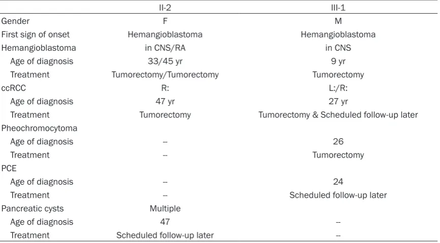
Hemangioblastoma in CNS/RA in CNS
Age of diagnosis 33/45 yr 9 yr
Treatment Tumorectomy/Tumorectomy Tumorectomy
ccRCC R: L:/R:
Age of diagnosis 47 yr 27 yr
Treatment Tumorectomy Tumorectomy & Scheduled follow-up later Pheochromocytoma
Age of diagnosis -- 26
Treatment -- Tumorectomy
PCE
Age of diagnosis -- 24
Treatment -- Scheduled follow-up later
Pancreatic cysts Multiple
Age of diagnosis 47
Treatment Scheduled follow-up later
[image:3.629.100.533.181.420.2]AAGCCTGAGA3’, reverse 5’CATCCGTTGATGTG- CAATGCG3’. All of the PCRs were performed in triplicate and data analysis was performed using the ΔΔCT method.
Statistical analysis
All statistical analyses were carried out using the statistical program SPSS, version 17.0. The non-parametric test was used to evaluate the deletions in and expression of the VHL gene between patients and normal individuals. The chi-square test was performed to analyze the relationship between deletion polymorphisms, Alu-related deletions and coding deletions. Pearson correlation analysis was utilized to evaluate the association between the length of the deletions, the length of the whole chro-mosome, and the number of Alu elements on each chromosome. All tests were two-sided and P≤0.05 was considered statistically signi-ficant.
Result
Germline VHL deletion
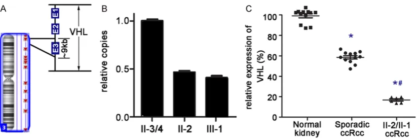
Two of the four subjects in this family were diag-nosed with VHL disease (Figure 1A, II-2 & III-1). The II2 was a 47 years old female with a history of surgery for hemangioblastomas of CNS and retina (Table 2). Imageological diagnosis as a routine test was performed and revealed a right renal tumor and multiple pancreatic cysts (Figure 1B, 1C). Patient III1 had similar symp-toms, with CNS hemangioblastomas and a
renal tumor, as well as pheochromocytomas and epididymal cystadenoma (Figure 1D-F). Both of the two patients underwent lapa- roscopic renal tumor resection, and pathologi-cal analysis revealed clear-cell renal cell car- cinoma.
After sequencing of the VHL gene, we did not identify any point mutations (missense muta-tion or small insermuta-tion and delemuta-tion) in II2 and III1. Array-CGH was further performed to detect potential fragment deletions. Focusing on chro-mosome 3p and the VHL gene (Figure 2A), a 9 kb deletion was detected and located in exon 3 of the VHL gene. Further Q-PCR showed that VHL copies of II3/4 were double than that of II2 and III1, as considered as VHL deletion occurred in the two patients (Figure 2B). VHL deletion also leaded to lower expression of pVHL in VHL associated ccRCCs than that in sporadic ccRCCs. (P=0.001), while expression of pVHL in these sporadic ccRCCs was also lower than that in corresponding adjacent normal tissue (P=0.001) (Figure 2C).
Coding deletion in genome
[image:4.629.104.528.81.222.2]Table 3A. Detected coding deletions in II2
Deletion Involved genes
Polymorphism Allele frequency <0.5% SMR3A, SMR3B, DMBT1, ZNF826P
Allele frequency: 0.5%-5% OR2T3, OR2T5, OR2G6, OR2T29, OR2T34, TUSC3
Allele frequency >5% SCAP, TRY6, PRSS2, OR4P4, OR4S2, OR4C6, TRIM49, ACOT1, SIGLEC14, LILRA3, SIRPB1
Novel deletion Diseased related MAP3K2, VHL, DACH1, PEX1
Unknown SLMAP, FLJ34503, NBPF6, EFHA2, FLT3, SLC15A4, KCNK10, PAK6, PLA2G4F, PDXDC1, PIR, KRTAP4-12, SLC25A17, MAGEA9, MAGE-A9B, RFPL1, ASB9
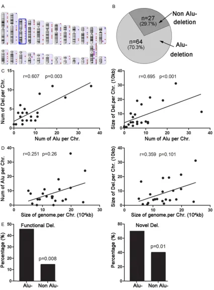
deletion on VHL, The CGH analysis together showed 91 deletions and 24.18% (22/91) were less than 8 K in size. Among these deletions, the deleted size ranged from 5 to 160 kb, and 35 of 91 were deletion polymorphisms listed in the Database of Genomic Variants (http://dgv-beta.tcag.ca/dgv/app/index.html), including 18 (51.4%) coding deletions.
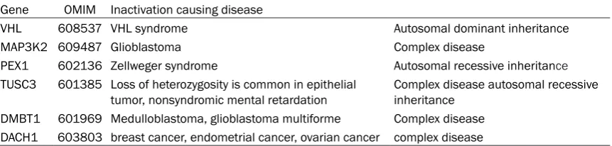
Moreover, there were 52 deletions located at 60 known genes across whole genome and 33 coding deletions at exon regions, indicating deteriorating the function of these involved 42 genes. Interestingly, dysfunction of 21 genes was reported as polymorphism in the data- base (datas from Database of Genomic Va- riants http://dgvbeta.tcag.ca/dgv/app/index. html and allele frequency from 1000 Genomes Consortium Phase 1) and deletions involved 11 genes were even common (allele frequency >5%) among population (Table 3A). Other dele-tions were at first detected Particularly, the dysfunction of 6 genes in novel or rare dele-tions was unequivocally related with different diseases (Table 3B). VHL is an autosomal domi-nant gene that contributed to the patients investigated. Two genes, PEX1 and TUSC3, were related with autosomal recessive inheri-tance (AR). Complete lack of PEX1 protein is associated with severe Zellweger syndrome. In an Iranian family, a homozygous deletion
involv-ing the first exon of TUSC3 gene was detected, while all unaffected parents of patients were heterozygous and all of the unrelated controls did not have this deletion. However, we did not know how the dysfunction of these genes would contribute to the phenotypes in this family. Alu elements distribution with DNA deletions
Regarding the breakpoints of the deletion, Alu elements appeared frequently through 1000 bp of the upstreams and downstreams. The Alu elements were detected at bilateral break-points in 32 of 91 deletions and unilateral in another 32 of 91 deletions (Figure 3B). In addi-tion, bilateral breakpoints of 4 deletions were located at homologous sequences and 1 dele-tion at a tandem repeat sequence.
As shown in Figure 3C, there was a liner corre-lation between the number of deletions and the number of Alu elements at each chromosome (r=0.607, P=0.003), as well as the sizes of dele-tions and the number of Alu elements at each chromosome (r=0.695, P<0.001). But, the size of the whole chromosome was not related to the number of Alu elements or the size of the deletions (Figure 3D). Moreover, 45.3% (29/64) of deletions mediated by Alu were coding dele-tions, and the percentage was significantly higher than that of deletions not mediated by Table 3B. Virulence genes involved with deletions
Gene OMIM Inactivation causing disease
VHL 608537 VHL syndrome Autosomal dominant inheritance MAP3K2 609487 Glioblastoma Complex disease
PEX1 602136 Zellweger syndrome Autosomal recessive inheritance TUSC3 601385 Loss of heterozygosity is common in epithelial
tumor, nonsyndromic mental retardation Complex disease autosomal recessive inheritance DMBT1 601969 Medulloblastoma, glioblastoma multiforme Complex disease
[image:6.629.101.532.241.344.2]ed with diseases, which has been largely ignored previously. In the present study, we identified 91 fragment deletions in a patient with VHL, suggesting that gene deletion is a relatively more common issue than has been previously considered, and it was consistent with other published studies [18, 19]. This study further showed that DNA deletions most often result from Alu element-mediated rear-rangements, which contribute to the novel mutations.
Regarding the patients with VHL syndrome, Array-CGH demonstrated a heterozygosis dele-tion on chromosome 3p25 where VHL gene locals. Familial VHL is a typical autosomal dom-inant heritable disease. Heterozygotes point mutation or germline deletion VHLleads to the inactivated allele and insufficient VHL protein [6]. The deletion investigated in this study has been confirmed to be associated with insuffi -cient VHL expression. The molecular genetic contribution to the clinical phenotype of VHL is: decreased pVHL led to an accumulation of HIF, overproduction of VEGF and Epo, and ultimately tumorigenesis.
Coding deletions are typically deleterious, such as the deletion in VHL. However, additional cod-ing deletions were detected without signifi -cance on disease, and such deletions are even considered to be a common issue. We found that 36.3% (33/91) of deletions detected in this patient were coding deletions. McCarroll et al. [18] found 10 expressed genes contained deletions and their research also demonstrat-ed that coding deletions are common in healthy individuals. Hinds et al. [19] found several dele-tions that resulted in loss of exons in genes in unrelated individuals.
The PEX1 [20, 21] and TUSC3 [22, 23] genes were previously demonstrated to be autosomal
quency varied across different races [18]. These findings highlight the functional dele -tions that generally exist in various popula-tions. The significance of coding deletions in genetic evolution and disease risk requires further clarification.
Another interesting finding of in the present study is that Alu elements are the most ele-ments involved in the genome deletions. The deletion in the VHL gene appeared to be the result of Alu-Alu recombination, as Alu ele-ments were detected frequently at both break-points. Of the 91 deletions, 64 (70.33%) were located within Alu elements. A liner correlation was found between the number of deletions and the number of Alu elements at each chro-mosome, as well as the sizes of the deletions and the number of Alu elements at each chro-mosome. An increasing number of studies are demonstrating that Alu elements are widely dis-tributed throughout the whole genome, and they are known to be hot spots for recombina-tion events. Alu element-mediated delerecombina-tions have been widely detected in various genes. Franke’s research demonstrated that 90% of deletions that involved the VHL gene were located in Alu elements [24]. Coutinho’s study also observed that Alu-Alu recombination medi-ated a large homozygous intragenic GNPTAB gene deletion in a mucolipidosis patient [25]. Our findings further provide evidence that Alu-Alu recombination is the main mechanism of mediating large deletions.
Conclusion
underlying pathogenesis is unclear. Moreover, investigating these fragment variations is help-ful for a better understanding of the universali-ty of coding deletions, as well the mechanism of DNA recombination by Alu-Alu elements. The findings of such studies enrich knowledge regarding the molecular mechanisms underly-ing large deletions of chromosomes, and enable us to recognize and identify such genet-ic phenomena.
Acknowledgements
The authors would like to thank all the mem-bers for assisting in guidance on PCR and Q-PCR procedure in Department of Medical Genetics, Nanjing University School of Medi- cine, Nanjing, China.
Disclosure of conflict of interest
None.
Address correspondence to: Huimei Chen, Depart- ment of Medical Genetics, Nanjing University School of Medicine, Nanjing, Jiangsu, China. E-mail: [email protected]; Hongqian Guo, Department of Urology, Nanjing Drum Tower Hospital, Medicine School of Nanjing University, Institute of Urology, Nanjing University, Nanjing 210008, Jiangsu, PR China. E-mail: [email protected]
References
[1] Bausch B, Jilg C, Glasker S, Vortmeyer A, Lutzen N, Anton A, Eng C and Neumann HP. Renal cancer in von Hippel-Lindau disease and related syndromes. Nat Rev Nephrol 2013; 9: 529-538.
[2] Lonser RR, Glenn GM, Walther M, Chew EY, Libutti SK, Linehan WM and Oldfield EH. von Hippel-Lindau disease. Lancet 2003; 361: 2059-2067.
[3] Latif F, Tory K, Gnarra J, Yao M, Duh FM, Orcutt ML, Stackhouse T, Kuzmin I, Modi W, Geil L and et al. Identification of the von Hippel-Lindau disease tumor suppressor gene. Science 1993; 260: 1317-1320.
[4] Iliopoulos O, Kibel A, Gray S and Kaelin WG Jr. Tumour suppression by the human von Hippel-Lindau gene product. Nat Med 1995; 1: 822-826.
[5] Schoenfeld A, Davidowitz EJ and Burk RD. A second major native von Hippel-Lindau gene product, initiated from an internal translation start site, functions as a tumor suppressor. Proc Natl Acad Sci U S A 1998; 95: 8817-8822.
[6] Kim WY and Kaelin WG. Role of VHL gene mu-tation in human cancer. J Clin Oncol 2004; 22: 4991-5004.
[7] Flamme I, Krieg M and Plate KH. Up-regulation of vascular endothelial growth factor in stromal cells of hemangioblastomas is correlated with up-regulation of the transcription factor HRF/ HIF-2alpha. Am J Pathol 1998; 153: 25-29. [8] Wiesener MS and Eckardt KU. Erythropoietin,
tumours and the von Hippel-Lindau gene: to-wards identification of mechanisms and dys-function of oxygen sensing. Nephrol Dial Transplant 2002; 17: 356-359.
[9] Collins ET. Intra-ocular growths (two cases, brother and sister, with peculiar vascular new growth, probably retinal, affecting both eyes). Trans Ophthalmol Soc UK 1894; 14: 141-149. [10] Maher ER and Kaelin WG Jr. von Hippel-Lindau
disease. Medicine (Baltimore) 1997; 76: 381-391.
[11] Tsutsumi H, Miyamoto C, Furuichi Y, Yoshiike M, Nozawa S and Iwamoto T. VHL tumor sup-pressor gene: its mutation and protein level in renal cell carcinoma. Oncol Rep 2003; 10: 1357-1361.
[12] Kondo K, Yao M, Yoshida M, Kishida T, Shuin T, Miura T, Moriyama M, Kobayashi K, Sakai N, Kaneko S, Kawakami S, Baba M, Nakaigawa N, Nagashima Y, Nakatani Y and Hosaka M. Comprehensive mutational analysis of the VHL gene in sporadic renal cell carcinoma: relation-ship to clinicopathological parameters. Genes Chromosomes Cancer 2002; 34: 58-68. [13] Banks RE, Tirukonda P, Taylor C, Hornigold N,
Astuti D, Cohen D, Maher ER, Stanley AJ, Harnden P, Joyce A, Knowles M and Selby PJ. Genetic and epigenetic analysis of von Hippel-Lindau (VHL) gene alterations and relationship with clinical variables in sporadic renal cancer. Cancer Res 2006; 66: 2000-2011.
[14] Stolle C, Glenn G, Zbar B, Humphrey JS, Choyke P, Walther M, Pack S, Hurley K, Andrey C, Klausner R and Linehan WM. Improved detec-tion of germline mutadetec-tions in the von Hippel-Lindau disease tumor suppressor gene. Hum Mutat 1998; 12: 417-423.
[15] Fruhman G and Van den Veyver IB. Applications of array comparative genomic hybridization in obstetrics. Obstet Gynecol Clin North Am 2010; 37: 71-85, Table of Contents.
[16] Sellner LN and Taylor GR. MLPA and MAPH: new techniques for detection of gene dele-tions. Hum Mutat 2004; 23: 413-419.
PEX1: phenotypes and PEX1 protein levels. Am J Hum Genet 2001; 69: 35-48.
[21] Reuber BE, Germain-Lee E, Collins CS, Morrell JC, Ameritunga R, Moser HW, Valle D and Gould SJ. Mutations in PEX1 are the most com-mon cause of peroxisome biogenesis disor-ders. Nat Genet 1997; 17: 445-448.
[22] Molinari F, Foulquier F, Tarpey PS, Morelle W, Boissel S, Teague J, Edkins S, Futreal PA, Stratton MR, Turner G, Matthijs G, Gecz J, Munnich A and Colleaux L. Oligosaccharyl- transferase-subunit mutations in nonsyndrom-ic mental retardation. Am J Hum Genet 2008; 82: 1150-1157.
type-phenotype correlations in VHL patients. Hum Mutat 2009; 30: 776-786.