Original Article
Adequacy rate comparison between liquid-based
cytology using SurePath versus conventional
smears in detecting thyroid malignancies
Xiaoyun Liu1*, Yun Cai1*, Zhixiao Wang1*, Dai Cui1, Hongqi Fan1, Lin Jiang1, Huanhuan Chen1, Ling Wei1, Qing Yao2, Yunsong Wu2, Rong Rong2, Zhihong Zhang2, Edmund S Cibas3, Erik K Alexander4, Tao Yang1, Xiaodong Wang1
Departments of 1Endocrinology, 2Pathology, The First Affiliated Hospital of Nanjing Medical University, Nanjing,
China; 3Department of Pathology, 4Thyroid Unit, Division of Endocrinology, Metabolism and Diabetes, Department of Medicine, Brigham & Women’s Hospital and Harvard Medical School, Boston, USA. *Equal contributors. Received December 22, 2015; Accepted March 20, 2016; Epub April 1, 2016; Published April 15, 2016
Abstract: Background: Controversy exists about the diagnostic value of liquid-based cytology (LBC) compared to conventional smears (CS). Most prior studies of LBC were performed using ThinPrep system. Few studies have ever compared the adequacy rate of SurePath with conventional smears. Methods: We performed a prospective com-parison of LBC using SurePath with CS in 304 thyroid nodules. Four needle sticks constituted a single nodule FNA, with 2 passes used for CS while the other 2 passes were used for SurePath. Cytopathologists separately read all samples, and all slides were reported using the Bethesda system for reporting thyroid cytology. The adequacy rate was compared between the CS and SurePath groups. Results: The adequacy rate for all solid nodules was 78.2% in CS group, significantly higher than 68.0% in the SurePath group (P=0.006). No significant difference was seen for mixed or cystic nodules. The adequacy rate using a combination of CS and SurePath in solid nodules was 86.4%, significantly higher than 78.2% in CS group (P<0.001). When excluding nodules less than 1 cm, the adequacy rate of CS for solid nodules was 83.5%, significantly higher than 71.3% in SurePath group (P=0.02). The adequacy rate of combination of CS and SurePath was 91.3% for solid nodules, significantly higher than 83.5% in CS group (P=0.04). Conclusion: Our study showed that LBC using SurePath is not superior to conventional smears. However, a combina-tion of both SurePath and CS may yield the most favorable adequacy rate compared to either process separately.
Keywords: Liquid-based cytology, thyroid cytology, conventional smear, adequacy rate, comparison, thyroid nod-ule, thyroid malignancy
Introduction
Thyroid nodules are increasingly common [1] and require further evaluation to rule out malig-nancy as suggested by various guidelines [2-6]. Cytological analysis of fine-needle aspiration (FNA) material is the primary and cost-effective modality for initial evaluation of a clinically rel-evant thyroid nodule [2-5, 7, 8]. Conventional smear (CS) for cytology diagnosis has been the main method for evaluation.
Since its introduction in 1996, liquid based cytology (LBC) has shown its advantages over conventional smears. Liquid-base preparations (LBP) were consistently devoid of obscuring ele-ments, and the cells were adequately
pre-other studies showed that LBC has its own dis-advantages, for example, higher non-adequacy rate [10], artifact which might lead to diagnostic pitfalls [9], missing of background factor which is essential clue for certain cases [11].
Controversies still exist about whether thyroid fine-needle aspirates (FNAs) should be pro-cessed solely with conventional smears or LBC [12]. Each has its own advantages and disad-vantages as described. As a result, the combi-nation of conventional smear with liquid-based preparations is often suggested [11] if LBC is available.
sons studies were using ThinPrep system, few has used another FDA-approved liquid based preparations, the SurePath system [13]. Nagarajan reported significantly higher per-centage of non-adequacy rate was found in LBP compared to conventional smears in a very large sample [10]. However, different nodules were included from the two different groups. Although the age, sex and nodule size were comparable between LBP and CS groups, the component and calcification within the nodule were not mentioned which were very important factors influencing the adquacy rate [14-16]. Additionally due to retrospective nature of this study, the result might not be applicable to other centers. Tripathy directly compared the diagnostic accuracy of LBP versus CS from the same sample [11]. However, only 18 cases of thyroid nodule were recruited in this study. Until now only two studies described LBP using SurePath system for thyroid cytology [13, 17]. One study compared the efficacy of SurePath vs. conventional smears in the FNA of thyroid nodules [17]. However, they used the remaining sample in the needle after direct smear for SurePath slides. This might constitute a source of inconsistency, since the proportion of remaining material in the total sample varied from time to time after making smears. To the best of our knowledge, no study has ever com-pared the LBP using SurePath and CS concern-ing the adequacy rate in separate sample from the same thyroid lesion after the publication of the Bethesda system in 2009 [18].
To further illustrate this issue, we implemented a direct comparison between LBP using SurePath system and conventional smears in a large sample from China.
Methods
Patients
We recruited 304 thyroid FNA specimens from 304 patients prospectively from Sep 1st 2014 to Jun 31st 2015 at the Department of Endocrinology in First Affiliated Hospital of Nanjing Medical University. The ultrasound-guided FNA biopsy was performed by an experi-enced radiologist (L.W.) and one of the endocri-nologists (X.L., Y.C., Z.W., D.C., H.F., L.J.). All the patients signed the written form consent before the procedure of thyroid FNA.
FNA procedure
The patient was placed in a supine position with a rolled towel behind the lower cervical spine to extend the neck. After the lesion is localized, the overlying skin is cleansed with 75% ethyl alcohol. A high-resolution (6-18 MHz, Esaote MYLAB 60 system, Italy) linear-array transducer, with a sterile cover placed over its head, was used for ultrasound examination and real-time guiding. The component of nodules was documented as hypoechogenecity (hypo), solid, cystic and mixed. The hypoechogenic area was diffusely hypoechogenic area under the ultrasound mimicking a nodule however the border was obscure which made precise mea-surement impractical. This situation was most-ly consistent with subacute thyroiditis or Hashimoto’s thyroiditis. All the ultrasound was performed by one radiologist who has more than 10 year experience of performing thyroid ultrasound (L.W.).
Cytopathology preparation and interpretation
All the samples were reported using the Bethesda System as follows: Nondiagnostic (ND), Benign (B), Atypia of Undetermined Significance (AUS), Suspicious for Follicular Neo- plasm (FOL), Suspicious for malignancy (SUS) and Malignant (M) [18]. Notably, the ND rate of each group was the primary concern of current study. For a thyroid FNA specimen to be satis-factory for evaluation (and benign), at least 6 groups of benign follicular cells are required, each group composed of at least 10 cells. Inadequate samples are reported as ND. We also appreciate several exceptions to the numeric requirement of benign follicular cells when abundant colloid or any atypia is present-ed [18]. A sparsely cellular specimen with abun-dant colloid is considered as B. Whenever there is atypia, the sample is not designated as ND. All the slides were reviewed by attending cyptopathologist (R.R.) first then confirmed by chief cytopathologist (Y.W. or Q.Y.). If any dis-crepancy occurred, final decision was made after the discussion with executive chief cyto-pathologist (Z.Z.). Cytological diagnosis was made only on the basis of each slide.
Final diagnosis was made as non-diagnostic when both CS and SP diagnosis were diag-nostic. If one of the two diagnoses was non-diagnostic, the final diagnosis was made according to the one with diagnostic cytology. If both were diagnostic but different, final diagno-sis was make according to the one with higher risk of malignancy recommended by the Bethesda system [18], as this was most likely to inform clinical decision making in the real-world context.
Statistical analysis
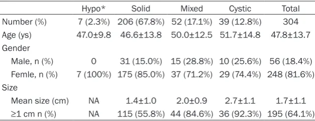
[image:3.629.99.413.92.213.2]Quantitative data we- re shown as mean ± SD, whereas numbers and percentage were provided for qualita-tive data. Quantitaqualita-tive data were compar- ed using independent samples T-test. Per- centages were com-pared using the χ2 test. All tests were 2-sided, and a P value <0.05 was considered Table 1. Baseline characteristics of the patients
Hypo* Solid Mixed Cystic Total
Number (%) 7 (2.3%) 206 (67.8%) 52 (17.1%) 39 (12.8%) 304 Age (ys) 47.0±9.8 46.6±13.8 50.0±12.5 51.7±14.8 47.8±13.7 Gender
Male, n (%) 0 31 (15.0%) 15 (28.8%) 10 (25.6%) 56 (18.4%) Femle, n (%) 7 (100%) 175 (85.0%) 37 (71.2%) 29 (74.4%) 248 (81.6%) Size
Mean size (cm) NA 1.4±1.0 2.0±0.9 2.7±1.1 1.7±1.1 ≥1 cm n (%) NA 115 (55.8%) 44 (84.6%) 36 (92.3%) 195 (64.1%) *Hypo was short for hypoechogenecity area which was diffusely hypoechogenic area under the ultrasound mostly consistent with subacute thyroiditis or Hashimoto’s thyroiditis. For these cases, the borders of these “nodules” were very obscure and precise measurement was impractical. Abbreviations: NA, not available.
statistically significant. Adequacy rate com- parisons were made using McNemar Test. Statistical analyses were performed with SPSS software, version 13.0 for Windows (SPSS Inc, Chicago, IL, USA).
This study protocol was reviewed and approved by the Institutional Review Board (IRB) of the First Affiliated Hospital of Nanjing Medical University. It was approved by the IRB for analysis.
Results
Baseline characteristics of the patients and
nodules
We recruited 304 nodules from 304 patients with average age at 47.8±13.7 years old and 81.6% being female. The mean size of the nod-ules was 1.7±1.1 cm with 67.8% being solid nodules (Table 1).
Final cytological diagnosis of the nodules with different components
The adequacy rate for all nodules in current study was 74.7% while 8.9% of the nodules were diagnosed as M and 13.2% were SUS. For nodules with solid component the adequacy rate was 86.4% and 12.6% were M and 19.4% were SUS. The non-diagnostic rate increased from 13.6% in nodules with solid component to 69.2% in nodules with cystic component (Table 2).
Adequacy rate comparison between conven
-tional smear, SurePath and combined meth -ods
showed in Table 3. As shown, discrepancy exist-ed between conventional smear and SurePath cytology. For all nodules, adequacy rate for con-ventional smears was 63.8% significantly high-er than in 57.2% in SurePath group. This was mostly attributable to solid nodules as the ade-quacy rate for solid nodules was 78.2% in CS group significantly higher than 68% in BD group, while for mixed, cystic nodules and hypo-echo-genic areas there was no significantly differ-ence between BD and CS groups. When com-paring the final cytology diagnosis with conven-tional smear, the final diagnosis rates were sig-nificantly higher than in convention smear group for all nodules, solid nodules, mixed nod-ules and cystic nodnod-ules (Table 4).
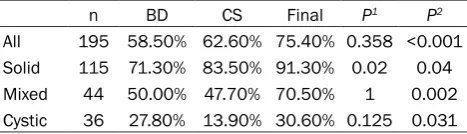
When excluding nodules less than 1 cm, no sig-nificant difference was found for all nodules using BD and CS method. For solid nodules, however, the adequacy rate was significantly higher in CS group than in BD group (Table 5). When comparing the final adequacy rate with
CS diagnosis, similar trend was found as for nodules with various components (Table 5).
Discussion
[image:4.629.98.532.94.172.2]Our prospective study showed that although less adequacy rate was found in the SurePath group compared to CS group especially for solid nodules ≥1 cm, combination of both methods would increase the adequacy rate even further on the basis of each method alone. Non-diagnostic rate in FNA has been a dilemma and an area of controversy. The exact frequency and mechanism of non-diagnostic cytology for thyroid FNA is Table 2. Nodule component and final cytological diagnosis
ND B AUS FOL SUS M Total
Hypo* 5 (71.4%) 1 (14.3%) 1 (14.3%) 0 0 0 7
Solid 28 (13.6%) 51 (24.8%) 58 (28.2%) 3 (1.5%) 40 (19.4%) 26 (12.6%) 206
Mixed 17 (32.7%) 26 (50.0%) 8 (15.4%) 0 0 1 (1.9%) 52
Cystic 27 (69.2%) 11 (28.2%) 1 (2.6%) 0 0 0 39
Total 77 (25.3%) 89 (29.3) 68 (22.4%) 3 (1.0%) 40 (13.2%) 27 (8.9%) 304
*Hypo was short for hypoechogenecity area which was diffusely hypoechogenic area under the ultrasound mostly consistent with subacute thyroiditis or Hashimoto’s thyroiditis. For these cases, the borders of these “nodules” were very obscure and precise measurement was impractical. Abbreviations: ND, Non-diagnostic; B, Benign; AUS, Atypia of Undetermined Significance or Follicular Lesion of Undetermined Significance; FOL, Follicular Neoplasm or Suspicious for a Follicular Neoplasm; SUS, Suspicious for Malignancy; M, Malignant.
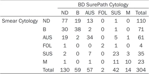
Table 3. Cytology Distribution among BD SurePath and conventional smear
BD SurePath Cytology ND B AUS FOL SUS M Total Smear Cytology ND 77 19 13 0 1 0 110
B 30 38 2 0 1 0 71
AUS 19 2 34 0 5 1 61
FOL 1 0 0 2 1 0 4
SUS 2 0 7 0 23 3 35
M 1 0 1 0 11 10 23
Total 130 59 57 2 42 14 304 Abbreviations: ND, Non-diagnostic; B, Benign; AUS, Atypia of Undeter-mined Significance or Follicular Lesion of UndeterUndeter-mined Significance; FOL, Follicular Neoplasm or Suspicious for a Follicular Neoplasm; SUS, Suspicious for Malignancy; M, Malignant.
[image:4.629.98.340.274.394.2]how-ever only 373 of 2587 patients (14.4%) had thy-roid cancer that measured ≥1 cm in greatest dimension, as determined by pathologic exami-nation [20]. In the report by Alexander [14], the non-diagnostic rate was 13% for all nodules. In sub-analysis the initial non-diagnostic rate for solid nodules was 8% which was similar to 8.7% for solid nodule with size ≥1 cm in our study. All these data suggest that when reporting the adequacy rate of a certain center, percentage of the nodules with different composition is needed. Otherwise the adequacy rate is not comparable. Other factors, for example, coarse or rim calcifications as well as proximity to important anatomic structures (such as the carotid artery or trachea) can limit the ability of the practitioner to obtain adequate tissue [15]. In some cases, no cause can be identified [15]. FNA volume and technique may also influence the adequacy rate. Our previous study showed that multiple passes using 25-gauge needle is superior to 22-gauge needle in obtaining enough sample for cytology evaluation [19]. Controversies exist regarding the lower size cut-off for thyroid FNA. The American Association
of Clinical Endocrinologists (AACE) and Korean Society of Thyroid Radiology (KSTR) guidelines [3, 5] recommend that nodules of any size with suspicious fea-tures undergo biopsy, whereas the ATA guidelines do not recommend biopsy of subcentimeter nodules unless the patient has a high-risk history [2]. In current study the adequacy rate for nodules <1 cm was 74.2% comparable to 75.4% in the nod-ules with size ≥1 cm (P=0.886) suggest-ing that the size had no major impact on the adequacy rate confirming previous result [21]. However, further study is needed regarding the clinical significance of performing FNA for nodules with size less than 1 cm.
[image:5.629.101.335.117.197.2]ATA guideline for thyroid nodules suggests repeating USFNA of these cytologically non-diagnostic nodules [6]. However, there is no specific recommendation or evidence on how and when this re-aspira-tion should be done to avoid the potential possibility for another non-diagnostic cytology diagnosis. A recent study from Brito [16] revealed that the most common approaches to increase the diagnostic yield were (1) use of suction with USFNA Table 4. Adequacy rate comparison between BD
SurePath and conventional smears (CS), Conventional smears and final diagnosis for all nodules
n BD CS Final P1 P2
All 304 57.20% 63.80% 74.70% 0.04 <0.001 Solid 206 68.00% 78.20% 86.40% 0.006 <0.001 Mixed 52 42.30% 48.10% 67.30% 0.678 0.002 Cystic 39 25.60% 15.40% 30.80% 0.289 0.031
Hypo 7 28.60% 28.60% 28.60% 1 1.00%
Note: P1 for adequacy rate comparison between BD SurePath and
conventional smears; P2 for adequacy rate comparison between final
cytology diagnosis and conventional smears; Abbreviations: BD, BD SurePath; CS, conventional smear.
Table 5. Adequacy rate comparison between BD SurePath and conventional smears (CS), Conventional smears and final diagnosis for nodules with size ≥1 cm
n BD CS Final P1 P2
All 195 58.50% 62.60% 75.40% 0.358 <0.001 Solid 115 71.30% 83.50% 91.30% 0.02 0.04
Mixed 44 50.00% 47.70% 70.50% 1 0.002
Cystic 36 27.80% 13.90% 30.60% 0.125 0.031 Note: P1 for adequacy rate comparison between BD SurePath and
conventional smears; P2 for adequacy rate comparison between
Final cytology diagnosis and conventional smears; Abbreviations: BD, BD SurePath; CS, conventional smear.
and (2) changing the targeted area of biopsy within the nodule. Few considered the patients’ preferences as an important driver for the man-agement of non-diagnostic USFNA. Finally, a molecular test for bypassing non-diagnostic USFNA was regarded as the most needed strat-egy for future research. How to improve the adequacy rate for thyroid FNA has been a major area for further research. Klooker demonstrat-ed that a significantly better diagnostic perfor-mance was achieved by using the screw needle compared to the conventional fine needle in cytology of thyroid nodules [22]. However, sam-pling technique using LBP has not been men-tioned in this study. As we may draw from our current study that including a combined meth-od using CS and SP might be one of the options for decreasing the non-diagnostic rate.
[image:5.629.100.334.298.365.2]performed after the conventional smear. There might be traditionally more bruising/bleeding on those later sticks which could compromise adequacy. Also, some patients are in pain, and the latter sticks are therefore shorter. A cross over design is needed to further eliminate the potential impact. Adequacy rate comparisons were made only between cytological diagnosis with different sample preparation methods instead of cytology diagnosis and histology diagnosis which might be the golden standard for comparison. The main purpose of current study was to compare the adequacy rate between different methods. The comparison with histology might be less demanded. However, we do believe that long term follow-up for surgical pathology is warrant for further illustrating this issue.
In conclusion, our study showed that LBC using SurePath is not superior to conventional smears. However, combined methods using both methods may further increase the ade-quacy rate for thyroid FNA than each method alone.
Acknowledgements
The study was supported by National Science Foundation of China (number 81270897) and grants from Jiangsu Health International Exchange Program (Xiaoyun Liu) and Inter- national Exchange Program sponsorship from Nanjing Medical University (Xiaoyun Liu), the Priority Academic Program Development of Jiangsu Higher Education Institutions (PAPD). This work has been presented at the 15th International Thyroid Congress as poster pre-sentation at Orlando, Florida, USA.
Disclosure of conflict of interest
None.
Address correspondence to: Drs. Tao Yang and Xiaodong Wang, Department of Endocrinology, The First Affiliated Hospital of Nanjing Medical Univer- sity, Jiangsu Province Hospital, 300 Guangzhou Road, Nanjing 210029, China. Tel: +86 138- 51498409; E-mail: [email protected] (TY); Tel: +86 13705176611; E-mail: [email protected] (XDW)
References
[1] Guo H, Sun M, He W, Chen H, Li W, Tang J, Tang W, Lu J, Bi Y, Ning G, Yang T, Duan Y. The
preva-lence of thyroid nodules and its relationship with metabolic parameters in a Chinese com-munity-based population aged over 40 years. Endocrine 2014; 45: 230-235.
[2] Cooper DS, Doherty GM, Haugen BR, Kloos RT, Lee SL, Mandel SJ, Mazzaferri EL, McIver B, Pacini F, Schlumberger M, Sherman SI, Steward DL, Tuttle RM. Revised American Thyroid Association management guidelines for patients with thyroid nodules and differenti-ated thyroid cancer. Thyroid 2009; 19: 1167-1214.
[3] Gharib H, Papini E, Paschke R, Duick DS, Valcavi R, Hegedüs L, Vitti P; AACE/AME/ETA Task Force on Thyroid Nodules. American Association of Clinical Endocrinologists, Associazione Medici Endocrinologi, and Euro- peanThyroid Association Medical Guidelines for Clinical Practice for the Diagnosis and Management of Thyroid Nodules. Endocr Pract 2010; 16 Suppl 1: 1-43.
[4] Levine RA. Current guidelines for the manage-ment of thyroid nodules. Endocr Pract 2012; 18: 596-599.
[5] Moon WJ, Baek JH, Jung SL, Kim DW, Kim EK, Kim JY, Kwak JY, Lee JH, Lee JH, Lee YH, Na DG, Park JS, Park SW; Korean Society of Thyroid Radiology (KSThR); Korean Society of Radiology. Ultrasonography and the ultra-sound-based management of thyroid nodules: consensus statement and recommendations. Korean J Radiol 2011; 12: 1-14.
[6] Haugen BR, Alexander EK, Bible KC, Doherty GM, Mandel SJ, Nikiforov YE, Pacini F, Randolph GW, Sawka AM, Schlumberger M, Schuff KG, Sherman SI, Sosa JA, Steward DL, Tuttle RM, Wartofsky L. 2015 American Thyroid Asso- ciation Management Guidelines for Adult Patients with Thyroid Nodules and Diffe- rentiated Thyroid Cancer. Thyroid 2016; 26: 1-133.
[7] Liu XY, Zhu LJ, Cui D, Wang ZX, Chen HH, Duan Y, Shen MP, Zhang ZH, Wang XD, Chen JW, Alexander EK, Yang T. Annual financial impact of thyroidectomies for nodular thyroid disease in China. Asian Pac J Cancer Prev 2014; 15: 5921-5926.
[8] Burman KD, Wartofsky L. Thyroid Nodules. N Engl J Med 2015; 373: 2347-2356.
[9] Mygdakos N, Nikolaidou S, Tzilivaki A, Tamiolakis D. Liquid Based Preparation (LBP) cytology versus Conventional Cytology (CS) in FNA samples from breast, thyroid, salivary glands and soft tissues. Our experience in Crete (Greece). Rom J Morphol Embryol 2009; 50: 245-250.
[11] Tripathy K, Misra A, Ghosh JK. Efficacy of liq-uid-based cytology versus conventional smears in FNA samples. J Cytol 2015; 32: 17-20. [12] Fischer AH, Clayton AC, Bentz JS, Wasserman
PG, Henry MR, Souers RJ, Moriarty AT. Performance differences between convention-al smears and liquid-based preparations of thyroid fine-needle aspiration samples: analy-sis of 47,076 responses in the College of American Pathologists Interlaboratory Com- parison Program in Non-Gynecologic Cytology. Arch Pathol Lab Med 2013; 137: 26-31. [13] Geers C, Bourgain C. Liquid-based FNAC of the
thyroid: a 4-year survey with SurePath. Cancer Cytopathol 2011; 119: 58-67.
[14] Alexander EK, Heering JP, Benson CB, Frates MC, Doubilet PM, Cibas ES, Marqusee E. Assessment of nondiagnostic ultrasound-guid-ed fine neultrasound-guid-edle aspirations of thyroid nodules. J Clin Endocrinol Metab 2002; 87: 4924-4927. [15] Alexander EK. Improving the approach to
non-diagnostic aspirates: learning from each other. Endocrine 2015; 49: 575-576.
[16] Brito JP, Castro MR, Dean DS, Fatourechi V, Stan M. Survey of current approaches to non-diagnostic fine-needle aspiration from solid thyroid nodules. Endocrine 2015; 49: 745-751.
[17] Jung CK, Lee A, Jung ES, Choi YJ, Jung SL, Lee KY. Split sample comparison of a liquid-based method and conventional smears in thyroid fine needle aspiration. Acta Cytol 2008; 52: 313-319.
[18] Cibas ES, Ali SZ. The Bethesda System for Reporting Thyroid Cytopathology. Thyroid 2009; 19: 1159-1165.
[19] Liu X, Zhu L, Wang Z, Cui D, Chen H, Wei L, Wu Y, Rong R, Wu Y, Yao Q, Zhang Z, Cibas ES, Alexander EK, Yang T. Comparison of two dif-ferent standards of care in detecting malig-nant thyroid nodules using thyroid fine-needle aspiration. Mol Clin Oncol 2015; 3: 682-686. [20] Yassa L, Cibas ES, Benson CB, Frates MC,
Doubilet PM, Gawande AA, Moore FD Jr, Kim BW, Nosé V, Marqusee E, Larsen PR, Alexander EK. Long-term assessment of a multidisci-plinary approach to thyroid nodule diagnostic evaluation. Cancer 2007; 111: 508-516. [21] Guo HQ, Zhang ZH, Zhao H, Niu LJ, Chang Q,
Pan QJ. Factors Influencing the Reliability of Thyroid Fine-Needle Aspiration: Analysis of Thyroid Nodule Size, Guidance Mode for Aspiration and Preparation Method. Acta Cytol 2015; 59: 169-174.