Original citation:
Alguel, Y., Cameron, Alexander, Diallinas, G. and Byrne, B.. (2016) Transporter
oligomerization : form and function. Biochemical Society Transactions, 44 (6). pp. 1737-1744.
Permanent WRAP URL:
http://wrap.warwick.ac.uk/86049
Copyright and reuse:
The Warwick Research Archive Portal (WRAP) makes this work of researchers of the University of Warwick available open access under the following conditions.
This article is made available under the Creative Commons Attribution 4.0 International license (CC BY 4.0) and may be reused according to the conditions of the license. For more details see: http://creativecommons.org/licenses/by/4.0/
A note on versions:
The version presented in WRAP is the published version, or, version of record, and may be cited as it appears here.
Transporter oligomerization: form and function
Yilmaz Alguel
1, Alexander D. Cameron
2, George Diallinas
3and Bernadette Byrne
11Department of Life Sciences, Imperial College London, London SW7 2AZ, U.K.;2School of Life Sciences, University of Warwick, Gibbet Hill Road, Coventry CV4 7AL, U.K.; and
3Department of Biology, National and Kapodistrian University of Athens, Panepistimioupolis 15781 Athens, Greece
Correspondence: Bernadette Byrne ([email protected])
Transporters are integral membrane proteins with central roles in the efficient movement of molecules across biological membranes. Many transporters exist as oligomers in the membrane. Depending on the individual transport protein, oligomerization can have roles in membrane trafficking, function, regulation and turnover. For example, our recent studies on UapA, a nucleobase ascorbate transporter, from Aspergillus nidulans, have revealed both that dimerization of this protein is essential for correct trafficking to the membrane and the structural basis of how one UapA protomer can affect the function of the closely associated adjacent protomer. Here, we review the roles of oligomerization in many particularly well-studied transporters and transporter families.
Introduction
The formation of transporter oligomers has been studied by a range of methods, includingfluorescence resonance energy transfer (FRET) [1], cross-linking [2], pull-downs [3] and co-immunoprecipitation [4], as well as biophysical characterization using, for example, size-exclusion chromatography–multiangle light scattering (SEC–MALS) [5]. While such approaches have effectively shown the formation of transporter oligomers, more recent investigations using dominant mutants and high-resolution structural studies have started revealing insights into the precise roles of oligomerization for transporter trafficking, function and regulation of function. This review uses many particularly well-studied transporters and transporter fam-ilies to provide a brief overview of current understanding of the roles of transporter oligomerization. There are additional examples where high-resolution structures have revealed that the interface between transporter protomers in an oligomeric arrangement forms the substrate-binding site and translocation channel. Examples include the small multidrug transporter, EmrE [6] and ABC transporters [7,8]. In these cases, oligomerization is responsible for generating the correct architecture for both substrate binding and transport. These transporters are not covered in this review. Additionally, we have not included transporters where the sole purpose of oligomerization seems to be for stability.
The nucleobase ascorbate transporters
In eukaryotes, transporters are assembled in the endoplasmic reticulum (ER) before being trafficked through COPII vesicles [9]. For some membrane proteins, the association of protomers into the correct quaternary arrangement seems to form a key quality control in this trafficking process [10]. This also seems to be true for the H+ uric acid–xanthine symporter, UapA, a nucleobase ascorbate transporter (NAT) from Aspergillus nidulans, which requires correct association of individual proto-mers into a dimer for effective exit from the ER and localization to the plasma membrane [5]. Mutations in TM7 appear to inhibit or impair oligomerization, preventing efficient trafficking to the membrane and increased turnover. The recent structure of UapA (see below) revealed that TM7 is not directly involved in the dimer interface, but structural changes in this region may indirectly inhibit dimer formation. The importance of oligomerization for correct trafficking to the plasma membrane has been reported for a range of other transporters, notably the neurotransmitter sodium symporter family (see below). However, it remains unclear precisely why oligomers need to form in the ER. The most plausible explanation is that the oligomeric arrangement requires the protomers to be correctly folded, with oligomer formation likely to act as a quality control, allowing only proteins in the fully
folded state to be trafficked. Interestingly, in the case of UapA, impaired trafficking can be overcome by co-expression of the TM7 mutants with wild-type (WT) UapA [5], suggesting, at least in this context, a domin-ant positive effect of the WT form.
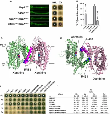
[image:3.595.136.521.179.583.2]In addition, dominant negative mutants of UapA have been described, which traffic effectively to the mem-brane but reduce the transport activity of co-expressed WT, strongly suggesting that this transporter also func-tions as a dimer (Figure 1a,b) [11]. The high-resolution structure of a thermostabilized form of UapA, trapped in the inward-facing conformation, has revealed further clues about the role that oligomerization has in
Figure 1. Role of oligomerization of UapA.
transport function [11]. UapA was crystallized as a closely associated dimer (Figure 1c,d), confirming the
earlierfindings [5]. There are many key residues in UapA involved in substrate selectivity [12–14]. The struc-ture revealed that one of these residues, Arg481, lies in close proximity to the binding site of the opposite pro-tomer, and it is likely to act as the last checkpoint, allowing efficient uptake of the native substrates, xanthine and uric acid, and not other related molecules. The size of the side chain is likely to be important here as muta-tion to the much smaller Gly allows uptake of adenine and hypoxanthine, substrates not transported by WT UapA (Figure 1e,f) [11]. While the structure highlighted the interdependency of the UapA protomers in sub-strate selectivity, it did not explain the dominant negative effects seen for the Q408E mutant. Thus, there are clearly other aspects of UapA protomer cross-talk that have yet to be elucidated. It is interesting to note that, in most crystal structures of dimeric transporters, the individual protomers adopt similar conformations. One exception to this is the recent structure of the CitS where an asymmetrical dimer is observed [15]. In this case, both outward- and inward-facing conformations can be seen in a single oligomer. While such arrangements appear to be stochastic, it is intriguing to ask whether such arrangements may be a co-ordinated part of the transport cycle. The UapA construct used for structure determination is a conformationally locked mutant; however, it is possible that, in the WT form, the two protomers can adopt different conformational states as seen in CitS. To date, there is no evidence that the CitS dimer is important for function.
The overall structure of UapA is similar to that of the anion exchanger 1 (AE1) [16]. Previous research had indicated that AE1 formed dimers [17], and this was confirmed in the high-resolution structure. Intriguingly,
although it seems likely that UapA and AE1 function using a very similar mechanism, dimer formation is markedly different in the two proteins. In the case of UapA, the buried interface is ∼6000 Å2, and it involves mainly interactions between TMs 12, 13 and the loop between the two TMs with TM13, particularly closed associated with the opposite protomer [11]. In contrast, the buried interface in the AE1 dimer is substantially smaller at∼1100 Å2, and only the extracellular tip of TM6 and the loop regions between TMs 5 and 6, 6 and 7 and 12 and 13 are involved in dimer formation [16]. It should be noted, however, that the same subdomain, the so-called gate domain, is involved in dimerization in both proteins. There are no data so far to support a role for dimerization in the function of AE1. It seems likely that the structural and functional differences of oligomerization observed are protein specific, even for transporters that operate in a very similar way. The UapA and AE1 proteins are in the inward- and outward-facing conformations, respectively, which may account for some of the observed differences. It should be noted that in the case of the sodium–proton antiporter, NapA, the dimer interface is very similar in the crystal structures of both the inward- and outward-facing con-formations [18,19], although it is not clear whether dimerization is important for function in this protein. It is still possible though that the variation between the UapA and AE1 dimer interfaces is the result of differences in the crystallization conditions used in each case and/or the fact that the AE1 structure lacks the large soluble N-terminal region.
The formation of the UapA dimer is largely mediated by hydrophobic interactions involving residues that are not particularly well conserved among NATs. The structure of the bacterial NAT, UraA, was described as a monomer, but analysis of the crystal contacts indicates that there are interactions between protomers. These result in the formation of a loose UraA dimer and involve similar regions of the protein as seen in the dimer interface of UapA and AE1 [20]. The interaction interface of UraA is much smaller than that of UapA, involv-ing only a handful of residues from TM13 and the loop between TMs 12 and 13 [20]. The functional
signifi-cance of dimerization of UraA, or indeed other members of the NAT family, remains to be determined.
The LeuT superfamily
export [24]. These studies are strongly suggestive that the activity of one protomer can influence the activity of
the associated molecule(s).
There is no clear structural motif associated with NSS transporter oligomerization. Leucine zipper motifs located in TM2 have been suggested as key in oligomerization of both DAT [22] and GAT-1 [21]. Several studies indicate the importance of the GXXXG motifs in dimerization of integral membrane proteins where the two Gly residues located on the same side of a transmembrane α-helix allow tight packing of protomers [25,26], and such a motif exists in TM6 of the NSS transporters. An additional oligomer interaction site has been suggested in TMs 11 and 12 of SERT [27].
It is interesting that although there is substantial evidence for oligomerization of both DAT and SERT [28], the crystal structures, to date, have failed to capture the oligomeric forms [29–31]. This might be due to the formation of relatively weakly associated oligomers unable to withstand the rigours of the extraction and isola-tion processes required for structural studies. This issue has also been highlighted for the recent structure of the monomeric form of an SLC26 family member, where loss of the oligomeric form was assumed to be a result of the harsh nature of the detergent treatment [32]. It remains to be seen if oligomerization is important for the function of SLC26. It is also worth noting that all the DAT and SERT structures available have been obtained in complex with antibody fragments. Antibody–transporter complex formation may disrupt oligomer interactions. Perhaps, approaches using fusion proteins of the individual protomers might facilitate trapping the oligomeric state of the NSS transporters.
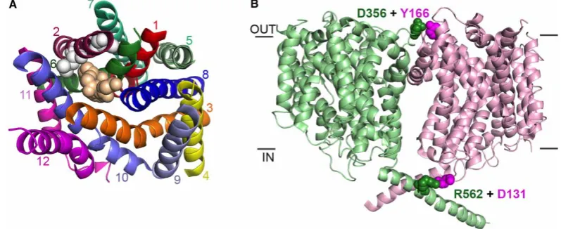
The monomeric structures of DAT [29,30] and SERT [31] reveal that both the Leu zipper motif and the GXXXG motif are buried away within the core of the protein, more likely to have roles in the architecture of the protomer than in mediating oligomerization (Figure 2a). Mutations in either of these regions may distort the protomer so much, that it can no longer form effective oligomers. Both TMs 11 and 12 are surface exposed, and they may have roles in oligomer formation (Figure 2a). Indeed, in the structure of SERT, TM12 is involved in the formation of a crystallographic dimer [31]. This is highly unlikely to represent a physiological arrangement of the molecules, however, as one protomer is rotated substantially compared with the other. This would put the soluble loop regions of one of the protomers into the membrane, a highly unlikely arrangement. TM12 may have roles in interactions with lipids as, in the structure, this region of the protein is associated with cholesterol hemisuccinate [31].
[image:5.595.126.525.464.627.2]Dimer structures of the bacterial NSS homologue, LeuT, are available however, and these reveal that dimer-ization is mediated by TM12 and TM9a as well as residues from the second extracellular loop [33]. However, there is no evidence as yet that LeuT functions as an oligomer.
Figure 2. Monomer versus oligomer transporter structures.
Studies on the bacterial osmoregulated betaine transporter BetP, a member of the betaine/choline/carnitine family, have shown that this protein forms a stable trimer [34]. Generation of mutants of the transporter reveals that the protein is still transport-active as an individual protomer, although the activity is reduced com-pared with that of the WT trimer [35]. However, the monomeric form is no longer responsive to osmotic stress, strongly suggesting that the trimer is key in detecting environmental change and activating the trans-porter in response to that change [35]. Recent structures have additionally highlighted that the interactions involved in trimer formation may play roles in the conformational changes associated with the transport cycle [36]. Both functions of the BetP trimer appear to be regulated by ionic interactions formed between the indi-vidual protomers [37] (Figure 2b). Intriguingly, as seen in CitS, the individual protomers in the BetP trimer can adopt different conformations [36].
The SWEET transporter family
SWEET transporters are responsible for uptake and distribution of mono- and disaccharides. Early studies on plant SWEET transporters using dominant negative mutants highlighted the importance of oligomerization for function [38]. These transporter proteins contain just seven transmembrane domains, and so it was thought that oligomerization was essential to form the functional translocating unit. However, the recent structure of the homotrimeric SWEET fromArabidopsis thaliana shows that the individual protomer forms the transloca-tion channel [39]. Further structure-based investigations revealed that functionally inactive mutations in the extracellular or intracellular gates of one protomer negatively affected the function of associated protomers [39]. It is possible that the lack of conformational change in one protomer inhibits movement in associated molecules. Thesefindings strongly suggest that SWEET homotrimers function through co-operativity between protomers; however, the precise molecular basis of this is yet to be revealed.
The major facilitator superfamily
Early studies on the bacterial major facilitator superfamily (MFS) transporter, LacS, gave the first indications that oligomerization may play a role in transporter function. Biophysical analyses had indicated that LacS was a dimer [40]. Co-expression of mutant non-functional transporter with functional transporter forms indicated co-operativity between the individual protomers of the dimer required for proton-dependent transport [41].
A dominant positive mutant has been described for the human proton-coupled folate transporter. Co-expression of the WT transporter with the mutant, which traffics effectively to the membrane but is transport-inactive, resulted in greater levels of activity than expression of WT alone [1]. Indeed, the level of activity observed was similar to that for co-expression of two differently tagged forms of WT transporter. This indicated a dominant positive effect whereby the active WT was able to recover the transport activity of the inactive mutant through direct association of the different transporter protomers [1].
Research on the plant phosphate transporter, Pht1, another MFS member, identified a mutant with expres-sion levels and localization similar to WT, but which both reduced oligomerization and increased transport activity (Vmax). Thesefindings strongly suggested that, in this case, oligomerization plays a regulatory role [42].
Oligomerization-dependent regulation has also been reported for the plant nitrate transporter, NRT1.1, which can act as either a low- or high-affinity transport system [43]. Crystal structures of the NRT1.1 revealed a dimeric form of the protein (Figure 3) [44,45]. Mutational studies indicated that phosphorylation of Thr101 resulted in reduced stability and increased transport activity compared with the unphosphorylated form [44]. Sun et al. [45] suggested that this was due to a change in the oligomeric status of the protein, and they carried out experiments, indicating that phosphorylation of Thr101 induced dissociation of the low-affinity dimer into high-affinity protomers (Figure 3). However, another plausible theory is that phosphorylation simply results in a minor structural change with the effect of changing the affinity [44]. Support for this theory came from the generation of a phosphomimic mutant of the equivalent residue in the related monomeric peptide transporter fromShewanella oneidensis, PepTso, which also decreased stability and increased transport activity [44].
The ammonium transporter/MEP/Rh transporter family
one protomer inhibits the activity of associated non-phosphorylated protomers [47,48]. Such regulatory mechanisms in the Pht1, NRT1.1 and the AMTs are likely to allow plants to rapidly and effectively adapt to changing environmental conditions.
Conclusion
It is clear that oligomerization is key to correct localization, function and regulation of different transporter proteins. Combinations of functional and structural studies have been particularly effective at providing insights into the molecular basis of functional cross-talk of transporter protomers in oligomeric arrangements. However, it is clear that we have only just scratched the surface of the precise roles of this important feature of many biologically and medically important transporter proteins.
Abbreviations
AE1, anion exchanger 1; AMTs, ammonium transporters; DAT, dopamine transporter; ER, endoplasmic reticulum; FRET,fluoresence resonance energy transfer; GAT1, GABA transporter 1; MFS, major facilitator superfamily; NATs, nucleobase ascorbate transporters; NSS, neurotransmitter sodium symporter;
SERT, serotonin transporter; WT, wild type.
Funding
This work was supported by the Biotechnology and Biological Sciences Research Council [grant BB/K017292/1].
Acknowledgements
The authors thank Dr Sotiris Amillis for the assistance in generatingFigure 1.
Competing Interests
The Authors declare that there are no competing interests associated with the manuscript.
References
1 Hou, Z., Kugel Desmoulin, S., Etnyre, E., Olive, M., Hsiung, B., Cherian, C. et al. (2012) Identification and functional impact of homo-oligomers of the human proton-coupled folate transporter.J. Biol. Chem.287, 4982–4995 doi:10.1074/jbc.M111.306860
2 Jess, U., Betz, H. and Schloss, P. (1996) The membrane-bound rat serotonin transporter, SERT1, is an oligomeric protein.FEBS Lett.394, 44–46 doi:10.1016/0014-5793(96)00916-7
3 Bartholomaus, I., Milan-Lobo, L., Nicke, A., Dutertre, S., Hastrup, H., Jha, A. et al. (2008) Glycine transporter dimers: evidence for occurrence in the plasma membrane.J. Biol. Chem.283, 10978–10991 doi:10.1074/jbc.M800622200
4 Pessino, A., Hebert, D.N., Woon, C.W., Harrison, S.A., Clancy, B.M., Buxton, J.M. et al. (1991) Evidence that functional erythrocyte-type glucose transporters are oligomers.J. Biol. Chem.266, 20213–20217 PMID:1939082
5 Martzoukou, O., Karachaliou, M., Yalelis, V., Leung, J., Byrne, B., Amillis, S. et al. (2015) Oligomerization of the UapA purine transporter is critical for ER-exit, plasma membrane localization and turnover.J. Mol. Biol.427, 2679–2696 doi:10.1016/j.jmb.2015.05.021
[image:7.595.136.520.77.201.2]6 Chen, Y.-J., Pornillos, O., Lieu, S., Ma, C., Chen, A.P. and Chang, G. (2007) X-ray structure of EmrE supports dual topology model.Proc. Natl Acad. Sci. USA104, 18999–19004 doi:10.1073/pnas.0709387104
Figure 3. Phosphorylation-dependent regulation of NRT1.1 fromArabidopsis thaliana(PDB: 4OH3).
7 Srinivasan, V., Pierik, A.J. and Lill, R. (2014) Crystal structures of nucleotide-free and glutathione-bound mitochondrial ABC transporter Atm1.Science
343, 1137–1140 doi:10.1126/science.1246729
8 Choudhury, H.G., Tong, Z., Mathavan, I., Li, Y., Iwata, S., Zirah, S. et al. (2014) Structure of an antibacterial peptide ATP-binding cassette transporter in a novel outward occluded state.Proc. Natl Acad. Sci. USA111, 9145–9150 doi:10.1073/pnas.1320506111
9 Barlowe, C., Orci, L., Yeung, T., Hosobuchi, M., Hamamoto, S., Salama, N. et al. (1994) COPII: a membrane coat formed by Sec proteins that drive vesicle budding from the endoplasmic reticulum.Cell77, 895–907 doi:10.1016/0092-8674(94)90138-4
10 Springer, S., Malkus, P., Borchert, B., Wellbrock, U., Duden, R. and Schekman, R. (2014) Regulated oligomerization induces uptake of a membrane protein into COPII vesicles independent of its cytosolic tail.Traffic15, 531–545 doi:10.1111/tra.12157
11 Alguel, Y., Amillis, S., Leung, J., Lambrinidis, G., Capaldi, S., Scull, N.J. et al. (2016) Structure of eukaryotic purine/H+symporter UapA suggests a role for homodimerization in transport activity.Nat. Commun.7, 11336 doi:10.1038/ncomms11336
12 Papageorgiou, I., Gournas, C., Vlanti, A., Amillis, S., Pantazopoulou, A. and Diallinas, G. (2008) Specific interdomain synergy in the UapA transporter determines its unique specificity for uric acid among NAT carriers.J. Mol. Biol.382, 1121–1135 doi:10.1016/j.jmb.2008.08.005
13 Kosti, V., Papageorgiou, I. and Diallinas, G. (2010) Dynamic elements at both cytoplasmically and extracellularly facing sides of the UapA transporter selectively control the accessibility of substrates to their translocation pathway.J. Mol. Biol.397, 1132–1143 doi:10.1016/j.jmb.2010.02.037 14 Diallinas, G. (2014) Understanding transporter specificity and the discrete appearance of channel-like gating domains in transporters.Front. Pharmacol.
5, 207 doi:10.3389/fphar.2014.00207
15 Wöhlert, D., Grötzinger, M.J., Kühlbrandt, W. and Yildiz, Ö. (2015) Mechanism of Na+-dependent citrate transport from the structure of an asymmetrical CitS dimer.eLife4, e09375 doi:10.7554/eLife.09375
16 Arakawa, T., Kobayashi-Yurugi, T., Alguel, Y., Iwanari, H., Hatae, H., Iwata, M. et al. (2015) Crystal structure of the anion exchanger domain of human erythrocyte band 3.Science350, 680–684 doi:10.1126/science.aaa4335
17 Casey, J.R. and Reithmeier, R.A. (1991) Analysis of the oligomeric state of Band 3, the anion transport protein of the human erythrocyte membrane, by size exclusion high performance liquid chromatography. Oligomeric stability and origin of heterogeneity.J. Biol. Chem.266, 15726–15737
PMID:1874731
18 Lee, C., Kang, H.J., von Ballmoos, C., Newstead, S., Uzdavinys, P., Dotson, D.L. et al. (2013) A two-domain elevator mechanism for sodium/proton antiport.Nature501, 573–577 doi:10.1038/nature12484
19 Coincon, M., Uzdavinys, P., Nji, E., Dotson, D.L., Winkelmann, I., Abdul-Hussein, S. et al. (2016) Crystal structures reveal the molecular basis of ion translocation in sodium/proton antiporters.Nat. Struct. Mol. Biol.23, 248–255 doi:10.1038/nsmb.3164
20 Lu, F., Li, S., Jiang, Y., Jiang, J., Fan, H., Lu, G. et al. (2011) Structure and mechanism of the uracil transporter UraA.Nature472, 243–246 doi:10.1038/nature09885
21 Scholze, P., Freissmuth, M. and Sitte, H.H. (2002) Mutations within an intramembrane leucine heptad repeat disrupt oligomer formation of the rat GABA transporter 1.J. Biol. Chem.277, 43682–43690 doi:10.1074/jbc.M205602200
22 Torres, G.E., Carneiro, A., Seamans, K., Fiorentini, C., Sweeney, A., Yao, W.-D. et al. (2003) Oligomerization and trafficking of the human dopamine transporter. Mutational analysis identifies critical domains important for the functional expression of the transporter.J. Biol. Chem.278, 2731–2739 doi:10.1074/jbc.M201926200
23 Zhen, J., Antonio, T., Cheng, S.-Y., Ali, S., Jones, K.T. and Reith, M.E.A. (2015) Dopamine transporter oligomerization: impact of combining protomers with differential cocaine analog binding affinities.J. Neurochem.133, 167–173 doi:10.1111/jnc.13025
24 Seidel, S., Singer, E.A., Just, H., Farhan, H., Scholze, P., Kudlacek, O. et al. (2005) Amphetamines take two to tango: an oligomer-based counter-transport model of neurotransmitter transport explores the amphetamine action.Mol. Pharmacol.67, 140–151 PMID:15615700
25 Arselin, G., Giraud, M.-F., Dautant, A., Vaillier, J., Brèthes, D., Coulary-Salin, B. et al. (2003) The GxxxG motif of the transmembrane domain of subunit e is involved in the dimerization/oligomerization of the yeast ATP synthase complex in the mitochondrial membrane.Eur. J. Biochem.270, 1875–1884 doi:10.1046/j.1432-1033.2003.03557.x
26 Overton, M.C., Chinault, S.L. and Blumer, K.J. (2003) Oligomerization, biogenesis, and signaling is promoted by a glycophorin A-like dimerization motif in transmembrane domain 1 of a yeast G protein-coupled receptor.J. Biol. Chem.278, 49369–49377 doi:10.1074/jbc.M308654200
27 Just, H., Sitte, H.H., Schmid, J.A., Freissmuth, M. and Kudlacek, O. (2003) Identification of an additional interaction domain in transmembrane domains 11 and 12 that supports oligomer formation in the human serotonin transporter.J. Biol. Chem.279, 6650–6657 doi:10.1074/jbc.M306092200 28 Anderluh, A., Klotzsch, E., Reismann, A.W.A.F., Brameshuber, M., Kudlacek, O., Newman, A.H. et al. (2014) Single molecule analysis reveals
coexistence of stable serotonin transporter monomers and oligomers in the live cell plasma membrane.J. Biol. Chem.289, 4387–4394 doi:10.1074/jbc.M113.531632
29 Penmatsa, A., Wang, K.H. and Gouaux, E. (2013) X-ray structure of dopamine transporter elucidates antidepressant mechanism.Nature503, 85–90 doi:10.1038/nature12533
30 Wang, K.H., Penmatsa, A. and Gouaux, E. (2015) Neurotransmitter and psychostimulant recognition by the dopamine transporter.Nature521, 322–327 doi:10.1038/nature14431
31 Coleman, J.A., Green, E.M. and Gouaux, E. (2016) X-ray structures and mechanism of the human serotonin transporter.Nature532, 334–339 doi:10.1038/nature17629
32 Geertsma, E.R., Chang, Y.-N., Shaik, F.R., Neldner, Y., Pardon, E., Steyaert, J. et al. (2015) Structure of a prokaryotic fumarate transporter reveals the architecture of the SLC26 family.Nat. Struct. Mol. Biol.22, 803–808 doi:10.1038/nsmb.3091
33 Yamashita, A., Singh, S.K., Kawate, T., Jin, Y. and Gouaux, E. (2005) Crystal structure of a bacterial homologue of Na+/Cl−-dependent neurotransmitter transporters.Nature437, 215–223 doi:10.1038/nature03978
34 Ressl, S., Terwisscha van Scheltinga, A.C., Vonrhein, C., Ott, V. and Ziegler, C. (2009) Molecular basis of transport and regulation in the Na+/betaine symporter BetP.Nature458, 47–52 doi:10.1038/nature07819
35 Perez, C., Khafizov, K., Forrest, L.R., Krämer, R. and Ziegler, C. (2011) The role of trimerization in the osmoregulated betaine transporter BetP.EMBO Rep.12, 804–810 doi:10.1038/embor.2011.102
37 Gärtner, R.M., Perez, C., Koshy, C. and Ziegler, C. (2011) Role of bundle helices in a regulatory crosstalk in the trimeric betaine transporter BetP.
J. Mol. Biol.414, 327–336 doi:10.1016/j.jmb.2011.10.013
38 Xuan, Y.H., Hu, Y.B., Chen, L.-Q., Sosso, D., Ducat, D.C., Hou, B.-H. et al. (2013) Functional role of oligomerization for bacterial and plant SWEET sugar transporter family.Proc. Natl Acad. Sci. USA110, E3685–E3694 doi:10.1073/pnas.1311244110
39 Tao, Y., Cheung, L.S., Li, S., Eom, J.-S., Chen, L.-Q., Xu, Y. et al. (2015) Structure of a eukaryotic SWEET transporter in a homotrimeric complex.
Nature527, 259–263 doi:10.1038/nature15391
40 Friesen, R.H.E., Knol, J. and Poolman, B. (2000) Quaternary structure of the lactose transport protein ofStreptococcus thermophilusin the detergent-solubilized and membrane-reconstituted state.J. Biol. Chem.275, 33527–33535 doi:10.1074/jbc.M004066200
41 Veenhoff, L.M., Heuberger, E.H.M.L. and Poolman, B. (2001) The lactose transport protein is a cooperative dimer with two sugar translocation pathways.
EMBO J.20, 3056–3062 doi:10.1093/emboj/20.12.3056
42 Fontenot, E.B., Ditusa, S.F., Kato, N., Olivier, D.M., Dale, R., Lin, W.-Y. et al. (2015) Increased phosphate transport ofArabidopsis thalianaPht1;1 by site-directed mutagenesis of tyrosine 312 may be attributed to the disruption of homomeric interactions.Plant Cell Environ.38, 2012–2022 doi:10.1111/pce.12522
43 Liu, K.-H. and Tsay, Y.-F. (2003) Switching between the two action modes of the dual-affinity nitrate transporter CHL1 by phosphorylation.EMBO J.22, 1005–1013 doi:10.1093/emboj/cdg118
44 Parker, J.L. and Newstead, S. (2014) Molecular basis of nitrate uptake by the plant nitrate transporter NRT1.1.Nature507, 68–72 doi:10.1038/nature13116
45 Sun, J., Bankston, J.R., Payandeh, J., Hinds, T.R., Zagotta, W.N. and Zheng, N. (2014) Crystal structure of the plant dual-affinity nitrate transporter NRT1.1.Nature507, 73–77 doi:10.1038/nature13074
46 Lanquar, V., Loqué, D., Hörmann, F., Yuan, L., Bohner, A., Engelsberger, W.R. et al. (2009) Feedback inhibition of ammonium uptake by a phospho-dependent allosteric mechanism inArabidopsis.Plant Cell21, 3610–3622 doi:10.1105/tpc.109.068593
47 Neuhäuser, B., Dynowski, M., Mayer, M. and Ludewig, U. (2007) Regulation of NH4 +
transport by essential cross talk between AMT monomers through the carboxyl tails.Plant Physiol.143, 1651–1659 doi:10.1104/pp.106.094243
48 Yuan, L., Gu, R., Xuan, Y., Smith-Valle, E., Loqué, D., Frommer, W.B. et al. (2013) Allosteric regulation of transport activity by heterotrimerization of