RESEARCH ARTICLE
PATTERN OF CARDIAC CONDUCTION DEFECTS – A HOSPITAL BASED STUDY
*Aadil Ashraf, Touseef ahmad Mir, Irfan Gul, Gazzanfar Ali and Javed Khan
Department of Medicine, Govt. Medical College, Srinagar, India
ARTICLE INFO ABSTRACT
Background: The interest in cardiac conduction system has focused primarily on its role as a predictor of mortality and coexistent cardiovascular diseases particularly hospitalized patients. Objectives: The study was undertaken to study the pattern of cardiac conduction defects in a tertiary care hospital.
Methodology: The study was conducted from 1st March 2012 to 31st august 2013 and included cases >20yrs of age presenting to OPD or admitted in SMHS hospital and showing some form of cardiac conduction defect on a standard 12 lead ECG. A total of 1710 cases were studied. A thorough medical history and meticulous physical examination was done and appropriate statistical methods applied to derive the results.
Results: Of the 1710 cases, 990(57.9%) were males and 720(42.1%) were females. Most of cases were seen in the age group of 70-79yrs (25.7%). The various cardiac conduction defects in decreasing order of frequency were: LBBB 501 (29.2%), LAHB 431 (25.2%), RBBB 308 (18.01%), Bifascicular block 173(10.1%), Complete Heart Block 104(6.08%), 2nddegree heart block 91(5.32), Trifascicular Block 48(2.8%), 1st degree Heart block 34(1.99%), SI SII SIII Syndrome 13(0.76%) and LPHB
7(0.4%).
Conclusion: LBBB is the most common conduction defect in a hospitalized setting. The cardiac conduction defects increase with advancing age and a close follow up on patients cardiac status should be maintained as they are linked with significant morbidity and mortality.
Copyright©2017, Aadil Ashraf et al.This is an open access article distributed under the Creative Commons Attribution License, which permits unrestricted use, distribution, and reproduction in any medium, provided the original work is properly cited.
INTRODUCTION
The heart is unique among the muscles of body in that it possesses the properties of automatic impulse formation and rhythmic contraction. Electrical impulse formation occurs within the conduction system of heart (Nova Golschalger and Mervin, 1989). Cardiac conduction defects are divided into two parts (John Merideth and Raymond Pruitt, 1973):
Atrioventricular (A-V) conduction disturbances; and
Bundle-branch block and intraventricular block.
The AV Blocks defined are (Hurst`s):
First degree heart Block.
Second degree heart Block:
Mobitz type I Mobitz type II
2:1 atrioventricular Block
*Corresponding author: Aadil Ashraf,
Department of Medicine, Govt. Medical College, Srinagar, India.
High grade atrioventricular Block
Third degree (Complete) heart Block.
Atrioventricular Dissociation.
The Bundle Branch Block and intraventricular defects include (John Merideth and Raymond Pruitt, 1973):
Right Bundle Branch Block (RBBB).
Left bundle branch Block (LBBB).
Left Anterior Fascicular Block (LAFB).
Left Posterior Fascicular Block (LPFB).
Right Bundle Branch Block combined with either Left Anterior Fascicular Block or Left Posterior Fascicular block.
The interest in Bundle Branch Blocks has focused primarily on its role as a predictor of mortality and coexistent cardiovascular diseases. Conduction defects are presenting increasingly common on electrocardiograms of adult population (John et al., 1982, 1978, 1979 & 1980) and in hospitalized patients they are reported to be about 10-15% (Fisch et al., 1957; Suarez et al., 1961; Nihalick et al., 1974) although this does not represent the actual prevalence of
ISSN: 0975-833X
International Journal of Current Research Vol. 9, Issue, 07, pp.54875-54879, July, 2017
INTERNATIONAL JOURNAL OF CURRENT RESEARCH
Article History: Received 27thApril, 2017
Received in revised form 22ndMay, 2017
Accepted 19thJune, 2017
Published online 31stJuly, 2017
Citation: Aadil Ashraf, Touseef ahmad Mir, Irfan Gul, Gazzanfar Ali and Javed Khan, 2017.“Pattern of cardiac conduction defects–A hospital based
study”,International Journal of Current Research, 9, (07), 54875-54879. Key words:
Cardiac conduction defects (LBBB,CHB, AV BLOCKS), Increasing age,
Male sex.
RESEARCH ARTICLE
PATTERN OF CARDIAC CONDUCTION DEFECTS – A HOSPITAL BASED STUDY
*Aadil Ashraf, Touseef ahmad Mir, Irfan Gul, Gazzanfar Ali and Javed Khan
Department of Medicine, Govt. Medical College, Srinagar, India
ARTICLE INFO ABSTRACT
Background: The interest in cardiac conduction system has focused primarily on its role as a predictor of mortality and coexistent cardiovascular diseases particularly hospitalized patients. Objectives: The study was undertaken to study the pattern of cardiac conduction defects in a tertiary care hospital.
Methodology: The study was conducted from 1st March 2012 to 31st august 2013 and included cases >20yrs of age presenting to OPD or admitted in SMHS hospital and showing some form of cardiac conduction defect on a standard 12 lead ECG. A total of 1710 cases were studied. A thorough medical history and meticulous physical examination was done and appropriate statistical methods applied to derive the results.
Results: Of the 1710 cases, 990(57.9%) were males and 720(42.1%) were females. Most of cases were seen in the age group of 70-79yrs (25.7%). The various cardiac conduction defects in decreasing order of frequency were: LBBB 501 (29.2%), LAHB 431 (25.2%), RBBB 308 (18.01%), Bifascicular block 173(10.1%), Complete Heart Block 104(6.08%), 2nddegree heart block 91(5.32), Trifascicular Block 48(2.8%), 1st degree Heart block 34(1.99%), SI SII SIII Syndrome 13(0.76%) and LPHB
7(0.4%).
Conclusion: LBBB is the most common conduction defect in a hospitalized setting. The cardiac conduction defects increase with advancing age and a close follow up on patients cardiac status should be maintained as they are linked with significant morbidity and mortality.
Copyright©2017, Aadil Ashraf et al.This is an open access article distributed under the Creative Commons Attribution License, which permits unrestricted use, distribution, and reproduction in any medium, provided the original work is properly cited.
INTRODUCTION
The heart is unique among the muscles of body in that it possesses the properties of automatic impulse formation and rhythmic contraction. Electrical impulse formation occurs within the conduction system of heart (Nova Golschalger and Mervin, 1989). Cardiac conduction defects are divided into two parts (John Merideth and Raymond Pruitt, 1973):
Atrioventricular (A-V) conduction disturbances; and
Bundle-branch block and intraventricular block.
The AV Blocks defined are (Hurst`s):
First degree heart Block.
Second degree heart Block:
Mobitz type I Mobitz type II
2:1 atrioventricular Block
*Corresponding author: Aadil Ashraf,
Department of Medicine, Govt. Medical College, Srinagar, India.
High grade atrioventricular Block
Third degree (Complete) heart Block.
Atrioventricular Dissociation.
The Bundle Branch Block and intraventricular defects include (John Merideth and Raymond Pruitt, 1973):
Right Bundle Branch Block (RBBB).
Left bundle branch Block (LBBB).
Left Anterior Fascicular Block (LAFB).
Left Posterior Fascicular Block (LPFB).
Right Bundle Branch Block combined with either Left Anterior Fascicular Block or Left Posterior Fascicular block.
The interest in Bundle Branch Blocks has focused primarily on its role as a predictor of mortality and coexistent cardiovascular diseases. Conduction defects are presenting increasingly common on electrocardiograms of adult population (John et al., 1982, 1978, 1979 & 1980) and in hospitalized patients they are reported to be about 10-15% (Fisch et al., 1957; Suarez et al., 1961; Nihalick et al., 1974) although this does not represent the actual prevalence of
ISSN: 0975-833X
International Journal of Current Research Vol. 9, Issue, 07, pp.54875-54879, July, 2017
INTERNATIONAL JOURNAL OF CURRENT RESEARCH
Article History: Received 27thApril, 2017
Received in revised form 22ndMay, 2017
Accepted 19thJune, 2017
Published online 31stJuly, 2017
Citation: Aadil Ashraf, Touseef ahmad Mir, Irfan Gul, Gazzanfar Ali and Javed Khan, 2017.“Pattern of cardiac conduction defects–A hospital based
study”,International Journal of Current Research, 9, (07), 54875-54879. Key words:
Cardiac conduction defects (LBBB,CHB, AV BLOCKS), Increasing age,
disease in the general population. This type of study has not been done in the state so far. Hence this study was undertaken in this Hospital to know the pattern of different cardiac conduction defects.
Aims
To Study the pattern of Cardiac Conduction Defects in Hospitalized Patients and to Study the age-sex distribution of Cardiac conduction defects.
MATERIALS AND METHODS
This study included all the patients admitted in medical and surgical wards of SMHS hospital with some form of ECG documented Cardiac conduction abnormality. The patients admitted in medical and surgical intensive care units were also included in the study. The patients were included in the study irrespective of the underlying illness. This study was conducted from a period of 1st of March 2012 to 31st of August 2013. A detailed History was taken from the patients regarding the presenting illness. A detailed history with regard to the underlying illness and drug history was also taken from the patients. A standard 12 lead ECG was taken with a paper speed of 25 mm/sec. In addition a one minute rhythm strip was also recorded. The different cardiac conduction defects were diagnosed using the definitive criteria (Kellkar and Elizabeth, 1996; Lawrence et al., 1971):
A-V block first degree: PR interval ˃200ms, each P
wave followed by QRS complex.
2nd Degree Block Type 1: Progressive P-R
prolongation leading to dropped beats.
2ndDegree Block Type 2: Herein dropped beats occur
but the P-R interval of all conducted beats is constant.
Complete Heart Block:
a) Regular PP interval. b) Regular RR interval. c) Varying PR interval.
d) PP interval less than RR interval.
Left Bundle Branch Block(LBBB):
a) QRS duration≥0.12 sec inany three standard leads. b) Dominant S wave in V1.
c) R duration 0.08 sec in lead V5 or V6. d) S duration 0.04 sec.
e) No Q or S wave in lead V5 or V6.
Right Bundle Branch Block(RBBB):
a) QRS duration˃120ms.
b) Secondary R` wave in right precordial leads V1 or V2.(usually rsR`,rsr`,rSR`).
c) Wide S Wave in leads 1, V5, V6(more than 40 ms).
Left Anterior Hemi Block (LAHB) :
a) Left axis deviation(usually between -45 and -90 degrees).
b) Small Q waves with tall R waves (= 'qR complexes') in leads I and aVL.
c) Small R waves with deep S waves (= rS complexes') in leads ll. III. aVF.
d) QRS duration normal or slightly prolonged (80-110 ms).
e) Prolonged R wave peak time in aVL > 45 ms. f) Increased QRS voltage in the limb leads.
Left Posterior Hemi Block (LAHB):
a) Right axis deviation (> +90 degrees).
b) Small R waves with deep S waves (= `rS complexes') in leads I and aVL.
c) Small Q waves with tall R waves (= complexes') in leads IL III and aVF.
d) QRS duration normal or slightly prolonged (80-1 10ms).
e) Prolonged R wave peak time in aVF. f) Increased QRS voltage in the limb leads. g) No evidence of right ventricular hypertrophy. h) No evidence of any other cause for right axis
deviation.
RBBB + LAHB: Mean QRS axis between -45° and
120° in presence of RBBB.
RBBB + LPHB: presence of RBBB and mean QRS
axis in frontal plane to about +120° with absence of right ventricular Hypertrophy.
Trifascicular Block:
a) Bifascicular Block + 1stdegree AV block. b) Bifascicular Block + 2nddegree AV block. c) RBBB + alternating LAFB/LPFB. d) Bifascicular block + 3rddegree AV block.
SISIISIIISyndrome:
a) Prominent S wave in all of the standard leads I,II, III.
b) S wave greater than the preceding R wave in atleast one of these leads.
c) S wave in lead II should be greater than S wave in lead III.
Statistical methods
Statistical testing was conducted with the statistical package for the social science system version SPSS 20.0.Continuous variables are presented as mean ± SD and categorized into groups; Categorical variables are presented as frequencies and percentage. Nominal categorical data between the groups were compared using Chi-square test or Fisher's exact test as appropriate. p˂0.05 was considered statistically significant.
[image:2.595.363.505.690.734.2]RESULTS
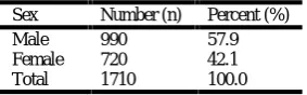
Table 1. Number and Percentage of Cases in Relation to Sex
Sex Number (n) Percent (%) Male 990 57.9 Female 720 42.1 Total 1710 100.0
Table 2. Distribution and percent of studied cases in relation to age
Age in years n Percent (%) 20-29 50 2.9 30-39 130 7.6 40-49 230 13.5 50-59 308 18.0 60-69 386 22.6 70-79 458 26.8 80-89 128 7.5 >90 20 1.2 Total 1710 100.0
[image:3.595.49.273.266.385.2]The number of studied cases increased with increase in age till 70-79 years of age; with maximum number of cases seen in the age group of 70-79 years: 458 (26.8%). Mean age was (61.5 ± 15.2) years.
Table 3. Distribution of studied cases in relation to age and sex
Age (years) Sex
Male Female Total
n % n %
20-29 35 70.0% 15 30.0% 50 30-39 75 57.7% 55 42.3% 130 40-49 146 63.5% 84 36.5% 230 50-59 140 45.5% 168 54.5% 308 60-69 233 60.4% 153 39.6% 386 70-79 279 60.9% 179 39.1% 458 80-89 70 54.7% 58 45.3% 128 >90 12 60.0% 8 40.0% 20 Total 990 57.9% 720 42.1% 1710
Among males, most of the studied cases were in 70-79 years age group: 279(28.18%) and among females most of the studied cases were in the age group of 70-79 years: 179(24.86%). Mean age among males was (62 ± 16) years with minimum and maximum age of (22 and 94) years respectively. Mean age among females was (62 ± 15) years with minimum and maximum age of 23 and 95 years respectively. There was no statistical significance between age of males and females (p=0.88). Males were more as compared to females in all the age groups except 50-59 year age group (54.5 % females vs 45.5 % males).
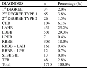
Table 4. The number and percentage of different cardiac conduction defects in the studied population
DIAGNOSIS n Percentage (%) 1stDEGREE 34 2.0%
2ndDEGREE TYPE 1 65 3.8%
2ndDEGREE TYPE 2 26 1.5%
CHB 104 6.1%
LAHB 431 25.2%
LBBB 501 29.3%
LPHB 7 0.4%
RBBB 308 18.0%
RBBB + LAH 161 9.4% RBBB + LPH 12 0.7% SI SII SIII 13 0.8%
TFB 48 2.8%
Total 1710 100.0%
Of the studied cases, the different cardiac conduction defects in decreasing order of frequency were:
LBBB 501 (29.2%).
LAHB 431(25.2%).
RBBB 308 (18.01%).
Bifascicular block 173(10.1%).
Complete Heart Block 104(6.08%).
2nddegree heart block type1 65(3.8%).
Trifascicular Block 48(2.8%).
1stdegree Heart block 34(1.99%).
2nddegree heart block type 2 26(1.5%).
SISIISIIISyndrome 13(0.76%). LPHB 7(0.4%).
LBBB was the commonest conduction defect in both the sexes; 27.7% in males and 31.5% in females. In all the conduction defects, males were more as compared to females except in LPHB where females outnumbered males (57 % vs 43%).There was no statistical significance between males and females with regard to the conduction abnormalities (p=0.58). Left bundle branch block (LBBB), the commonest conduction defect in the studied cases showed increase in frequency with increase in age till 70-79 years of age. Maximum cases occurred in 70-79 years of age: 174(34.7%). Mean age for LBBB was 67 ± 12 years with minimum and maximum case seen at ages of 30 and 94 years respectively. Left anterior hemiblock (LAHB) had maximum cases in the age group of 70-79 years: 118 (27.4%). Mean age for LAHB was 59 ± 14 years with minimum and maximum case seen at ages of 23 and 87 years respectively. Right bundle branch block (RBBB) occurred maximum in the age group of 40-49 years: 72 (23.4%). Mean age for RBBB was 54 ±16 years with minimum and maximum case seen at ages of 22 and 89 years respectively.
Bifascicular block (RBBB + LAH, RBBB + LPH) occurred maximum in the age group of 70-79 years of age: 56 (32.3%). Mean age for RBBB + LAH was 65 ± 12 years with minimum and maximum case seen at ages of 36 and 94years respectively and Mean age for RBBB + LPH was 62 ± 12 years with minimum and maximum case seen at ages of 36 and 79 years respectively. Complete Heart Block (CHB) was common in the older age groups and maximum number of cases occurred in age group of 70-79 years: 47 (45.2%). Mean age for CHB was 76 ± 8 years with minimum and maximum case seen at ages of 58 and 93 years respectively.
2ndDegree Type 1 occurred maximum in the age group of 40-49 years: 20 (30.8%). Mean age for 2ndDegree Type 1 was 47 ± 11 years with minimum and maximum case seen at ages of 26 and 76 years respectively. Trifascicular block (TFB) was common in the older age groups with maximum cases occurring in the age group of 70-79 years : 23(47.9%). Mean age for TFB was 77 ± 9 years with minimum and maximum case seen at ages of 64 and 95 years respectively.
1stDegree heart block was common in the younger age groups and maximum cases belonged to the age group 30-39 years: 18 (52.9%). Mean age for 1stDegree Heart block was 32 ± 5 years with minimum and maximum case seen at ages of 24 and 44 years respectively.
2nddegree type 2 occurred maximum in the age group of 50-59 years:11(42.3 %).Mean age for 2ndDegree Type 2 was 57 ± 13 years with minimum and maximum case seen at ages of 33 and 85 years respectively.
[image:3.595.76.252.565.701.2]Left posterior hemiblock (LPHB) occurred maximum in the age group of 50-59 years: 3(42.9%). Mean age for was 57 ± 9 years with minimum and maximum case seen at ages of 44 and 70 years respectively.
DISCUSSION
Cardiac conduction abnormalities vary with population, age ( being lowest in young and highest in elderly), from symptomatic to asymptomatic and from male to female. We conducted this hospital based study on 1710 patients to find out the pattern of the cardiac conduction defects.
Sex Distribution
In our study we found males contributed 57.9% and females contributed 42.1 % of the studied cases with a male to female ratio of 1.4: 1 which is in conformity with the male predominance found by Gupta et al., 1996 and De-Bacquer D
et al., 1995; Arvo et al., 1996 found male: female ratio of
2.1:1 and Lone, found male: female ratio of 1.2:1.
We found male predominance in all the cardiac conduction defects except LPHB.
Age distribution
In our study, the number of cases increased with increase in age which is in conformity with other studies like Wani, 1983; Lone; Charles et al., 1981; Prata et al., 1993; Arvo et al., 1996. The maximum number of cases were seen in the age group of 70-79 years in conformity with the findings of Lawrence V. Perlman et al., 1971. However, in our study the frequency of cases decreased after 80 years which may be due to less number of patients admitted in the hospital. Mean age in our study was 61.5 ± 15 years which was comparable to the mean age found by Okmen et al., 2000 (63.5 ± 11) years.
Age-sex distribution
[image:4.595.134.457.74.230.2]In our study, both males and female cases showed increased frequency till 80 years of age. Maximum number of cases in both sexes were found in the age group of 70-79 years which was in conformity the findings of Pooja et al., 2012 and Table 5. the number and percentage of different cardiac conduction defects in relation to sex
Diagnosis Sex
Male Female
n Row % Column % n Row % Column % 1stDegree 23 67.6% 2.3% 11 32.4% 1.5%
2ndDegree Type 1 42 64.6% 4.2% 23 35.4% 3.2%
2ndDegree Type 2 13 50.0% 1.3% 13 50.0% 1.8%
CHB 65 62.5% 6.6% 39 37.5% 5.4%
LAHB 257 59.6% 26.0% 174 40.4% 24.2% LBBB 274 54.7% 27.7% 227 45.3% 31.5%
LPHB 3 42.9% 0.3% 4 57.1% 0.6%
RBBB 171 55.5% 17.3% 137 44.5% 19.0% RBBB + LAH 99 61.5% 10.0% 62 38.5% 8.6% RBBB + LPH 6 50.0% 0.6% 6 50.0% 0.8% SI SII SIII 7 53.8% 0.7% 6 46.2% 0.8%
TFB 30 62.5% 3.0% 18 37.5% 2.5%
Total 990 57.9% 100.0% 720 42.1% 100.0%
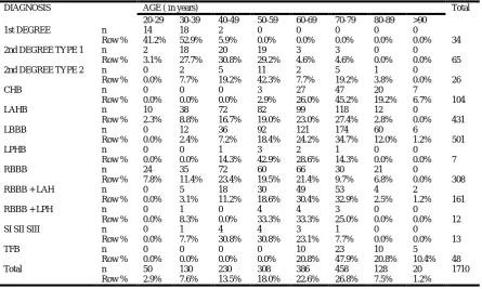
Table 6. The number and percentage of different cardiac conduction defects in relation to the age in the studied population
DIAGNOSIS AGE ( in years) Total
20-29 30-39 40-49 50-59 60-69 70-79 80-89 >90
1st DEGREE n 14 18 2 0 0 0 0 0
Row % 41.2% 52.9% 5.9% 0.0% 0.0% 0.0% 0.0% 0.0% 34
2nd DEGREE TYPE 1 n 2 18 20 19 3 3 0 0
65 Row % 3.1% 27.7% 30.8% 29.2% 4.6% 4.6% 0.0% 0.0%
2nd DEGREE TYPE 2 n 0 2 5 11 2 5 1 0
26 Row % 0.0% 7.7% 19.2% 42.3% 7.7% 19.2% 3.8% 0.0%
CHB n 0 0 0 3 27 47 20 7
104 Row % 0.0% 0.0% 0.0% 2.9% 26.0% 45.2% 19.2% 6.7%
LAHB n 10 38 72 82 99 118 12 0
431 Row % 2.3% 8.8% 16.7% 19.0% 23.0% 27.4% 2.8% 0.0%
LBBB n 0 12 36 92 121 174 60 6
501 Row % 0.0% 2.4% 7.2% 18.4% 24.2% 34.7% 12.0% 1.2%
LPHB n 0 0 1 3 2 1 0 0
7 Row % 0.0% 0.0% 14.3% 42.9% 28.6% 14.3% 0.0% 0.0%
RBBB n 24 35 72 60 66 30 21 0
308 Row % 7.8% 11.4% 23.4% 19.5% 21.4% 9.7% 6.8% 0.0%
RBBB + LAH n 0 5 18 30 49 53 4 2
161 Row % 0.0% 3.1% 11.2% 18.6% 30.4% 32.9% 2.5% 1.2%
RBBB + LPH n 0 1 0 4 4 3 0 0
12 Row % 0.0% 8.3% 0.0% 33.3% 33.3% 25.0% 0.0% 0.0%
SI SII SIII n 0 1 4 4 3 1 0 0
13 Row % 0.0% 7.7% 30.8% 30.8% 23.1% 7.7% 0.0% 0.0%
TFB n 0 0 0 0 10 23 10 5
48 Row % 0.0% 0.0% 0.0% 0.0% 20.8% 47.9% 20.8% 10.4%
Total n 50 130 230 308 386 458 128 20 1710
[image:4.595.75.521.267.534.2]Risteard Mulchay et al., 1968 and Lawrence V. Perlman et al., 1971. In contrast to our study, Gupta et al15reported maximum prevalence in males in age group of 40-49 years and 50-59 years in females. Lone, found maximum prevalence in males and females in age groups of 45-54 and 55-64 years respectively.
Conduction defects
In our study, we found the different cardiac conduction defects in the decreasing order of frequency as:
LBBB 501 (29.2%).
LAHB 431 (25.2%).
RBBB 308 (18.01%).
Bifascicular block 173(10.1%).
Complete Heart Block 104(6.08%).
2nddegree heart block type1 65(3.8%).
Trifascicular Block 48(2.8%).
1st degree Heart block 34(1.99%).
2nddegree heart block type 2 26 (1.5%).
SISIISIIISyndrome 13(0.76%). LPHB 7(0.4%).
Wani, 1983 showed in a hospital based study the following sequence:
RBBB(32%) > LBBB=LAH(25%) >BFB(17%). Najar, 1986 reported the following sequence:
LBBB(40%)>LAHB(32%)>RBBB+LAHB(18%)>RBBB(10) which was in conformity with our study.
REFERENCES
Arvo, J.O. et al. 1996. Major Electrocardiographic Abnormalities among American Indians aged 45-74 years(The Strong Heart Study). Am J Cardiol., 78:1400-1405.
Assantachai, P. et al. 2002. An electrocardiographic survey of elderly Thai people in the rural community. J Med Assoc
Thai., Dec 85(12) : 1273-1279.
Charles, F. 1981. Electrocardiograms in the aged: An independent Marker of heart disease, Am J Of Med., 70:4-5. De Bacquer et al. 1995. Prevalence and correlates of ECG abnormalities in the adult Belgian population, J Electrocardiol., 28(I): I-II.
Fisch et al. 1957. The Electrocardiogram in persons over 70 years. Geriatrics, 12:616-620.
Gupta, R., Sharma, S. 1996. Prevalence of asymptomatic electrocardiographic abnormalities in a rural population.
JAPI, 44(II):775-777.
Harrisons Principles of Internal Medicine. 18thEd. Hurst`s The Heart; 11thEdition p 893-895.
John H Mc Anulty et al ; Natural History of High risk Bundle Branch Block. N Eng J Med. 1982 ; 137:302.
John H Mc Anulty et al. 1978. A prospective study of sudden
cardiac death in “High risk” Bundle Branch Block.N Eng J Med., 299:209-215
John Merideth, Raymond D Pruitt, 1973. Disturbances in Cardiac Conduction and their Management. Circulation, 47:1098-1107.
John, F.S. et al. 1979. Newly acquired Right Bundle Branch Block. The Framingham Study. Ann Intern Med., 90:303. John, F.S. et al. 1980. Newly acquired Right Bundle Branch
Block.The Framingham Study. Ann Intern Med., 92:37-44. Kellkar, P.N., Elizabeth, J. 1996. Intraventricular conduction
disturbances. JAPI, 44(6):402-406.
Lawrence V. Perlman et al. 1971. An epidemiologic study of first degree atrioventricular Block in Tecumseh, Michigan. Chest, 59: 40-46.
Najar, M.S. 1986. Clinical profile of intraventricular conduction defects in hospitalized patients in Kashmir, Thesis submitted of university of Kashmir, 1-72.
Nihalick, M.H. and Fisch, et al. 1974. The electrocardiographic findings in the aged. Am Heart J., 87:117.
Nova Golschalger and Mervin J. Goldman, 1989. Priciples of Clinical Electrocardiogrpahy.13th Edition, Prestine Hall International Inc.
Pooja Hingorani et al. 2012. Indian J Med Res., 135, March, 322-330.
Prata, S.P., Cunha, D.F., Prata, S.C., Nogucira, N. 1993. Prevalence of electroardiographic Abnormalities in 2000 aged and nonaged chagastic patients. Arq Brass Cardiol., 60(6):369-72.
Risteard, M., Noel, H., Brain, M. 1968. Aetiology of Bundle Branch Block, Brit Heart J., 30:34.
Siedell JC. Obesity, 2000. Insulin Resistance and diabetes-world epidemic. Br. J., 183:5-8.
Suarez, R.M. et al. 1961. The Electrocardiogram in the aged. J
Am Geriatrics Soc., 91: 645.
Upshaw, C.B. Jr. 2004. Comparison of the prevalence of first degree atrioventricular block in African–American and in Caucasian patients: an electrocardiographic study III, J
Natl Med Assoc., Jun ; 96(6): 756-760.
Wani, B.A., Zargar, A.H. 1983. Pattern of Heart Block in Kashmir- An electrocardiographic Analysis in 3600 Hospitalized patients. Paper presented during llnd National conference on Cardiac Pacing, Calcutta March 3-5.