Rap1 GTPases. (Under the direction of Carla Mattos).
Ras and Rap1 are monomeric GTPases in the Ras subfamily of the Ras superfamily that
despite high sequence homology, have distinct diverse functions in the cell including
involvement in cell proliferation and cell adhesion. GTPases undergo conformational
changes associated with the transition from GTP to GDP predominately in the switch I and
switch II regions. Switch I and switch II are dynamic regions that can take many different
conformations. Here we examine the effect that small molecules can have on the
equilibrium between different switch II conformations. We show that binding of calcium
acetate in Ras to an allosteric site results in a catalytically active conformation that we call
“allosteric on”. We also show that binding of DTT or DTE in the interlobal region result in a
different Ras catalytically inactive conformation we call “allosteric off”. This conformation
was also seen in oncogenic Ras mutants.
The intrinsic hydrolysis mechanism of Ras is currently debated. Based on the allosteric on
structure we propose a two water model. Our proposed mechanism can be supported by
examining the protonation state of the active site residues. We have pursued neutron
crystallography as a means to visualize hydrogen positions in Ras to support our hypothesis
of intrinsic hydrolysis. Deuteration and perdeuteration are utilized in neutron
crystallography to improve the signal to noise ratio in the neutron data collected. Resulting
crystals were grown to a large volume and when mounted on a neutron beam diffraction
spots were detected. This proves feasibility of collecting neutron diffraction data on Ras.
Rap1, as the closest homologue to Ras in the Ras superfamily, provides a comparison
protein for specificity determinants in Ras. The structure of Rap1 alone has not been
solved. Additionally, specific analysis of Rap1 small molecule binding has also not been
presented that examine small molecule binding to Rap1 using the Multiple Solvent Crystal
Structures (MSCS) technique. Combined with the MSCS data of Ras, these data can
© Copyright 2011 by Genevieve Anne Holzapfel
by
Genevieve Anne Holzapfel
A dissertation submitted to the Graduate Faculty of North Carolina State University
in partial fulfillment of the requirements for the degree of
Doctor of Philosophy
Biochemistry
Raleigh, North Carolina
2012
APPROVED BY:
_______________________________ ______________________________
Carla Mattos Robert Rose
Committee Chair
________________________________ ________________________________
DEDICATION
This thesis is dedicated to my husband Joe who is always willing to lend support if I need it
BIOGRAPHY
Genevieve was born and spent her early childhood in the suburbs outside of Chicago. Her
parents were divorced very early in her life. Her father worked various jobs in the financial
market. Her mother had various careers including respiratory therapist and social worker.
The financial situation was always tight; however, Genevieve’s grandparents were generous
and provided a house for her to live in throughout her childhood. While in the Chicago
area, Genevieve attended a wonderful elementary school that exposed her to many things
including Argonne National Lab and Fermi Lab. After elementary school Genevieve moved
to Colorado, a big change from the metro area. Colorado was a great place to spend the
remainder of her youth. While in high school Genevieve was able to secure a student
position with the United States Department of Agriculture as a biological lab aide. This
provided a glimpse into what working in a laboratory environment is like and also provided
an alternative to working as a hostess. Attending high school near Colorado State
University provided an opportunity to enroll in college classes while still in high school.
Genevieve attended CSU and graduated in 2003 with a degree in biochemistry and a minor
in chemistry. Genevieve received the Peg Baker memorial scholarship provided initially by
Spear, Kellog and Logan which was later Goldman Sachs. While in school, Genevieve had
the opportunity to study abroad to the University of Glasgow in Scotland. This opportunity
provided a new perspective. After graduating college Genevieve moved to North Carolina
to attend NCSU and study with Carla Mattos. She was awarded an NIH biotechnology
training fellowship for her second and third years in graduate school. Here she met her
ACKNOWLEDGMENTS
I would like to acknowledge my advisor Carla Mattos for her mentoring and support during
even the worst of times. I would also like to acknowledge my committee for the assistance
and advice. Special thanks for Greg Buhrman and Paul Swartz for teaching me the basics of
crystallography and for many helpful discussions. I would also like to acknowledge past and
present students that have helped with parts of the work presented included Kelly
TABLE OF CONTENTS
LIST OF TABLES ... vii
LIST OF FIGURES ... viii
CHAPTER 1... 1
Introduction ... 1
Ras Effectors ... 3
Rap Effectors ... 5
Structural Regions of Ras GTPases ... 6
Ras Hydrolysis ... 9
Rap Hydrolysis ... 10
Posttranslational Modification and Cellular Localization ... 13
Ras Oncogenesis ... 14
MSCS/FT-Map Analysis of Ras... 16
CHAPTER 2... 17
Allosteric Modulation of Ras Positions Q61 for a Direct Role in Catalysis ... 17
Introduction ... 18
Materials and Methods ... 20
Results ... 20
Discussions ... 28
Contribution ... 33
CHAPTER 3... 34
Small Molecules Selectively Shift the Equilibrium Between On and Off Allosteric States in Ras-GppNHp Crystals ... 34
Introduction ... 34
Results ... 38
Conclusions ... 55
Materials and Methods ... 57
Introduction ... 59
Methods ... 72
Results ... 77
Discussion... 93
CHAPTER 5 Structural Analysis of Uncomplexed Rap1A ... 99
Introduction ... 99
Experimental Procedures ... 104
Results ... 107
Discussion... 122
CHAPTER 6 The Multiple Solvent Crystal Structures Method Performed on Rap1a ... 128
Introduction ... 128
Methods ... 134
Results and Discussion ... 136
LIST OF TABLES CHAPTER 2
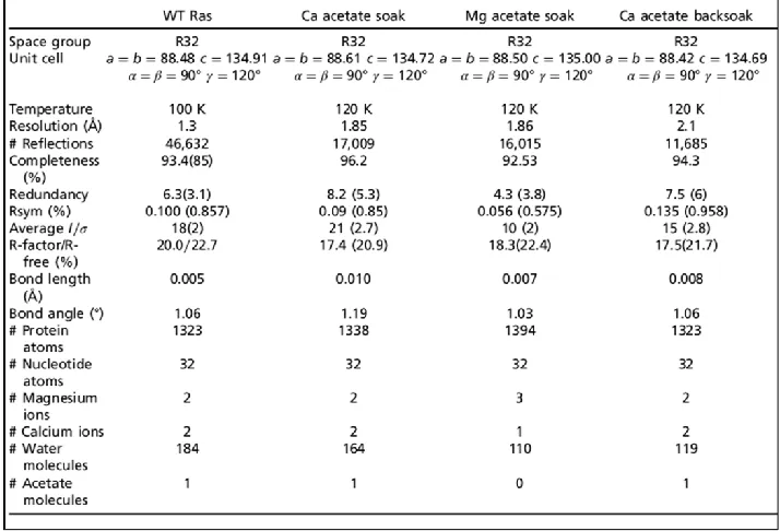
Table 2.1: Data collection and refinement statistics are presented for the four
structures discussed in this article ... 21
CHAPTER 3
Table 3.1: Data refinement and collection statistics for Chapter 3 ... 40
CHAPTER 4
Table 4.1: Data collection and refinement statistics for perdeuterated Ras crystals
grown in D2O conditions in the P3221 space group ... 81
Table 4.2: Data collection and refinement statistics are presented for several
structures of perdeuterated Ras in the R32 space group... 87
CHAPTER 5
Table 5.1: Data collection and refinement statistics for Rap1A bound to GTP or
GppNHp ... 108
CHAPTER 6
Table 6.1: Data collection and refinement statistics are presented for the Rap1A
LIST OF FIGURES CHAPTER 1
Figure 1.1: Phylogenic tree of the Ras superfamily ... 2
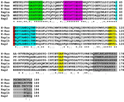
Figure 1.2: Sequence alignment of the Ras subfamily members H-Ras, N-Ras, K-Ras, Rap1a, Rap1b and Rap2 from homo sapien ... 3
Figure 1.3: Ribbon diagram of the structural regions in Ras ... 7
Figure 1.4: Active site comparison of Ras-RasGAP ... 11
CHAPTER 2 Figure 2.1: The allosteric switch in Ras ... 23
Figure 2.2: H-bonding networks 1 and 2 linking the allosteric site to Q61 ... 25
Figure 2.3: Switch I and switch II in the well-ordered active site in Ras-GppNHp 26 Figure 2.4: Allosteric site in structures resulting from soaked crystals ... 27
Figure 2.5: Proposed mechanism of intrinsic hydrolysis in Ras ... 29
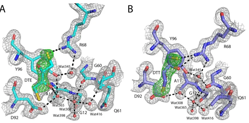
CHAPTER 3 Figure 3.1: Difference density for bound DTE and DTT in the switch II/ helix 3 pocket ... 43
Figure 3.2: Allostreric off active site comparison ... 44
Figure 3.3: View of the allosteric site in the Ras High PEG CaAcetate soaked structure (885) ... 45
Figure 3.4: Soak Flow Chart ... 47
Figure 3.5: Mixed conformation electron density ... 48
Figure 3.6: Zinc cyclen soaked switch II/helix 3 pocket ... 52
Figure 3.7: Allosteric “on” active site comparison ... 55
CHAPTER 4 Figure 4.1: Cartoon of Ras state 1 and state 2 ... 61
Figure 4.2: Density map difference between deuteration levels ... 70
Figure 4.4: Neutron Laue diffraction patterns for Ras at different exposure lengths
... 69
Figure 4.5: SDS-PAGE separation of perdeuterated Ras... 80
Figure 4.6: Room temperature data set of Ras in P3221 crystal form ... 82
Figure 4.7: The active site of the perdeuterated Ras grown in the P3221 crystal form collected at room temperature ... 83
Figure 4.8: Pre-switch I variation ... 84
Figure 4.9: Pre-switch I hydrogen bonding networks ... 85
Figure 4.10: Room temperature comparison of Ras in the R32 crystal form ... 91
Figure 4.11: Room temperature electron density ... 92
Figure 4.12: Perdeuterated active and allosteric site conformations ... 94
Figure 4.13: Room temperature perdeuterated conformations ... 95
CHAPTER 5 Figure 5.1: The active site of Raps in complex with Raf ... 103
Figure 5.2: Chromatogram of Rap1 run on a QHP column ... 105
Figure 5.3: Global comparison of Rap1a ... 110
Figure 5.4: T61 interacts with the catalytic water molecule ... 111
Figure 5.5: Pre-bridging and bridging water molecule conformations ... 114
Figure 5.6: T61 replaces the bridging water molecule ... 115
Figure 5.7: A comparison of the conformations adopted by switch II in Rap1 .. 117
Figure 5.8: Comparison of Rap1 helix 3 and helix 4 residues with Ras in the allosteric on state ... 118
Figure 5.9: Two different conformations of loop 7 are observed in the presented Rap1 structures ... 119
Figure 5.10: Calcium Acetate binding site ... 122
Figure 6.2: Electron density for GOL molecules placed in the Rap1 soaked structure
... 143
Figure 6.3: The binding sites seen in the Rap1-GOL structure are compared to the
data obtained in the Ras MSCS set ... 143
CHAPTER ONE
Introduction:
The Ras superfamily is a family of GTPases which act as molecular switches in a wide variety
of signal transduction pathways. This superfamily is comprised of the Ras, Rho, Rab, Arf,
and GNA subfamilies (Figure .1) [2]. Members of the Ras superfamily undergo
conformational changes upon transition from the GTP-bound active form to GDP-bound
inactive form [11]. These changes are greatest at the switch I (residues 30-40) and switch II
(residues 60-73) regions near the active site and allow for protein binding partners to
distinguish between GTP and GDP bound protein. In the GTP bound form a conformational
change occurs which activates the GTPase to interact with downstream effector molecules.
The transition from GDP to GTP is facilitated by Guanine Nucleotide Exchange Factors
(GEFs) which bind to the GDP-bound form. This interaction loosens the active site and
allows for the release of GDP. Once GDP is released, the GTPase binds GTP, as it is
encountered at higher concentration in the cell [12]. To turn the signal off, GTP is
hydrolyzed to GDP. GTPases possess an intrinsic GTP hydrolysis rate that is typically slow.
The binding of GTPase Activating Proteins (GAPs) speeds up the intrinsic hydrolysis rate.
Maintaining the proper balance between an active or inactive GTPase is essential in the cell
because the balance is used to filter, amplify or time upstream signals [13].
The Ras subfamily is comprised of several members including Ras, Rap, Ral, R-Ras, and Rheb
[2] (Figure .1). Ras has served as a model protein to compare proteins in both the Ras sub
and superfamily owing to it being characterized early and to its prominent role in about 30%
of human cancers. Ras has three isoforms, H-Ras, K-Ras, and N-Ras which are sequentially
similar (>95%) with much of the variation occurring at the C-terminus. Rap1 is the closest
homolog to Ras having a >50% sequence identity. Rap consists of Rap1a, Rap1b, Rap2a,
Rap1a and Rap1b is also high at >90% and like the isoforms of Ras most of the variation
occurs in the C-terminal hyper variable region (HVR) (Figure 1.2). Rap1 and Ras have
effector binding regions that have a high sequence similarity (switch I). The similarity
between Ras and Rap1 makes Rap1 a good model for determining Ras specificity.
Ras Effectors:
Ras has many known effectors with which it interacts in a GTP dependent manner including
Raf, PI3K, RalGDS, RASSF (NORE1), Rin1, Tiam, Af6, PLCε, PKCζ and functionally related
isoforms. Fortunately, there is structural information available for Ras effector complexes
NORE1A-RBD (K302D) [16], and Raps-RafRBD [17] having been solved by X-ray
crystallography. The Ras binding domain (RBD) is a putative binding region for Ras on the
effector protein. There are three classifications of RBD: Raf and Tiam1; PI3K; and the Ras
association (RA) domain in RalGDS and AF6. While switch I is implicated in the binding of
all known Ras effectors, secondary binding sites are possible as revealed by mutagenesis,
NMR, and X-ray crystallography studies. For example, RafRBD binds switch I, but the Ras
cystine rich domain (CRD) also interacts with Ras at a region composed of residues
immediately preceding and following switch I [18]. PI3K, RalGDS and NORE1A all form
interactions with switch II as well as switch I.
Much focus has been placed on the role of Ras in Raf activation as part of the
Mitogen-Activated Protein Kinase (MAPK) signal cascade. This pathway exists in all eukaryotes and
leads to cell differentiation, proliferation, survival, apoptosis and thus is implicated in
oncogenesis [19]. Ras is activated in this pathway where it binds and activates Raf, a MAPK
Kinase Kinase (MAPKKK). Raf in turn phosphorylates and activates MEK (MAPKK). The
signal is then propagated through MEK phosphorylation and activation of ERK (MAPK) which
once activated can interact with transcription factors. The Ras-Raf interaction has a high
affinity of 3.5nM [20]. This is compared with the weaker affinity of PI3K and RalGDS for Ras,
which is in the µM range [14, 21]. Another Ras effector with a high affinity is NORE1A,
which functions as a tumor suppressor. Though NORE1A has a low Kon similar to weak Ras
protein binders, it has a slow Koff of 0.1s-1 which gives it a similar affinity for Ras as that of
Raf [16]. RasGAP (p120) has an affinity for Ras in the µM range similar to RalGDS and PI3K.
Both Raf and NORE1A have a much higher affinity for Ras than does RasGAP. This casts
doubt as to whether GAP could compete for Ras binding with high affinity proteins such as
Rap1 Effectors:
Rap1 has similar effector binding regions to Ras and can interact with some of the same
effector proteins including Raf, RASSF, PI3K and RalGDS [23]. Rap1a, like Ras, gained
notoriety for its ability to bind Raf. It was shown that Rap1 (originally named Krev-1) was able to revert K-Ras transformed NIH3T3 cells [24]. It was proposed that Rap1 acted as an
antagonist to Ras by binding Raf and sequestering away. However, more recent studies
with either full-length transfected or endogenous Rap1 do not show that Rap1 is able to
affect the functioning of Raf in vivo [25]. Given that Ras and Rap1 are localized to different areas of the cell it is possible that though both are capable of binding the same proteins
those interactions are regulated by factors associated with spatial localization.
Much study of Rap1 now focuses on its role in cell-cell adhesion and cell-cell junction
formation involving interactions with effector proteins such as AF-6, Krit1, RAPL (a RASSF
splice form), Riam, RacGEFs (Tiam1 and Vav2) and RhoGAPs (RA-RhoGAP and Arap3) [26].
Through these interactions, Rap1 plays an important role in the specialized cells of the
cardiovasculature reviewed in Jeyaraj et al [27]. Using knockout mice, animal models and experiments in cells it has been shown that Rap1 has a significant role in blood vessel
formation and permeability, platelet aggregation, and cardiac myocyte growth and survival
[28-31]. It was also shown that Rap1 is involved in migration, adhesion, or development of
hematopoietic cells [27, 32, 33]. Information on the involvement of Rap1 in disease
progression in the cardiovasculature is currently being investigated [27]. While much
structural information is available for Ras in complex with effectors, there is little
information available for Rap1 or Rap2. The Rap1-Raf structure is the only effector complex
Structural Regions of Ras GTPases:
Ras GTPases consist of a catalytic core (1-166) and a HVR (167-189) that is post
translationally modified. The catalytic core consists of six β strands, five α helices and 10
loop regions [34]. Ras can be split into two global regions; the N-terminal lobe 1 (residues
1-86) and the C-terminal lobe 2 (87-171) (Figure 1.) [35]. Lobe 1 consists of the active site
components including switch I, switch II, the P-loop and most of the nucleotide binding
pocket. This lobe can be termed the effector lobe as it contains the sites of protein-protein
interaction. Lobe 2 contains the membrane-interacting portion of the protein and can be
termed the allosteric lobe [5]. Amongst Ras isoforms the effector lobe is 100% conserved
and the allosteric lobe has 95% sequence identity. There is also higher sequence identity in
effector lobe compared with the allosteric lobe between Ras and Rap1. The important
structural regions in Ras, discussed below, are highlighted in Figure 1..
The Ras family contains two highly dynamic regions, switch I and switch II which undergo
conformational changes upon GDP/GTP binding [34]. Switch I, comprised of residues
(30-40), is the primary effector binding region and is involved in downstream effector binding as
well as interaction with GAPs. This region is highly similar in Ras and Rap1 with the only
variation seen at residues 30 (Ras has a Asp and Rap has a Glu) and 31 (Ras has a Glu and
Rap has a Lys). The similarity in the primary effector binding region makes it unsurprising
that Ras and Rap1 are able to interact with many of the same downstream effectors. It is
Switch I functions not only in facilitating interactions with downstream effectors, but it also
comprises part of active site with the P-loop and switch II. Switch I has been shown through
NMR to adopt two states, state 1 and state 2, where state 1 corresponds to conformations
where Y32 is away from the nucleotide in what we call an “open conformation” and state 2
is where Y32 is found in near the nucleotide in a “closed conformation” [37, 38]. It has Figure 1.3: Ribbon diagram of the structural regions in Ras. Structure of H-Ras bound to the
been shown that the ratio between state 1 and state 2 varies in the Ras family. The state 1
population is 36±2% in H-Ras, 93±2% in M-Ras and 5±1% in Rap1 [39]. In both Rap1 and Ras
state 2 is the state that interacts with downstream effectors [17]. State 1 is the Ras switch I
conformation adopted in the Ras-RasGAP complex [40]. Conversely, in the Rap1-RapGAP
complex switch I is in the state 2 conformation [41]. It was proposed that Y32 is a
nucleotide sensing residue and that it helps stabilize switch I in the state 2 conformation
when bound to GTP [13]. Another switch I residue, T35, is a conserved residue that makes
direct interactions with both the ɣ-phosphate and the active site Mg2+ and is necessary for
stabilization of switch I in the GTP-bound form. Mutation of T35 to Ser or Ala results in a
disordered switch I as observed in NMR experiments [37].
Switch II is comprised of residues 60-73 which NMR shows adopts multiple conformations
[42]. This is reflected in Ras crystals structures, where switch II tends to either be stabilized
by crystal contacts or disordered. While switch I is the primary effector binding region,
switch II can also interact with effectors such as NORE1A, PI3K and RalGDS. Switch II also
forms interactions with GAP and with GEF proteins. Residue 64 (Ras Y64, Rap1 F64) is
important in binding to downstream effectors for both Ras and Rap1. In Ras and Rap1
mutation of these residues affects binding to NORE1A [16] and to their respective GAPs [41,
43]. It is also proposed that residue 64 is a specificity determinant for GAP binding Ras or
Rap1. Like switch I, switch II undergoes conformational changes upon GTP binding. G60 is a
nucleotide sensing residue with its backbone amide forming a hydrogen bond to the
ɣ-phosphate. Another important switch II residue is residue 61 which is crucial for hydrolysis
in Ras [44]. This residue varies in Ras and Rap1 where in Ras residue 61 is a Gln and in Rap1
residue 61 is a Thr. The importance of this residue is detailed in the Ras Hydrolysis and Rap Hydrolysis sections below.
The P-loop (10-17) and nucleotide binding pocket complete the nucleotide binding regions.
nucleotide through interactions with the backbone amides of residues 14-17. The negative
charge is also partially stabilized by a positively charged magnesium ion. The coordination
of this conserved magnesium ion involves residues S17 and T35, the β and ɣ-phosphates,
and two water molecules stabilized by D33, T58, and D57. Magnesium is required for
hydrolysis and can affect the affinity for GTP or GDP. Mutation of P-loop residues G12 and
G13 are commonly seen in Ras oncogenesis and result in disruption of the active site. The
guanine nucleotide binding pocket is comprised of residues 116-119 (NKXD) and 145-147
(SAK). The three nucleotide binding regions are connected through hydrogen bonding
involving N116 [45].
Ras Hydrolysis:
Hydrolysis in Ras proceeds through two mechanisms: intrinsic and GAP-stimulated. The
intrinsic mechanism is slow and is dependent on Q61, where mutation of Q61 reduces the
intrinsic rate of hydrolysis by ten-fold [44]. Residue Q61 is also important in GAP stimulated
hydrolysis. Both the mechanism of intrinsic and GAP stimulated hydrolysis has been
debated for years. Evidence now supports a loose, dissociative-like transition state for both
intrinsic and GAP stimulated hydrolysis [46]. It was observed in the canonical P3221 crystal
form and in the Ras-RasGAP structure that Q61 interacts with the catalytic water molecule
(Figure 1.4). The positioning of Q61 in the P3221 space group lead to the proposal that Q61
activates a catalytic water molecule for nucleophilic attack on the ɣ-phosphate of GTP [47].
However, Gln is a weak base for such a function. Given that Q61 is the only base in the
vicinity of the catalytic water molecule, it was proposed that GTP can act as a general base
and abstract a proton from the catalytic water molecule [48, 49].
Our lab solved Ras in the R32 space group where switch I is in a state 2 conformation and
developed an alternative mechanism of intrinsic hydrolysis [50]. In this mechanism the O1G
molecule. This proton is stabilized by a bridging water molecule located between Y32 and
the ɣ-phosphate. This mechanism was based on a closed Y32 conformation seen in effector
complexes. Y32 would serve to help position the bridging water molecule and it would
accept a H-bond from the bridging water molecule making this water molecule more prone
to accept a H from the ɣ-phosphate.
In Rho, Y32 has been shown to be important in GAP stimulated hydrolysis where mutation
to Ala, Phe, Ser, Glu, or Lys reduces the rate of hydrolysis [51]. In both Rho and Ras Y32
(Y34 in Rho) is away from the active site in the GAP complex allowing for insertion of the
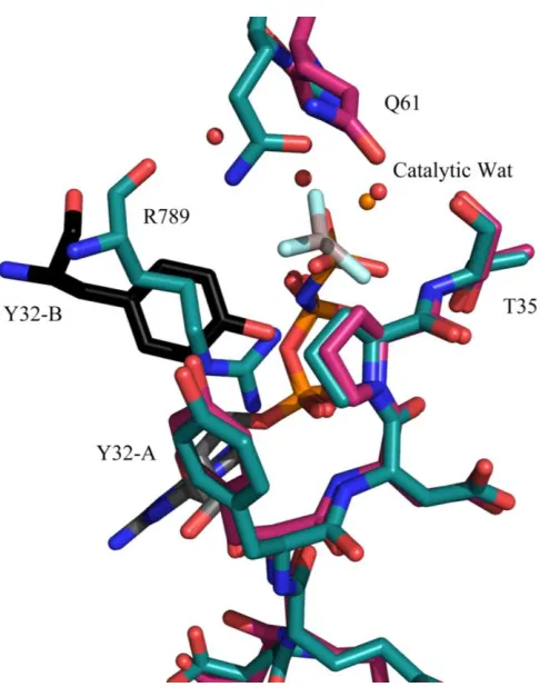
Arginine (Arg) finger (R789) [43]. Y32 participates in hydrophobic interactions with Ras
residues P34, I36 and Y64 with RasGAP residues L902 and L910 [52]. In the P3221 crystal
form Y32 is similarly away from the active site in a state 1 conformation and interacts with a
symmetry related nucleotide. The insertion of a symmetry related Y32 into the active site
overlays with the position of the Arg finger in the Ras-RasGAP complex (Figure 1.4). In GAP
stimulated hydrolysis the Arg finger helps stabilize the negative charge that develops
between the β and ɣ-phosphate as the transition state is formed [46]. Kinetic isotope
experiments suggest a change in electrostatic potential of the leaving group oxygen atoms
when Q61 is mutated to His [46]. This suggested Q61 has a role as an active site stabilizer.
The Ras-GAP complex structure shows Q61 interacting with both the catalytic water and the
backbone of the Arg finger. This lead to the postulation that Q61 may help orient both the
catalytic water and the Arg finger for hydrolysis [46].
Rap Hydrolysis:
Rap has a Q61T substitution when compared to Ras which gives Rap a slow rate of intrinsic
hydrolysis similar to the rate observed in the Ras Q61T mutation [53]. While overall little is
known about the mechanism of intrinsic hydrolysis in Rap1, mutational analysis provides
hydrolysis rate similar for that observed in Ras [53]. Hydrolysis experiments suggest that
T61 is not involved in the intrinsic hydrolysis reaction. Mutation to either T61L or T61A
does not significantly alter the intrinsic hydrolysis rate [53]. Unlike in the case of Ras,
mutation of residue 61 in Rap1 does not change the rate of GAP stimulated hydrolysis,
though mutation of residue 61 was shown to affect the affinity of the Rap1-Rap1GAP
complex [53]. GAP stimulated hydrolysis also proceeds differently in Rap1 than in Ras.
Rap1 specific GAPs contain several Arg residues, however, mutation of these residues
affects binding between Rap1 and Rap1GAP and not the hydrolysis rate. Instead RapGAPs Figure 1.4: Active site comparison of Ras-RasGAP. Ras bound to GppNHp solved in the P3221 (PDB code: 1ctq warm pink) space group compared to the conformation in the Ras-RasGAP complex with GDP and AlF3 (PDB code: 1wq1 teal). Y32 is swung away from the active site (Y32-A) allowing for insertion of the Arg finger, R789, or Y32 from a symmetry related molecule, Y32-B (black).
employ an Asn (D290) which was termed the Asn thumb, keeping with the Ras terminology.
This Asn thumb in Rap is inserted in a different orientation from the Arg finger in Ras and
overlays the position of Q61 in Ras in the Ras-RasGAP complex. It is postulated that the Asn
thumb in Rap replaces Q61 in Ras and performs a similar function, in orienting the catalytic
water molecule in the active site [54]. The structure of Rap1 in complex with GAP also
demonstrates one other major structural difference when compared to Ras. In Rap1 in
complex with GAP, Y32 is in direct interaction with the ɣ-phosphate (state 2). This is
contrary to Ras where Y32 was swung away from the active site to allow insertion of the Arg
finger. The position of Y32 in Rap1 would sterically clash with an inserted Arg finger.
Mutation of Y32 also affects Rap1 GAP stimulated hydrolysis [41]. The Y32F mutant shows a
two-fold reduction in hydrolysis rate. The Y32A mutant has a significantly impaired
hydrolysis rate. It was postulated that Y32 helps stabilize the state 2 conformation of switch
I in Rap1 necessary for GAP binding and that the hydroxyl group of Y32 is essential for this
function [41]. In early structures of Rap2 an active site where switch I was stabilized in the
state 2 conformation was revealed and it was proposed that Y32 was also a nucleotide
sensitive residue that helps stabilize switch I in Rap [13].
Interestingly, certain Ras GAPs can also stimulate Rap1 GTP hydrolysis [55]. A series of
mutagenesis experiments sought to examine how Ras GAP stimulated Rap1 GTP hydrolysis
in the absence of Q61 [56]. It was shown that Arg finger from GAP1IP4BP (a Ras GAP) was not
essential for complex formation with Rap1 but did significantly affect the rate of GTP
hydrolysis. This indicates that in Rap1 the Arg finger from GAP may be playing a similar role
as it does in Ras hydrolysis and that Rap1 would subsequently be in the state 1
conformation to allow for insertion of the Arg finger. Mutagenesis experiments also
examined the impact of Asn and Gln residues from GAP in the active site region. These
mutations did not affect the hydrolysis rate, indicating that Ras GAP stimulation of Rap1
the binding of RasGAP may stabilize Rap1 in a conformation that promotes hydrolysis,
although the specific details of that conformation is currently elusive.
Post Translational Modification and Cellular Localization:
Amongst both Ras and Rap isoforms much of the sequence diversity occurs at the
C-terminal HVR that is post translationally modified and is thought to be related to the
proteins cellular localization [57]. Ras was originally observed at the plasma membrane
[58], although more recent work has established its presence in some internal cellular
membrane systems [59]. Rap1 is predominantly localized perinuclearly in the Golgi
apparatus and late endosomes [60, 61]. Rap1 can also be located at small levels at the
plasma membrane [62]. Proper targeting of Ras to the plasma membrane is important for
proper cellular function including plasma membrane recruitment and activation of both Raf
and RalGDS [63-65]. It was originally proposed that Rap1 could compete with Ras for these
proteins. While experimental evidence clearly shows in vitro that Rap1 shares effector proteins with Ras (RalGDS, Raf for example) there is less evidence to support these
interactions in vivo. This is thought to be a result of the different cellular localization of Rap1 and Raf [26].
The differences in Rap1 and Ras cellular localization is at least in part due to differences in
post-translational modification. In the process of post-translational modification in Ras a
farnesyl group and in Rap1 a geranylgeranyl group is added to a Cys on the C-terminus. The
addition of the different moieties in K-Ras and in Rap1 is sufficient in conjunction with the
poly-basic region at the C-terminus HVR in both proteins for proper membrane targeting.
In H-Ras an additional palmityl group is required for proper plasma membrane targeting as
H-Ras lacks a C-terminal poly-basic region. The process of Rap1 targeting is less understood
than Ras targeting. It was discovered that residues 85-89 (found in a loop between β sheet
Rap1 residues 85-89 to mimic Ras resulted in a ubiquitously expressed protein. The reverse
substitution where H-Ras residues 85-89 were substituted for Rap1 resulted in a chimera
protein that was perinuclearly expressed and not located at the plasma membrane despite
proper post translational modification. How this sequence functions in maintaining the
perinuclear distribution in Rap1 despite the post translational modification and poly basic
region is not understood, but it may involve protein-protein interactions [61].
Ras Oncogenesis:
Ras is mutated in around 30% of human cancers including mutations in three of the most
lethal cancers, (pancreatic (90%), lung (30%) and colon (50%)) [66]. The high incidence of
Ras mutants in cancer makes Ras a prime target for drug targeting. This therapy, however,
remains elusive. There are three mutations in Ras that account for almost 97-99% of Ras
mutations in cancer, G12V, G13V, and Q61L [67]. These mutations affect the hydrolysis of
GTP. The G12V and Q61L mutations are GAP insensitive (where the rate of GTP hydrolysis
is unaffected by the addition of GAP). In the G12V mutant, GAP insensitivity likely results
from steric hindrance that prevents proper the proper active site conformation for catalysis,
including placement of the Arg finger [43, 68]. To better understand mutation of position
61 seventeen Q61 mutants were created. All mutation had a similar 10-fold reduction in
intrinsic hydrolysis rate, however, there was large variation in transformation efficiencies
(1000X) [44]. The results showed that Q61E, Q61P and Q61G were poorly transforming.
The moderately transforming mutants were Q61T, Q61W, and Q61N. Strongly transforming
mutations included Q61L, Q61R, Q61K, Q61A, Q61C [44].
Work from our lab examined the different structural properties of Ras Q61 mutants with
varying transformation efficiencies. The structures of Q61L, Q61V, Q61K (strongly
transforming), Q61I (moderately transforming) and Q61G (weakly transforming; which was
symmetry of the R32 space group. The Ras conformation seen in this space group is similar
to the structure seen in the Raps-Raf complex and a Ca2+ ion is positioned to take the place
of K84 in Raf. Crystal contacts stabilized switch I in the closed conformation (state 2).
Switch II, on the other hand, was free of crystal contacts and was disordered in the wild
type structure. In the wild type structure a water molecule (wat 189) between Y32 and the
γ-phosphate was observed and was termed the bridging water molecule. This water
molecule was seen in the Raps-Raf complex but not in the Rap-Raf complex. The weakly
transforming Ras mutant, Q61G, revealed an active site that was similar to wild type.
Switch II was also similarly disordered [50].
The moderately and strongly transforming mutants adopted a different conformation
where switch II was ordered outside of crystal contacts. There was no bridging water
molecule present and Y32 moved making a direct interaction with the nucleotide. This
conformation was stabilized by hydrophobic interactions between Y32, P35, I36, L/V/K/I61
and Y64 and overlayed with the conformation seen in Ran-Importin-β. The Ran-Importin-β
complex has been shown to impair hydrolysis of GTP and thus the conformation of the
highly and moderately transforming mutants was designated to be noncatalytic [70]. Here
a correlation between the stabilization of a noncatalytic conformation and transformation
efficiencies was observed. Since it is thought that mutation of Q61 in Ras works through
the Raf pathway the effect of Raf was examined. It was shown that both wild type and
Q61L in the absence of Raf hydrolyzed GTP as shown by low levels of Ras-GTP at the end of
the experimental time frame, assayed by the amount of complex with Raf as visualized by
gel filtration. In wild type Ras addition of Raf before or after incubation with Ras-GTP
resulted in similar results. In Q61L, however, addition of Raf during hydrolysis incubation
MSCS/FT-Map Analysis of Ras:
The Multiple Solvents Crystal Soaks (MSCS) is a technique where different crystals of a
protein are soaked in various organic solvents. Typically, organic solvents are quite
damaging and, therefore, the crystals are crosslinked before transfer into organic solvents.
This technique provides insight into protein binding sites, plasticity, and hydration. In
model systems such as elastase and RNAse A organic solvent positions overlay with known
“hot spots” or protein interaction sites [71, 72]. In RNAse A hot spots in the active site
overlayed with the binding site for known inhibitors. The ability of MSCS to pick out binding
site hot spots makes this technique valuable in gathering information that could be used in
drug design. FT-Map is a computational version of MSCS where molecular probes are
clustered at druggable sites based on their free energy of interaction with the protein [73].
Like MSCS this technique can also garner information on sites of protein-protein interaction.
Both MSCS and FT-Map were performed on Ras and gave complimentary results [5]. In the
MSCS set, those structures solved in the presence of trifluoroethanol, hexane,
cyclopentanol, and glycerol show an ordered of switch II [5]. The ordering of switch II
overlayed well with the noncatalytic conformation seen in the highly and moderately
transforming mutants as well as the Ran-Importin-β complex. We are interested in
advancing the MSCS technique to determine if differences in organic solvent binding can be
observed between two highly similar proteins. Rap1 in comparison to Ras would provide
such a comparison given the high homology between the two proteins, similarity in binding
CHAPTER TWO
Allosteric Modulation of Ras Positions Q61 for a Direct Role in Catalysis
Greg Buhrman, Genevieve Holzapfel, Susan Fetics, and Carla Mattos
PNAS. 2010 March 16; 107(11): 4931–4936.
Summary:
Ras and its effector Raf are key mediators of the Ras/Raf/MEK/ERK signal transduction
pathway. Mutants of residue Q61 impair the GTPase activity of Ras and are found
prominently in human cancers. Yet the mechanism through which Q61 contributes to
catalysis has been elusive. It is thought to position the catalytic water molecule for
nucleophilic attack on the γ-phosphate of GTP. However, we previously solved the structure
of Ras from crystals with symmetry of the space group R32 in which switch II is disordered
and found that the catalytic water molecule is present. Here we present a structure of
wild-type Ras with calcium acetate from the crystallization mother liquor bound at a site remote
from the active site and likely near the membrane. This results in a shift in helix 3/loop 7
and a network of H-bonding interactions that propagates across the molecule, culminating
in the ordering of switch II and placement of Q61 in the active site in a previously
unobserved conformation. This structure suggests a direct catalytic role for Q61 where it
interacts with a water molecule that bridges one of the γ-phosphate oxygen atoms to the
hydroxyl group of Y32 to stabilize the transition state of the hydrolysis reaction. We propose
that Raf together with the binding of Ca2+ and a negatively charged group mimicked in our
structure by the acetate molecule induces the ordering of switch I and switch II to complete
Introduction:
The Ras/Raf/MEK/ERK signaling pathway is the most well studied of five known mitogen
activated protein kinase (MAPK) cascades involved in the mediation and timing of signaling
events in the cell [74]. This pathway is activated by Ras GTPase in response to extracellular
signals and is involved in the control of cell proliferation, differentiation, and survival [75].
In its resting state Ras is bound to GDP and is in a conformation in which it does not interact
with Raf or other effector proteins [76]. Guanine nucleotide exchange factors facilitate the
release of GDP [77]. Once the more abundant GTP binds, the Ras switch I (residues 30–40)
and switch II (residues 60–76) regions become poised for interaction with effector proteins,
leading to the propagation of signal transduction cascades. Ras has a low intrinsic rate of
GTPase activity that is enhanced by 3–5 orders of magnitude in the presence of GTPase
activating proteins (GAPs), resulting in depletion of Ras-GTP as the switch is turned off [43].
The biochemical properties of Ras and its oncogenic mutants have been well characterized
in the absence of Raf or other factors [78, 79], and numerous structures of wild-type and
oncogenic Ras mutants have been used to study the possible mechanisms through which
Ras becomes defective in its ability to hydrolyze GTP [80-84]. The switch regions in these
structures, solved from the crystal form with symmetry of space group P3221, are
modulated by crystal contacts to resemble the switch I and switch II conformations found in
the Ras/RasGAP complex [43, 85]. Since the structure of this complex has elucidated the
mechanism through which GAP enhances the GTPase activity of Ras [43], it seems
reasonable, given its similarity to the canonical structure of the uncomplexed form, that it
could also serve as a framework for the mechanism of intrinsic hydrolysis in Ras [47, 86].
Based on this assumption, a mechanism for intrinsic hydrolysis has been proposed with a
two-water model: The γ-phosphate of GTP abstracts a proton from W189, which in turn
activates the catalytic W175 for nucleophilic attack on the γ-phosphate during the
More recently, we proposed an alternative mechanism based on the structure of Ras from
crystals with symmetry of space group R32 [50], where switch I and water molecules in the
active site are as observed in the Raps*/Raf complex [36] and switch II is unhindered by
crystal contacts. It is clear that the R32 crystal form is an excellent mimic for the Ras active
site in the complex with Raf [50] and could model events that may occur in this complex in
the absence of GAPs. Raf interacts with Ras-GTP through two domains: the Ras-binding
domain (RBD) and the cystein-rich domain (CRD) [87]. The crystal structure of Raf-RBD in
complex with Raps-GppNHp shows the conserved switch I residue Y32 with its hydroxyl
group interacting with a γ-phosphate oxygen atom through a bridging water molecule [88],
exactly as we see in our structure [50]. In our proposed mechanism a proton from the
catalytic water molecule is shuttled via the γ-phosphate to the water molecule bridging the
phosphate to Y32 (which we have also named W189 due to its proximity to the
γ-phosphate, although it does not overlap with the position of W189 in the P3221 crystal
form) and is eventually delivered to the GDP leaving group. The fact that switch II is
disordered with no electron density for Q61 means that a key catalytic residue was left out
of this mechanism. In the present paper, we resolve this issue with analysis of a crystal
structure of wild-type Ras in which switch II is ordered through an allosteric switch. We
show that the remote allosteric site binds Ca2+ but not Mg2+ in the crystals. The result is a
more complete picture of our proposed mechanism of intrinsic hydrolysis in Ras, and a
paradigm shift for future studies of regulation in the Ras/Raf//MEK/ERK pathway.
*
Raps refers to the Ras homologue Rap with the switch I mutations E30D,K31E that results in a switch I region
Materials and Methods:
Ras-GppNHp Crystallization
The Ras-GppNHp protein solution consisted of 20 mg/mL protein in a stabilization buffer
(20mM Hepes pH 7.5, 50mM NaCl, 20mM MgCl2, and 10mM dithioerythritol (DTE). Crystals
were grown at 18°C for 1 week using sitting drop crystallization trays containing 5μL protein
solution and 5μL reservoir solution. The reservoir solution was composed of 500μL of
200mM calcium acetate hydrate, 20%w/v PEG 3350, and 0.05%
n-Octyl-β-D-glucopyranoside (NOG) diluted with 50μL of stabilization buffer. Synchrotron data were
collected at the Southeast Regional Collaborative Access Team (SER-CAT) beamline at the
Advanced Photon Source (APS).
Soaks in Calcium Acetate and Magnesium Acetate Solutions
For the calcium acetate and magnesium acetate soaks, Ras-GppNHp crystals grown as
above were transferred to a solution containing 10mM Hepes pH 7.5, 30%w/v PEG 3350,
10mM MgCl2, 25mM NaCl, and either 200mM Ca(OAc)2 or 200mM Mg(OAc)2 and flash
frozen after 1 or 2h for data collection. After soaking in Mg(OAc)2 for 3 days, a crystal was
then transferred back to the solution containing 200mM Ca(OAc)2 and soaked for 24h
before data collection. Datasets for the soaked crystals were collected on our home
MAR345 area detector mounted on a Rigaku RuH3R rotating anode generator.
Results:
H-Ras with 23 residues truncated at the C-terminus is used for the structural studies and
includes the entire catalytic domain, residues 1–166 (referred to as Ras throughout the
grown in 200mM calcium chloride, with symmetry of the space group R32 [50]. More
recently we found that the crystals grew larger if calcium acetate was used instead of
calcium chloride, and the resulting model revealed structural features that are explored
here. Wild-type Ras-GppNHp was solved in the presence of calcium acetate to 1.3Å
resolution from a crystal with R32 symmetry. Crystals removed from the mother liquor and
soaked either in 200mM calcium acetate or 200mM magnesium acetate yielded structures
solved to 1.85 and 1.86Å resolution, respectively, and crystals transferred to calcium
acetate after a magnesium acetate soak resulted in a 2.1Å resolution structure. Data
collection and refinement statistics for the four structures are presented in Table 2.1.
Structure of Wild-Type Ras in the Presence of Calcium Acetate Reveals Allosteric Modulation of Switch II
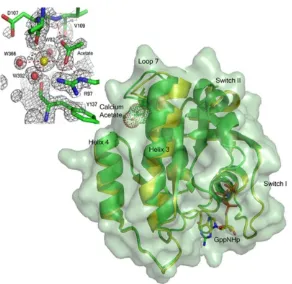
The overall structure of wild-type Ras in the presence of 200mM calcium acetate is similar
to that in the presence of 200mM calcium chloride (PBD ID code 2RGE), with one striking
exception: There is a shift of loop7, helix 3, and the C-terminal end of switch II, which
culminates in the ordering of the N-terminal portion of switch II and placement of Q61 in
the active site near the bridging water molecule that interacts with Y32 and the O1G atom
of GppNHp (Figure 2.1). This shift is modulated by the binding of calcium acetate at a site
remote from the catalytic center. The structural elements involved in the shift include the
C-terminal end of helix 3 (residues 98–103) and the sequentially adjacent loop 7 (residues
104–108). Anchoring residues on either end of this shifted region interact directly with the
bound acetate molecule: The side chain of R97 interacts with one of the acetate oxygen
atoms and the main chain amide of V109 donates an H-bond to the other (Figure 2.1).
Accompanying the shift in residues 98–108 is also a shift in residues 69–75 comprising the
C-terminal portion of switch II, with R68 nestled between helix 3 and residues 60–67 at the
N-terminal portion of the switch.
The Ca2+ ion binds between helix 4 and loop 7. It is hexacoordinated, interacting closely with
one of the acetate oxygen atoms, three water molecules, W82, W392, W366, and the main
chain carbonyl oxygen atoms of D107 and Y137, which are opposite each other in the
coordination sphere (Figure 2.1). The observed shift appears to be a result of a decrease in
distance between these two residues due to their involvement in coordinating the Ca2+ ion.
While the carbonyl of Y137 on helix 4 changes positions only slightly, D107 shifts
significantly toward Y137, bringing with it loop 7 and the C-terminal portion of helix 3. The
shift creates a set of conditions leading to formation of two connected H-bonding networks,
network 1 and network 2 described below, that have the effect of ordering switch II and
hydrolysis reaction. These H-bonding networks comprise the elements of an allosteric
switch, which in the present structure is in the “on” conformation, and which is “off” in our
previously published structures of wild-type Ras and of the RasQ61L oncogenic mutant [50].
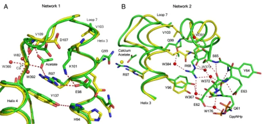
Network 1 includes the calcium acetate with R97 at the center. It involves H94, R97, E98,
and K101 (on the side of helix 3 facing away from switch II), the side chains of D107 (loop 7),
and Y137 (helix 4) (Figure 2.2A). This network, consisting entirely of protein atoms and the calcium acetate, clusters in a space that is largely occupied by crystallographic water
molecules in the structure of Ras in the presence of calcium chloride. With the helix 3
facing switch II are in position to contribute, along with switch II residues M72, Y71, D69,
R68, S65, E62, and several water molecules, to network 2 centered around R68 (Figure
2.2B). This leads to an extension of the α-helix found in the C-terminal half of switch II. The extended switch II helical structure places key elements important in ordering the
N-terminal half of the switch, from which the side chain of catalytic residue Q61 extends. For
example, S65 in the ordered switch II conformation makes a good H bond with the main
chain carbonyl group of E62, while its carbonyl group connects to Q99 on helix 3 through
water molecule W391 (Figure 2.2B). Q99 in turn links to R68 through W384. Surprisingly, the side chain of E62, which is disordered in all other available structures of Ras, is very well
ordered and interacts through water molecule W176 with the side chain oxygen atom of
Q61. In addition to interacting with the side chain of E62 through W176, the side chain of
Q61 H-bonds to the active site water molecule, W189, through its other lone pair electrons.
Water molecule W176 also links Q61 to the side chain of R68 as well as to helix 3 residues
D92 and Y96 through an H-bonding network involving W9, W28, and W367 (Figure 2.3).
While the side chain of Q61 is involved in this network, its carbonyl group is bridged to the
side chain of R68 through water molecule W372. Due to resonance stabilization involving
the aromatic ring, the Y32 hydroxyl oxygen atom has its two lone pairs confined to the
plane of the ring. Thus, in addition to its interaction with the active site bridging water
molecule W189, it is involved in a Hydrogen bonding network through which it is linked to
the side chain of N86 at the beginning of helix 3. The C-terminal end of helix 3 is at the
center of the allosteric switch. This network is shown in detail in Figure 2.3. While the side
chain of Q61 is involved in this network, its carbonyl group is bridged to the side chain of
R68 through water molecule W372. The net effect of all these interactions is that Q61 is
positioned through both backbone and side chain H bonds that place it in the active site and
that connect it through networks 1 and 2 to the remote allosteric site, occupied in our
The active site features reported for the wild-type Ras structure solved in the presence of
calcium chloride [50] are also observed in the calcium acetate bound structure, with the
added feature of the ordered switch II with placement of the Q61 near the bridging water
molecule W189. Thus, in both structures, all of switch I is in the same conformation as
found in the Raps/Raf-RBD complex with Y32 closed over the nucleotide and interacting
with it through the bridging water molecule. Two water molecules interact closely with the
Y32 side chain oxygen atom: On one side is W396, which links to N86 at the N-terminal end
of helix 3 through an H-bonding network involving W162 and W101, and on the other is the
bridging water molecule, W189, which in turn H-bonds to the O1G atom of the γ-phosphate
of GppNHp (Figure 2.3). The T35 residue maintains its well-known interactions within the
active site: Its side-chain hydroxyl group coordinates the Mg2+ ion and its main-chain
addition to donating an H bond to the carbonyl oxygen atom of T35 and to the O1G atom of
the nucleotide analogue as observed in the calcium chloride condition, in the presence of
calcium acetate the catalytic water molecule also accepts an H bond from the backbone N
atom of the Q61 residue, which in our previously published structure (PBD ID code 2RGE) is
disordered.
Ras Binds Ca2+ but not Mg2+ at the Allosteric Site
As an initial test of the specificity of the allosteric site for Ca2+, crystals grown in the
presence of calcium acetate were removed from the mother liquor and soaked in
stabilization solutions containing either 200mM calcium acetate or 200mM magnesium
acetate. The soak in 200mM calcium acetate is a control to make sure that a transfer to
between calcium acetate and magnesium acetate solutions that are otherwise identical.
Figure 2.4A and B shows the allosteric binding site in the two resulting structures. The structure soaked in calcium acetate is the same as that described above from crystals taken
directly from the mother liquor, with clear electron density for the calcium acetate in the
allosteric site. The soak in magnesium acetate, however, resulted in a structure with an
“empty” allosteric site and with an alternate conformation for R97 typical of the off state of
the allosteric switch, overlapping with the acetate binding site as seen in crystals grown in
calcium chloride. Although there are other features of the off state, such as an alternate
position of Y96 relative to R68 and a small shift of loop 7 toward the off state, the full shift is
not made, probably due to constraints of the crystal packing. Crystals soaked in magnesium
acetate and then transferred back to calcium acetate show a bound Ca2+ ion and acetate
molecule as seen in Figure 2.4C, regaining all of the features of the on state of the allosteric switch.
Discussion:
Proposed Mechanism of Intrinsic Hydrolysis
It is now established that intrinsic hydrolysis in Ras occurs through a loose, dissociative-like
transition state (TS) with significant concerted character, as does the GAP-catalyzed
reaction [89]. This does not rule out a mechanism in which the GTP itself receives a proton
from the catalytic water molecule [90]. Within these parameters, we propose a general
outline for catalysis deduced from our structure of the ground state of Ras with an activated
allosteric switch, augmenting the mechanism we previously published [50]. As the reaction
proceeds, a proton is shuttled from the catalytic water molecule via the O1G atom of the
γ-phosphate of GTP to the bridging water molecule, which is particularly prone to accept an H
bond, as it can donate H bonds to both Y32 and Q61 (Figure 2.5). This would promote
development of a partial positive charge in the active site near the β-γ bridging oxygen
atom of GTP to stabilize a dissociative-like TS creating a counterpart, albeit weaker, to the
arginine finger provided by GAPs. This is the key element lacking in our previously published
mechanism. It is consistent with kinetic isotope effect experiments in which it was
concluded that Q61 must mediate stabilization of negative charges on the leaving group
oxygen during the reaction [89]. We envision that as the reaction proceeds, W189 moves
toward the β-γ bridging oxygen atom of GTP and away from the Q61 side chain, which can
then flip to interact directly with the γ-phosphate in a manner analogous to that observed
in the Ras/RasGAP structure [43]. Indeed the ordered structure of switch II in our
Ras-GppNHp model is not so different from that observed in the complex with GAP, and a flip of
the Q61 side chain would put it within reach of the γ-phosphate. This process would release
the allosteric switch, resulting in a disordered switch II, ultimately leaving more room for
the release of the inorganic phosphate product at the end of the reaction. The allosteric
switch that we propose leads to enhanced catalysis is consistent with recently published
residues 66–74 and helix 3/loop 7 residues 93–110 in Ras-GTP, but not in Ras-GDP [91].
Correlated or “breathing” motions involving switch II and helix 3 have also been observed
experimentally by NMR [92].
Reconsidering the Relevance of Intrinsic Hydrolysis in Ras
Ras is a protein that has long been considered to undergo conformational changes
associated with regulation strictly due to the state of the bound nucleotide, where it can
interact with effectors in the GTP-bound but not in the GDP-bound state. We have
uncovered an additional level of modulation of switch II (and along with it catalytic residue
therefore the timing of interaction with Raf. We propose that the allosteric switch is not
general to all effectors, but is activated by Raf, which interacts with Ras through switch I
[88, 93]. In addition to leaving a free switch II in contrast to RasGAP and other effectors that
bind both switches, Raf is unique in that its 3.5nM affinity for Ras [20] is 3 orders of
magnitude stronger than the micromolar affinity observed for Ras/RasGAP [94] and for Ras
interaction with effectors such as PI3K [14] and RalGDS [21]. Given that GAPs and effectors
have overlapping binding sites [95], GAPs must displace the effectors in order to turn off the
GTPase signaling and directly control the timing of the Ras/effector interaction. Since
RasGAP, PI3K and RalGDS have similar affinities to Ras, it is likely that GAP-catalyzed
hydrolysis is the primary mechanism of negative regulation of pathways involving these
effectors. However, it is unlikely that GAPs can outcompete Raf at the concentrations found
in cells1 [22]. It has been suggested that the role of RasGAP in regulating the Ras/Raf
pathway could be to deplete the pool of available free Ras and that Raf could somehow
increase the GTP hydrolysis rate [95, 96]. However this has not been part of the discourse in
the literature for the last 15 years, given the slow intrinsic hydrolysis rate of Ras and the
lack of rate enhancement observed in vitro in the presence of Raf. Given our discovery of
the allosteric switch, this possibility must be seriously reconsidered and investigated
further. We propose a model in which the binding of Raf is controlled by the state of the
bound nucleotide through interaction of the RBD, but the timing of interaction is modulated
allosterically at the remote site by Ca2+ in conjunction with another cellular factor
containing a negatively charged group, mimicked by the acetate in our structure.
The discovery of an allosteric switch in Ras provides insight into the mystery associated with
the function of Q61 in intrinsic catalysis and the slow rate of hydrolysis observed for Ras in
the absence of GAPs. It suggests a unique mechanism by which the binding of Raf and
simultaneous association of a negatively charged ligand at the remote allosteric site could
increase hydrolysis rates to biologically relevant levels in the presence of Ca2+, promoting
1
attenuation of the signaling in Ras/Raf-associated pathways. It appears that in order to have
a fully formed active site for intrinsic hydrolysis of GTP, Ras needs to have both switch I and
switch II in highly ordered conformations. Full stabilization of switch I can be achieved with
the binding of Raf. Both in free Ras and in the complex with Raf, however, switch II is likely
disordered in the absence of the allosteric switch [92, 93]. In this situation the correct
placement of Q61 for catalysis would be expected to be a slow, rate-determining step in the
hydrolysis reaction, leading to the previously observed hydrolysis rates, where the binding
of Raf alone would not be expected to have an effect. However, the simultaneous binding of
Raf at switch I, placing the hydroxyl group of Y32 in position to interact with the bridging
water molecule and enable the allosteric switch, and a natural ligand at the remote
allosteric site (perhaps a membrane phospholipid or carboxylate group) in the presence of
Ca2+ could stabilize switch II through the mechanism we described above involving
H-bonding networks 1 and 2. The completion of the active site would then no longer be a
rate-determining step in the reaction, leading to an increase in intrinsic hydrolysis rate. Calcium
has been shown to be one of the activators of the Ras/Raf/MEK/ERK pathway, promoting
increases in Ras-GTP levels leading to the recruitment of Raf to the membrane [97].
Furthermore, we show here that the allosteric site binds Ca2+, but not Mg2+, supporting a
specific role for calcium as we propose. It is conceivable that the timing of the Ras/Raf
interaction is affected by the higher Ca2+ levels with binding of a membrane negatively
charged group to the allosteric site once the complex with Raf is in place. An increase in Ca2+
concentration has already been shown to inactivate the Ras/Raf/MEK/ERK pathway in at
least two different instances [98, 99]. Although the balance between the activating and
inactivating roles of Ca2+ with respect to the Ras/Raf pathway is still not well understood, it
is possible that the allosteric switch may be one of the means through which this balance is
achieved by direct action of Ca2+ on Raf-bound Ras at the membrane.
The idea that a molecule at or near the membrane may act in conjunction with Ca2+ to
lipid-modified Ras in a 1,2-dimyristoylglycero-3-phosphocholine bilayer, showing that in
Ras-GTP the catalytic domain makes more extensive interactions with the membrane
phospholipids than in Ras-GDP, and is positioned such that the allosteric site near loop 7 is
adjacent to the membrane [100]. The computational model shows that Ras-GTP interacts
with phospholipid head groups at residues R128, R135, and Q165, anchoring helix 4 to the
membrane. This is supported experimentally in cells by mutation of the two arginine
residues to alanine, which results in a decrease in neurite outgrowth due to impaired signal
transduction [100]. In our crystal structures of Ras-GppNHp, helix 4 is nestled against
extensive crystal contacts, with R135 making a salt bridge with the terminal carboxylate
group of a symmetry-related molecule. Thus, the stabilization of helix 4 by the membrane
appears to be mimicked to some extent in crystals with symmetry R32. This may be an
important element of the allosteric switch, as helix 4 contains Y137 (only two residues away
from R135) that coordinates to the Ca2+ ion through its carbonyl group. Lack of this
stabilizing interaction in solution may prevent the activation of the allosteric switch,
accounting for the fact that we have been unable to measure an increase in the intrinsic
hydrolysis rate by adding calcium acetate to the solutions in which the hydrolysis
experiments are performed in vitro. If our assessment is correct that the crystal contacts
serve a similar function to that of the membrane as far as stabilizing Y137 on helix 4, it
appears that the structural components that lead to a highly ordered switch II and
placement of Q61 in the active site are sensitive to both placement of Ras against the
membrane in a manner that happens only in the GTP-bound form [100] and to the binding
of Raf, which is necessary for the stabilization of Y32. In this situation Y32 could serve as a
venue through which the binding of Raf is imposed as a requirement in the activation of Ras
in lieu of activation by GAPs, ensuring that the membrane-bound Ras-GTP does not engage
in futile hydrolysis of GTP to GDP before it has a chance to recruit Raf from the cytoplasm
Contribution:
This article was published in Jan of 2010. I optimized crystallization of H-Ras that gave a
high resolution 1.3Å that was deposited with a PDB code 3k8y. This structure was an
improvement over previous data sets of WT Ras grown in CaAcetate based upon which the
analysis for this part of the paper was made. As in previous structure, this structure clearly
showed electron density for the allosteric on state including the bound CaAcetate and the
side chain of Q61, but to a higher resolution which showed some of the details more clearly.
I also performed a set of soaks designed to probe the specificity of the allosteric site for
CHAPTER THREE
Small Molecules Selectively Shift the Equilibrium Between On and Off Allosteric States in
Ras-GppNHp Crystals.
Genevieve Holzapfel, Greg Buhrman and Carla Mattos
Summary:
We have recently described an allosteric binding site in Ras where binding of calcium
acetate in this site resulted in a shift in helix 3 and loop 7. The resulting shift allowed for a
disorder to order transition in the active site of Ras GTPase to what we term the
catalytically active conformation. Earlier we also reported that the highly and moderately
transforming mutants stabilize a conformation of Ras in a catalytically inactive
conformation. Organic solvent soaks of Ras using the multiple solvent crystal soaks method
established several cluster sites in the interlobal region. Here we examine the binding of
DTT or DTE to a site in the interlobal region referred to as the switch II/helix 3 pocket.
Binding of DTT or DTE is associated with a disorder to order transition of switch II to the
catalytically inactive conformation seen in the Ras oncogenic mutants. We use the small
molecules acetate and DTT or DTE to selectively shift the equilibrium between different Ras
conformations in the crystal environment.
Introduction:
Ras is a small monomeric GTPase that functions as a molecular switch in signal transduction
[101, 102]. It is involved in cell proliferation, apoptosis, and multiple cellular functions that
play critical roles in the tumorigenesis of a variety of human cancers [103, 104]. It is
biochemical and cell biology studies [44, 50, 78, 79, 105, 106] as well as structural biology
experiments primarily using the GTP analog GppNHp to obtain the activated state [5, 80, 82,
83, 85, 107, 108]. Ras is anchored to the membrane via an isoprenyl group as well as other
posttranslational modifications at the C-terminus [59, 109] and is normally activated
through cell surface receptors [110]. When bound to GTP, Ras propagates its signal by
interacting with effector proteins such as Raf [93], phosphoinositide-3-kinase (PI3K) [14],
Ral guanine nucleotide Dissociation Stimulator (RalGDS) [21], NOR1A [16] and many others
[59]. Once GTP is hydrolyzed to GDP on Ras, interaction with effectors is no longer favored
and signaling is turned off. The levels of Ras-GTP are kept in check by the opposing actions
of guanine nucleotide exchange factors (GEFs) that catalyze the loading of GTP [77], and of
GTPase activating proteins (GAPs) that increase the intrinsically slow GTPase activity of Ras
for timely depletion of Ras-GTP [43]. The active site residues are situated primarily in the
so-called switch I, switch II and the phosphate binding loop (P-loop) comprised of residues
30-40, 60-76 and 10-17 respectively [111]. Oncogenic mutations interfere with the ability of
Ras to hydrolyze GTP, resulting in a prolonged signal that promotes uncontrolled cell growth
[104]. RasG12V and RasQ61L have received the greatest attention for being two of the most
frequent point mutants commonly found in human cancers [66, 112, 113].
The accepted view of the Ras cycle implies the absolute necessity of GAPs to increase
hydrolysis rates in vivo, as the intrinsic hydrolysis rates measured in vitro are too slow to be biologically relevant [78]. However, it has been known for many years that the RasG12P
mutant is insensitive to GAPs, yet has a normal intrinsic hydrolysis rate and a
non-transforming phenotype [80]. Furthermore, the effector protein Raf binds to Ras with an
affinity that is a thousand fold greater than the affinities of GAPs for Ras, and GAP would
have to displace Raf for binding [20, 94, 114]. This is not the case for effectors such as PI3K
and RalGDS that have affinities comparable to that of GAP [14, 115]. We have recently
discovered an allosteric switch in Ras where binding of calcium acetate in crystals with
![Figure 1.1: Phylogenic tree of the Ras superfamily. The Ras superfamily consists of five subfamilies, Rho (green) GNA (brown), ARF (yellow), Rab (cyan), and Ras (magenta), shown as an unrooted phylogenetic tree ( From Colicelli 2004) .[2]](https://thumb-us.123doks.com/thumbv2/123dok_us/1726273.1220266/15.612.123.510.216.548/phylogenic-superfamily-superfamily-consists-subfamilies-unrooted-phylogenetic-colicelli.webp)