The Journal of Laryngology & Otology
http://journals.cambridge.org/JLOAdditional services for
The Journal of Laryngology & Otology:
Email alerts: Click hereSubscriptions: Click here Commercial reprints: Click here Terms of use : Click here
Cochlear reimplantation
Shakeel R. Saeed, Richard T. Ramsden, Christopher Hartley, Timothy J. Woolford and Paul Boyd The Journal of Laryngology & Otology / Volume 109 / Issue 10 / October 1995, pp 980 985
DOI: 10.1017/S0022215100131810, Published online: 29 June 2007
Link to this article: http://journals.cambridge.org/abstract_S0022215100131810
How to cite this article:
Shakeel R. Saeed, Richard T. Ramsden, Christopher Hartley, Timothy J. Woolford and Paul Boyd (1995). Cochlear reimplantation. The Journal of Laryngology & Otology, 109, pp 980985 doi:10.1017/S0022215100131810
Request Permissions : Click here
Cochlear reimplantation
SHAKEEL R. SAEED, F.R.C.S., RICHARD T. RAMSDEN, F.R.C.S., CHRISTOPHER HARTLEY, F.R.C.S. TIMOTHY J. WOOLFORD, F.R.C.S., PAUL BOYD, P H . D .
Abstract
Since its inception in 1988 the Cochlear Implant Programme in Manchester has successfully implanted 69 adults and 23 children. Of these 92 procedures, three patients have undergone revision surgery with the insertion of either a new implant or re-positioning of the existing device. We examine the circumstances that lead to the need for reimplantation in these patients, discuss the technical aspects of revision surgery together with the functional results of such procedures.
Key words: Cochlear implant; Re-operation
Introduction
Cochlear implantation is now an established means of providing auditory perception to certain children or adults with profound or total bilateral sensorineural deafness. As the global experience in this procedure has increased, sporadic reports have emerged describing technical difficulties, electrode misplacement and re-implantation, often as part of a general review of surgical complications (Webb et ah, 1991; Cohen et ai, 1993). The timing of revision surgery, technical ease, histopathological cochlear changes and effects on subsequent auditory function are now beginning to be defined (Jackler et al., 1989).
This paper describes three cases in which revision surgery was technically feasible. Two patients suffered device failure and the third case was reimplanted for electrode slippage. In these three instances reimplantation was no more difficult than the primary procedure. In all cases performance with the implant was at least as good with the reimplanted device as the original.
Case reports Case 1
A 43-year-old man was referred for assessment of his suitability for cochlear implantation. He had been rendered totally blind due to retrolental fibropt&K^a following the administration of oxygen at birth. There had been no history of associated otological problems and indeed he had worked as a piano tuner until the age of 40 years when he had developed a rapid deterioration in the hearing in his left ear. Two weeks later he suddenly lost the hearing in the right ear, leaving him with poor but aidable hearing. Two years later he lost his residual hearing, becoming entirely dependent on his wife and his laptop Braille conversion unit for communication.
Following clinical, radiological, audiometric and psycho-logical assessment, the patient underwent implantation of the right ear with a Nucleus device, with successful insertion of all 22 active electrodes and nine supporting bands. Post-operatively the patient developed a low grade
wound infection which settled with antibiotics and a transorbital X-ray on the sixth day confirmed satisfactory positioning of the implant (Figure 1).
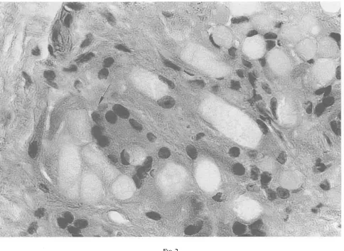
Four weeks later the implant was 'switched on' and during the subsequent weeks the patients showed a remarkably good result in terms of his audiological performance with a Bamford-Kowal-Bench (BKB) sen-tences score of 76 per cent. However, six weeks post-implantation the patient experienced sudden device failure and the only factor of note in the history was the exposure of the patient to a mains electric shock the day before whilst his wife was attempting to repair a domestic appliance! Assessment of device integrity confirmed its failure and the patient was thus reimplanted with a similar implant three weeks later. The new electrode array went into the scala tympani as easily as the original device with the insertion of 22 electrodes and all nine supporting bands. At operation a smooth capsule of fibrous tissue was found around the receiver/stimulator with granulation tissue in the mastoid bowl. Histological examination of the fibrous tissue showed chronic inflammation with a giant cell reaction to strands of refractile foreign material (Figure 2). We believe this represents a macrophage response to the ionomeric cement used to secure the electrode array in the mastoid cavity. Babighian (1992) observed a similar response to extruded bone cement used to reconstruct bony canal wall defects in chronic ear surgery.
Since the revision surgery the patient has suffered a brief episode of rotatory vertigo and sensitivity to electrical stimulation which settled with symptomatic treatment. The implant has continued to function well and the patient now has open set recognition and is even able to gain useful musical information. His BKB scores were 94 per cent at nine months and 88 per cent at 18 months. The original implant was evaluated by the manufacturer and this showed a failure of one of the capacitors in the receiver/ stimulator circuit, but they were reluctant to incriminate the electrical shock sustained by the patient.
From the University Department of Otolaryngology, Manchester Royal Infirmary, Manchester. Accepted for publication: 24 June 1995.
CLINICAL RECORDS 981
FIG. 1
Case 1: post-operative transorbital X-ray confirming correct electrode position.
Case 2
This 51-year-old lady suffered a bilateral hearing loss following an episode of rubella at the age of 15 years. She subsequently developed a rapid deterioration in her residual hearing, first on the left side and then the right in her fourth decade, rendering her entirely dependant on lip reading. Following standard audiovestibular, radiologi-cal, physical and psychological evaluation she was implanted with a Nucleus 22 channel device into the right ear with insertion of all 22 active electrodes. Five weeks later the implant was switched on and the patient reported some perception of sound.
During the following 12 months the patient's audio-ogical performance fluctuated despite a satisfactory :linical appraisal and radiological confirmation of elec-:rode position. At three months post-implantation her BKB scores were only two per cent on electrical itimulation rising to 37 per cent with the addition of lip •eading. After due consideration and consultation with the mplant manufacturer the patient was reimplanted, 14 nonths after the original procedure. At operation an ngrowth of fibrous tissue between the magnet and the )uter table of the skull was noted with marked new bone ormation around the subcortical channel for the proximal :lectrode array. This bone was carefully drilled away and emoval of fibrous tissue from the posterior tympanotomy illowed the implant to be delivered, with straightforward nsertion of the new implant, the scala tympani being videly patent. The original implant was returned to the Nucleus Group for analysis which confirmed that the mplant function was unstable at higher temperatures, with :omplete loss of function at 50°C. Histology of the fibrous
tissue removed at revision surgery showed granulation tissue, metaplastic bone formation and a foreign body reaction to suture material.
Since reimplantation, the patient's audiological perfor-mance has varied slightly. Her BKB scores have improved to 53 per cent on electrical stimulation and 98 per cent with the addition of lip reading. The relevance of the temperature instability of the original implant is open to debate.
Case 3
A 55-year-old woman was assessed for her suitability for cochlear implantation. She had become progressively deaf over a 10-year period following her third pregnancy at the age of 28 years. Until recently this had been associated with attacks of vertigo which had subsided spontaneously and apart from a family history of otosclerosis there were no causative factors implicated. Examination suggested a profound deafness with good lip reading. Pure tone audiometry confirmed a vibrotactile response at the lower frequencies at thresholds of 100 dB and higher, bilaterally. No benefit was obtained with a conventional hearing aid. High resolution CT scanning showed normal middle and inner ear anatomy and round window stimulation revealed a good dynamic range on the right side. Following a psychological and audiological assess-ment and after discussion with the patient, implantation of the right ear was offered.
FIG. 2
Case 1: histological section of tissue removed from the mastoid cavity at revision surgery. There is evidence of a multinucleate giant cell reaction to strands of refractile foreign material (arrowed) which represent particles of ionomeric bone cement.
front of the round window niche. No particular problem was encountered during insertion although it was noted that the electrode array was somewhat more springy than the Nucleus device to which the surgeon was accustomed. The intraoperative impedances were satisfactory in five of the six electrodes as well as the two earth electrodes (Table I). A transorbital plain X-ray on the fifth day however showed the implant to be incorrectly positioned with the electrode wire seemingly running across the middle ear cleft (Figure 3). This was reflected in the post-operative testing in that apart from electrode 5, the impedances had risen by a factor of between 1.7 (electrode 6) and 6.25 (electrode 4). The absolute values are shown in Table II.
The procedure was revised four weeks later a n d ^ t operation the proximal electrode array was found to be in position with intact ionomeric bone cement in the mastoid
TABLE I
CASE 3: INTRAOPERATIVE ELECTRODE IMPEDANCES (KILO OHMS). VALUES LESS THAN 2 0 K OHMS ARE CONSIDERED NORMAL. THE IMPEDANCE MEASUREMENTS ARE THUS ALL SATISFACTORY APART FROM ELECTRODE 5. THE ACTIVE ELECTRODE MEASUREMENTS ARE WITH REFERENCE TO ELECTRODES 7 AND 8 (BURIED IN THE TEMPORALIS MUSCLE) AND AN EXTERNAL GROUND REFERENCE ON
THE PATIENT'S ARM
Arm Ground 8
7 1 3.0 2.6 3.0
2 3.4 2.8 3.3
Active 3 3.8 3.2 3.9
4 3.0 2.4 2.9
electrodes
5
38.3 45.7 37.6
6 8.6 4.1 4.5
7 2.1
8 1.7
cavity and posterior tympanotomy. Removal of this and the inflammatory tissue reaction from the tympanotomy confirmed that the electrode system was running across the promontory towards the eustachian tube orifice. Histology of the fibrous tissue confirmed the presence of granulation tissue and post-inflammatory fibrosis. The cochleostomy and proximal basal turn were also obliterated by a fibrous plug which was meticulously dissected out and the electrode system was reimplanted into an otherwise patent scala tympani with satisfactory insertion of five of the six electrodes.
FIG. 3
Case 3: post-operative transorbital X-ray showing electrode to be incorrectly positioned.
CLINICAL RECORDS 983
TABLE II
CASE 3: POST-OPERATIVE ELECTRODE IMPEDANCES (KILO OHMS). WHILST THE VALUES REMAIN ACCEPTABLE, THEY ARE HIGHER THAN
THE INTRAOPERATIVE MEASUREMENTS, SHOWN IN TABLE 1
Ground Arm 8 1 8.6 9.4 2 11.7 12.3 Active electrodes 3 14.0 14.6 4 14.4 15.0 5 16.2 16.8 6 15.3 15.9 7 8.7 8 3.6
Post-operative impedance testing (Table III) and radiology (Figure 4) were satisfactory and the patient is now progressing well following her 'switch-on'. Her pre-operative lip reading BKB score was 18 per cent. Using the implant to augment her lip reading skills the patient's BKB score was 81 per cent.
Discussion
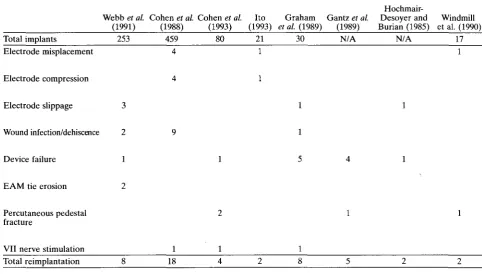
The cases described in this paper concur with the literature in that the three most likely reasons for revision surgery with reimplantation are wound dehiscence (usually with infection), device failure and electrode misplacement (Webb et al., 1991). Table IV summarizes the instances and reasons for device reimplantation in the reports examined.
Electrode related problems
Failure to insert the electrode array into the scala tympani or subsequent extrusion as described in our third case has been documented in the literature (Cohen et al., 1988; Ito, 1993; Kessler and Schindler, 1993). The most frequent error is inadvertent implantation of a hypotym-panic air cell and this is more likely to occur if the round window niche is not clearly identified. This may occur in experienced hands if there is fibrous or bony obliteration of the niche (and therefore reliance on the position of the oval window as a landmark), but it may also occur if a less experienced surgeon undertakes an inadequate posterior tympanotomy (Cohen et al., 1988) In Case 3 the operation proceeded uneventfully and the electrode array was clearly seen to be passing through the cochleostomy. We can only surmise that the electrode extruded sometime between the end of the surgery and the post-operative X-ray on the fifth day, coming to lie in the middle ear cleft. Certainly, the intraoperative impedance testing was satisfactory (Table I). A factor that may be significant is the greater springiness of the electrode array of the Ineraid device when compared to the Nucleus device. Our implantees routinely undergo a transorbital X-ray for electrode position on the fifth post-operative day and there is a case therefore for intraoperative radiology if there is any doubt about the electrode position at the time of surgery (Windmill et al., 1990).
Electrode compression and electrode slippage have also been described in the review of implant complications in the American, Melbourne and Hannover series (Webb et al., 1991). The effects of scar tissue around the extracochlear component of the array may account for
TABLE III
CASE 3: IMPEDANCES (KILO OHMS) AFTER CORRECT POSITIONING OF THE ELECTRODE ARRAY
Arm Ground 8 7 1 3.0 4.0 8.5 2 3.0 4.0 8.5 Active 3 3.3 4.3 8.8 4 3.7 4.7 9.2 electrodes 5 6 3.1 2.9 7 7.4 8 2.8 FIG. 4
Case 3: transorbital X-ray following revision surgery. The electrode array is now in a satisfactory position.
electrode slippage (Hochmair-Desoyer and Burian, 1985) but the critical time seems to be during fixation of the array and receiver/stimulator. The earlier reports studied refer to the use of Dacron® or silk ties to secure the implant (Graham et al., 1989) but more recently we have found the use of glass ionomer bone cement to be more satisfactory. The cement is placed at intervals around the electrode array as it courses round the mastoid bowl and is also applied to the margins of the receiver/stimulator. The cement hardens in a matter of minutes, provides secure fixation and is considerably easier and quicker than using Dacron® ties which may cause local necrosis of the overlying scalp flap (Ramsden et al., 1992) and have been associated with external meatal skin erosion (Webb et al., 1991).
Wound dehiscence
Wound related problems leading to explantation of the device can be avoided by careful planning of the incision and receiver/stimulator sites, meticulous handling of the flap and closure of the incision without tension (Cohen et al., 1988). The post-auricular scalp receives its blood supply from four potential sources: the posterior auricular artery, occipital artery, superficial temporal artery and the dermal plexus (Harris and Cueva, 1987). The anteriorly based C-shaped incision may compromise the predominant supply to this flap if the base is insufficiently broad, if the inferior limb extends beyond the lower margin of the pinna or if the patient has undergone previous surgery through a conventional post-auricular incision (Cohen et al., 1988). Webb et al. (1991) noted that, allowing for the fact that the C-shaped incision had been used in significantly greater numbers in the American series, it was associated with a higher incidence of flap necrosis. The inverted U-shaped incision has proved satisfactory in the Melbourne series but resulted in flap-related problems in the Hannover cases and was therefore superseded by the extended endaural incision (Webb et al., 1991). The extended endaural incision has been the approach of choice in our series with no major flap related morbidity.
Device failure
Hochmair-Webb et al. Cohen et al. Cohen et al. Ito Graham Gantz et al. Desoyer and Windmill (1991) (1988) (1993) (1993) et al. (1989) (1989) Burian (1985) et al. (1990)
Total implants 253 459 80 21 30 N/A N/A 17
Electrode misplacement 1
Electrode compression
Electrode slippage 3
Wound infection/dehiscence 2
Device failure 1
EAM tie erosion
Percutaneous pedestal fracture
VII nerve stimulation
Total reimplantation 18
design, manufacturing process (Case 2 in this paper) or user events such as the electrical shock sustained by
Casel.
Examination of complication reports allows a number of conclusions to be drawn. Firstly, device failure may occur irrespective of the type of device utilized (Gantz et al., 1989). However, early device designs were plagued with a higher failure rate due to problems with hermetic seals and individual electronic components (Hochmair-Desoyer and Burian, 1985; Conway and Boyle, 1989; Graham et al., 1989) and serial improvements in design lead to a corresponding reduction in the failure rate. Finally, with the exception of the now obsolete 3M device, implant failure tends to occur early in the life of the device and, as an example, the cumulative survival for the Nucleus 22 implant is greater than 97 per cent after six years (von Wallenberg et al., 1993).
Surgical and histological findings
The technical aspects of removing an indwelling electrode array with insertion of a new implant has been addressed to a certain degree by a number of implant centres. Several constant findings emerge in the reports studied. Firstly, that part of the array that courses the mastoid bowl is inevitably surrounded by intense fibrosis and dissection to release the electrode may prove difficult (Gantz et al., 1989; Windmill et al., 1990). Secondly, this fibrotic reaction extends into the posterior tympanotomy and posterior mesotympanum (Jackler et al., 1989) necessitating meticulous dissection in this area to avoid damage to the facial nerve. All three cases described in this paper exhibited these findings at the time of revision surgery. Removal of the Ineraid device (Case 3) proved particularly difficult due to involvement of the ball electrodes with such fibrosis whereas the Nucleus device, which has a smooth array could be delivered more readily. Difficulties with removal of the Ineraid array had been
noted previously by Gray (personal communication) describing how on removal, two of the ball electrodes were sheared off the supporting band and were left in the cochlea.
In the cochlea itself, a variable degree of osteoneogen-esis may be found in the basal scala tympani but more interestingly, the intracochlear electrode is surrounded by a fibrous sheath. The fibrous cuff remains in situ when the original electrode is removed and facilitates insertion of the new array, as long as the latter is implanted immediately. The cuff guides the new electrode into a similar position and therefore recreates the orientation of the original device. If for some reason a new device cannot be implanted at the time of explantation the electrode array should be divided at the cochleostomy leaving the intracochlear portion as a stent (Jackler et al., 1989), thus preventing collapse of the fibrous cuff and subsequent difficulty in electrode insertion.
With regard to the effects of electrode insertion on the cochlear neuronal elements, Fayad et al. (1991) noted that the insertion of an electrode into the scala tympani is most likely to traumatize the anterior basal turn as the array hits the outer cochlear wall and turns towards the modiolus. This results in strial and spiral ligament disruption and degeneration of any residual Organ of Corti and peripheral dendrites. Interestingly, no corresponding loss of ganglion cells was noted and therefore implant function was not affected. These findings concur with experimental reimplantation in cats undertaken by Jackler et al. (1989) in that the basal Organ of Corti showed some degeneration but the population of spiral ganglion cells remained intact.
Auditory performance
Cases 1 and 2 described in this paper showed no
CLINICAL RECORDS 985
electrode. These findings are consistent with the observa-tions of a number of reports studied which also found that reimplanted patients continue to fare as well or better than prior to the insertion of the second device (Hochmair-Desoyer and Burian, 1985). More specifically, Gantz et al. (1989) analysed the audiological performance of five patients undergoing reimplantation for device failure. On their own battery of audiological tests all five patients maintained or improved upon their original performance scores. Interestingly, two of these patients had been changed from a single to multichannel device. It would seem therefore that reimplantation can be accomplished without adverse physical or audiological consequences.
Conclusions
The continuing evolution of cochlear implant design and reliability coupled with advances in surgical technique should reduce the incidence of device replacement. However, should the need to reimplant arise, the current evidence suggests that a revision procedure can be accomplished without great technical difficulty and a favourable outcome can be expected in terms of the patient's audiological performance.
Acknowledgements
We wish to express our gratitude to Dr E. W. Benbow, Honorary Consultant Pathologist, for his advice and provision of the histological photograph. We are also grateful to the Department of Medical Illustration, Manchester Royal Infirmary, for their help with the illustrations.
References
Babighian, G. (1992) Use of a glass ionomer cement in otological surgery. A preliminary report. Journal of Laryngology and Otology 106: 954-959.
Cohen, N. L., Hoffman, R. A., Stroschein, M. (1988) Medical or surgical complications related to the nucleus multi-channel cochlear implant. Annals of Otology, Rhinology and Laryngology 97 (suppl. 135): 8-13.
Cohen, N. L., Waltzman, S. B., Fisher, S. G. (1993) A prospective, randomized study of cochlear implants. New England Journal of Medicine 328: 233-237.
Conway, M. J., Boyle, P. (1989) Design of the UCH/RNID cochlear implant system. Journal of Laryngology and Otology 103 (Suppl. 18): 4-10.
Fayad, J., Linthicum, F. H., Jr., Otto, S. R., Galey, F. R., House, W. F. (1991) Cochlear implants: histopathological findings related to performance in 16 human temporal bones. Annals of Otology, Rhinology and Laryngology 100: 807-811.
Gantz, B. J., Lowder, M. W., McCabe, B. F. (1989) Audiological results following reimplantation of cochlear implants. Annals of Otology, Rhinology and Laryngology 98: 12-16.
Graham, J. M., East, C. A., Fraser, J. G. (1989) UCH/RNID single channel cochlear implant: surgical technique. Journal of Laryngology and Otology 103 (Suppl 18): 14-19. Harris, J. P., Cueva, R. A. (1987) Flap design for cochlear
implantation: avoidance of a potential complication. Laryngoscope 97: 755-757.
Hochmair-Desoyer, I. J., Burian, K. (1985) Reimplantation of a molded scala tympani electrode: impact on psychophysical and speech discrimination abilities. Annals of Otology, ^Rhinology and Laryngology 94: 65-70.
Ito, J. (1993) Considerations of cochlear implant surgery. Clinical Otolaryngology 18: 108-111.
Jackler, R. K., Leake, P. A., McKerrow, W. S. (1989) Cochlear implant revision: effects of reimplantation on the cochlea. Annals of Otology, Rhinology and Laryngology 98: 813-820.
Kessler, D. K., Schindler, R. A. (1993) Progress with a multi-strategy cochlear implant system: the Clarion.Third Inter-national Cochlear Implant Conference, Innsbruck, Austria, April 4-7.
Ramsden, R. T., Herdman, R. C , Lye, R. H. (1992) Ionomeric bone cement in neuro-otological surgery. Journal of Laryngology and Otology 106: 949-953.
Von Wallenberg, E. L., Brinch, J., Money, D. K., West, R., Avunduk, K. (1993) Comparative reliability of cochlear implants. Advances in Otorhinolaryngology 48: 79-84. Webb, R. L., Lehnhardt, E., Clark, G. M., Laszig, R., Pyman,
B. C , Burkhard, K. H. G. (1991) Surgical complications with the cochlear multiple-channel cochlear implant: experience at Hannover and Melbourne. Annals of Otology, Rhinology and Laryngology 100: 131-136. Windmill, I. M., Martinez, S. A., Nolph, M. B., Eisenmenger,
B. A. (1990) Surgical and nonsurgical complications associated with cochlear prosthesis implantation. American Journal of Otology 11: 415-420.
Address for correspondence: Mr S. R. Saeed, F. R.C.S.,
University Department of Otolaryngology, Manchester Royal Infirmary,