ScholarWorks @ Georgia State University
ScholarWorks @ Georgia State University
Public Health Theses School of Public Health
Spring 5-12-2012
Could Low Vitamin D Status Explain the Increased Rates of
Could Low Vitamin D Status Explain the Increased Rates of
Hypertensive Disorder in Pregnancy in the US Population and in
Hypertensive Disorder in Pregnancy in the US Population and in
Non-Hispanic Black Women? An Examination of NHanes
Non-Hispanic Black Women? An Examination of NHanes
2001-2006
2001-2006
Michelle V. Leander-Griffith Georgia State University
Follow this and additional works at: https://scholarworks.gsu.edu/iph_theses
Recommended Citation Recommended Citation
Leander-Griffith, Michelle V., "Could Low Vitamin D Status Explain the Increased Rates of Hypertensive Disorder in Pregnancy in the US Population and in Non-Hispanic Black Women? An Examination of NHanes 2001-2006." Thesis, Georgia State University, 2012.
https://scholarworks.gsu.edu/iph_theses/221
TITLE PAGE
COULD LOW VITAMIN D STATUS EXPLAIN THE INCREASED RATES OF HYPERTENSIVE DISORDERS IN PREGNANCY IN THE US POPULATION AND IN
NON-HISPANIC BLACK WOMEN? AN EXAMINATION OF NHANES 2001-2006
by
Michelle Leander–Griffith B.Sc., University of Toronto
A Thesis Submitted to the Graduate Faculty of Georgia State University in Partial Fulfillment of the
Requirements for the Degree
COULD LOW VITAMIN D STATUS EXPLAIN THE INCREASED RATES OF HYPERTENSIVE DISORDERS IN PREGNANCY IN THE US POPULATION AND
IN NON-HISPANIC BLACK WOMEN? AN EXAMINATION OF NHANES 2001-2006
by Michelle Leander - Griffith
Approved:
__________________________________________ Committee Chair
__________________________________________ Committee Member
__________________________________________ Committee Member
ABSTRACT
Michelle Leander-Griffith.
Could Low Vitamin D Status Explain the Increased Rates of Hypertensive Disorders in Pregnancy in the US Population and in non-Hispanic Black Women? An Examination of
NHANES 2001-2006 (Under the direction of Dr. Ike Okosun)
Background: The incidence of Hypertensive Disorders in Pregnancy (HDP) is
increasing in the US and is linked to serious long and short-term health problems for both mother and fetus. Vitamin D has been shown to have direct influence on
molecular pathways involved in pregnancy. However a link between vitamin D status and HDP in Pregnant women has not been established.
Objectives: The purpose of this study is to determine (1) the association between
vitamin D deficiency and the occurrence of (HDP) and (2) whether non-Hispanic Black women (NHB) are at greater risk for HDP due to low vitamin D status.
Methods: Pregnant females in the National Health and Nutrition Examination
Survey (NHANES) study from 2001 to 2006 were used in this study. Participant’s response to interview questions and laboratory results were taken into account to determine HDP status. Logistic regression was used to determine the association between vitamin D status and HDP.
Results: Pregnant women with low vitamin D status (25(OH)D < 20ng/ml) were
1.123 (95%CI: 0.808-1.56) times more likely to have HDP compared to women who were vitamin D sufficient. This association was not significant. NHB women did not show a significant increased risk for HDP.
Conclusions: Low vitamin D status during pregnancy may lead to an increased risk
for Hypertensive Disorders in Pregnancy. However more research on larger sample size is needed to determine the true extent of the association of vitamin D status with HDP in the general population and that of non-Hispanic Black women.
In presenting this thesis as a partial fulfillment of the requirements for an advanced degree from Georgia State University, I agree that the Library of the University shall make it available for inspection and circulation in accordance with its regulations governing materials of this type. I agree that permission to quote from, to copy from, or to publish this thesis may be granted by the author or, in his/her absence, by the professor under whose direction it was written, or in his/her absence, by the Associate Dean, College of Health and Human Sciences. Such quoting, copying, or publishing must be solely for scholarly purposes and will not involve potential financial gain. It is understood that any copying from or publication of this dissertation which involves potential
financial gain will not be allowed without written permission of the author.
All theses deposited in the Georgia State University Library must be used in accordance with the stipulations prescribed by the author in the preceding statement.
The author of this thesis is:
Student’s Name: _____ MICHELLE V LEANDER-GRIFFITH_____________
Street Address: _________________________________________________
City, State, and Zip Code: _________________________________________
The Chair of the committee for this thesis is:
Professor’s Name: ____Dr. Ike Okosun______________________________
Department: _________ ______________________
College: _________ Institute of Public Health ________________________
Georgia State University
P.O. Box 3995 Atlanta, Georgia 30302-3995
Users of this thesis who not regularly enrolled as students at Georgia State University are required to attest acceptance of the preceding stipulation by signing below. Libraries borrowing this thesis for the use of their patrons are required to see that each user records here the information requested.
NAME OF USER ADDRESS DATE TYPE OF USE
(EXAMINATION ONLY OR
MICHELLE V LEANDER-GRIFFITH
EDUCATION
Master of Public Health, Georgia State University May 2012 (Prevention Sciences)
B.Sc. Biochemistry, University of Toronto November 1997
WORK EXPERIENCE
Research Assistant Sept 1999 - Present
Morehouse School of Medicine
Process Analyst Jan 1998 – Mar 1999
Brookhaven National Lab, Upton New York
Research and Teaching Assistant May 1995 – May
1997
University of Toronto, Scarborough Ontario
PUBLICATIONS
Mou, L., Lankford-Turner, P., Leander, M., Bissonnette, R., Donahoe, R., & Royal, W. (2004). RXR-induced TNF-[alpha] suppression is reversed by morphine in activated U937 cells. Journal of neuroimmunology, 147(1-2), 99-105. Royal III, W., Leander, M., & Bissonnette, R. (2005). Retinoid-induced mu opioid
receptor expression by phytohemagglutinin-stimulated U937 cells. Journal of neurovirology, 11(2), 157-165.
Royal III, W., Leander, M., Chen, Y., Major, E., & Bissonnette, R. (2004). Nuclear receptor activation and interaction with morphine. Journal of
Table of Contents
List of Figures ... vi
List of Tables ... iv
Chapter I – Introduction ... 1
Epidemiology of HDP ... 2
Vitamin D ... 3
Disparity of Preeclampsia ... 3
Link between vitamin D and preeclampsia ... 4
GAPS in the Literature ... 4
Purpose of Study ... 5
Chapter II – Literature Review ... 6
Main Clinical Disorders of HDP ... 7
Epidemiology of HDP ... 9
Preeclampsia ... 10
Pathogenesis of Preeclampsia and the Implication of Vitamin D ... 12
Previous research on Vitamin D and Preeclampsia ... 16
Vitamin D and the Disparity in Adverse Maternal Outcomes ... 21
What is Vitamin D ... 22
Classical and Non classical actions of Vitamin D ... 25
Vitamin D in Pregnancy ... 26
Prevalence of vitamin D insufficiency ... 27
Gaps in Research ... 28
Conclusion ... 28
Chapter III - Methods ... 30
Study Purpose ... 30
Data Source ... 30
Study Participants ... 31
Confounding Variables ... 34
Statistical Analysis ... 35
Chapter IV – Results ... 36
Strengths and Limitations ... 50
Recommendations ... 50
Conclusion ... 51
List of Figures
Figure 1. Shows how vitamin D is synthesized in the skin. ... 15
Figure 2. The absorption of UVA and UVB rays by the skin. ... 243
List of Tables
Table 1. BasicCharacteristics of the study population from NHANES 2001 to 2006………….39
Table 2. Distribution of independent variables by the presence and absence of HDP…40
Table 3. Distribution of independent Variables by the Vitamin D status………… ……41
Table 4. Association with Low vitamin D and the selected independent variables with HDP……… ………..42
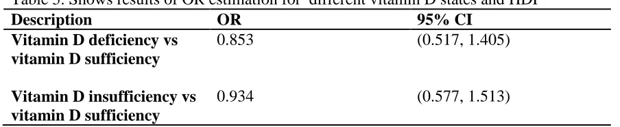
Table 5. Shows results of OR estimation for different vitamin D states and HDP… …..43
Chapter I – Introduction
About 5-10% of all pregnancies in the United States are affected by hypertensive
disorders in pregnancy (HDP), which contributes substantially to maternal mortality
(Wagner et al., 2007). HDPs encompass a spectrum of conditions such as preeclampsia,
eclampsia, and gestational as well as chronic hypertension (Wagner et al., 2007). The
incidence of hypertensive disorders during deliveries has increased from 67.2 per 1000
deliveries in 1998 to 81.4 per 1000 deliveries in 2006, and is associated with an enhanced
burden of severe obstetric morbidity (Kuklina et al., 2009). This trend is alarming as a
growing body of evidence confirms that HPD impacts public health beyond the apparent
immediate increased risk of maternal morbidity and mortality (Bilhartz and Bilhartz,
2010). In addition to latent maternal risk for heart disease, women with preeclampsia
have neonates who are often pre-term and of low birth weight (Bilhartz and Bilhartz,
2010). According to the Hospitalization Utilization Project (HCUP) Nationwide Patient
Survey, the economic burden of HDP in the US for 2003 was a staggering $2.3 billion
(AHRQ.gov, 2012), with approximately 204, 868 pregnant women admitted to hospital.
Risk factors for HDP include chronic hypertension, obesity, multifetal gestation,
history of preeclampsia, gestational diabetes, extremes of gestational age, and
pre-existing medical conditions (such as chronic hypertension, diabetes melitius, renal
disease thrombophilias, black race, low SES, low vitamin D and some maternal
infections) (Saibi et al., 2005; Mostello et al., 2002). Women with gestational
hypertension are at a higher risk of progressing to severe hypertension, preeclampsia or
hypertension, ischemic heart disease and stroke in the long term (Bellamy et al., 2007).
Most of our understanding of hypertensive disorders in pregnancy, and the associated
severe obstetric complications, is informed by studies of preeclampsia and eclampsia
(Zang et al., 2003).
Epidemiology of HDP
According to a 2006 report of the World Health Organization, hypertension in
pregnancy is said to be responsible for 16% of all deaths in developed countries, 9% in
Africa and Asia and over 25% in Latin America and the Caribbean (Khan et al., 2006).
Most often, 10 to 20% of Gestational hypertension progresses to preeclampsia, which is
one of the most common causes of maternal and fetal morbidity and mortality (Hauth et
al., 2000; Saudan et al., 1998).
Preeclampsia, one of the most severe forms of HDP, affects about 5% to 7% of all
pregnancies, and places women at a greater risk for placental abruption, cerebral
hemorrhage, acute renal failure, disseminated intravascular coagulation, pulmonary
edema, circulatory collapse and eclampsia, which itself can lead to seizures, coma and
death, and are at increased risk for delayed growth, low birth weight and preterm birth
(American Academy of Family Physicians, 2006).
Non-Hispanic Black women are at an increased risk for HDPs, as confirmed by
longitudinal studies in NC and NY (Miranda et al., 2010; Tanaka et a., 2007; Samadi et
al. 1996). One potential cause of this disparity is vitamin D, which has direct influence on
molecular pathways involved in the pathogenesis of preeclampsia and the high
preeclampsia and data suggesting that maternal vitamin D deficiency may increase the
risk of preeclampsia and fetal growth restriction (Bodnar et al., 2010), further
investigation is warranted in understanding the role of vitamin D in HDP.
Vitamin D
Vitamin D is a pleiotrophic secosteriod hormone that is primarily known for its
role in bone metabolism and mineral homeostasis (Grundman et al., 2011). Until recently,
Vitamin D deficiency was well known for its role in causing rickets, which leads to
softening of bones and the occurrence of multiple fractures in children. Vitamin D has no
biological activity on its own; however, we now know that a hormonal form with diverse
biological activities is generated when vitamin D is converted to 1α, 25dihydroxyvitamin
D (1, α 25(OH)2D (Shin et al., 2010). Evidence also exist that vitamin D is able to
regulate key target genes associated with implantation of the embryo, tropoblast invasion
and implantation tolerance (Evans et al., 2004).
In recent years, increased attention has been focused on vitamin D because of the
report by Shin et al. (2010). This report found that, depending on country of residence
and other factors, 20 -85% of pregnant women are deficient in vitamin D, a lack that has
been linked to the non-classical actions of this hormone: preeclampsia, insulin resistance,
and gestational diabetes mellitus (Shin et al., 2010).
Disparity of Preeclampsia
Previous research highlights an intractable disparity of maternal health outcomes
between African American women and Caucasian women in the US (Bodnar et al.,
at greater risk for the most severe HDP-related complications (Kozak and Lawerence,
1999). This occurs despite the fact that extensive research on HDP and other aspects of
prenatal care has led to no substantial improvements in predicting or preventing the
disorder (Saibi et al., 2005).
Link between vitamin D and preeclampsia
The link between vitamin D and preeclampsia has been observed as early as 2007
by Bodnar et al., who claimed that, a 50nmol/l decrease in 25(OH)D3 concentration
doubles the risk of preeclampsia I pregnant women. The link between vitamin D and
preeclampsia was documented again by Robinson and colleagues (2011), who found that
vitamin D may influence fetal growth through placental mechanisms. Furthermore, a
study examining the correlation between mid-gestation vitamin D deficiency and the risk
of severe preeclampsia, reported that women who developed preeclampsia had lower
mid-gestation concentrations of maternal 25OHD compared to controls (Baker et al,
2010).
GAPS in the Literature
Most research findings from the past five years implicate vitamin D in
preeclampsia; however studies linking vitamin D to other hypertensive disorders in
pregnancy, is lacking. Even studies that link vitamin D and preeclampsia lack sufficient
power and nation-wide representation and cannot negate the effects of a single location.
Significance – Determining whether vitamin D deficiency is, indeed, related to HDP, will
inform actions needed to remedy such a deficiency. This in turn will be instrumental in
Results of this study may provide insight as to whether or not vitamin D supplementation
in pregnant women could prevent adverse outcomes for the mother as well as the fetus.
Since HDPs include preeclampsia, which is a well-known and studied disorder, literature
on preeclampsia will be used in this study to inform many aspects of HDP.
Purpose of Study
This cross-sectional study aims to determine (1) the association between vitamin
D deficiency and the occurrence of Hypertensive Disorders in pregnancy and (2) whether
African American women are at greater risk for preeclampsia due to their low levels of
vitamin D. These associations are important since in addition to being a major contributor
to maternal and fetal morbidity and mortality, HPD also causes short and long term
Chapter II – Literature Review
HDPs affect about 10% of all pregnancies in the US and contribute significantly
to maternal mortality (Wagner et al., 2007). These disorders encompass a spectrum of
conditions such as preeclampsia, eclampsia, and gestational as well as chronic
hypertension (Wagner et al., 2007). This definition is supported by the National High
Blood Pressure Education Program Working Group on High Blood Pressure in
Pregnancy and includes eclampsia/preeclampsia superimposed on chronic hypertension,
gestational hypertension and chronic hypertension (Report of the National High Blood
Pressure Education Program Working Group on High Blood Pressure in Pregnancy,
2000). Mild hypertension and gestational hypertension are the most common of the HDPs
(Kuklina et al., 2009). Preeclampsia is thought to be responsible for 18% of all maternal
deaths in the US (Preeclampsia Foundation, 2006) and in 2002 was responsible for 56
maternal deaths per 100,000 live births (National Vital Statistics, 2004) and 71 neonatal
deaths per 100,000 live births (American College of Obstetricians and Gynecologists,
2005).
A 2008 study using the National Hospital Discharge Survey (NHDS) found
significant increases of preeclampsia and gestational hypertension, by 25 and 84%,
respectively, in the period between 1987 and 2004 (Wallis et al., 2008). Additional
support of this trend has found that hypertensive disorders during delivery have increased
significantly, from 67.2 per 1000 deliveries in 1998 to 81.4 per 1000 deliveries in 2006,
thus enhancing the burden of severe obstetrical morbidity that is associated with the
and 184%, respectively, from 1987 to 2004 using the National Hospital Discharge Survey
(Wallis et al., 2008): they also point out thata change in clinical guidelines for gestational
hypertension in the 1990’s may have led to an exaggeration in the rates of the disorder.
Main Clinical Disorders of HDP
HDPs are comprised of the following clinical disorders: namely chronic
hypertension, gestational hypertension, pre-eclampsia and chronic hypertension with
superimposed preeclampsia. Precise definition of these conditions remains a major
challenge in studying them (Hutcheon et al., 2011).
Preeclampsia consists of new-onset hypertension and proteinurea after 20 weeks of
gestation, and is a multi-systemic pregnancy-specific disorder (Bodnar et al., 2007). The
etiology of preeclampsia is unknown, but we know that it is unique to human pregnancy
(Saibi et al., 2005). The hypertension portion of the disorder is characterized by systolic
blood pressure of at least 140mm Hg, diastolic pressure of 90mm Hg, measured on two
occasions, at least 4-6 hours apart, following the 20th week of gestation in women who
are otherwise normotensive (Saibi et al., 2005). Proteinuria, on the other hand, is
characterized by the excretion of more than 300mg of protein in the urine every 24 hours
(Saibi et al., 2005). At a physiological level, preeclampsia is characterized by an
abnormal vascular response to the implantation of the placenta, which is linked to
increased systemic vascular resistance, increased platelet aggregation, activation of the
coagulation system, and endothelial dysfunction (Report of the National High Blood
Chronic Hypertension is defined as pre-existing hypertension, which is detected before
pregnancy, or prior to 20 weeks of gestation (Report of the National High Blood Pressure
Education, 2000); it is also defined as hypertension that is first diagnosed after 20 weeks
gestation and continues past 12 weeks postpartum (Hutcheon et al., 2011). In the US, this
condition affects 1 to 4% of women aged 18 – 29 years old, and 5 to 15% of women 30 to
39 years old from 1999 to 2004) (Cutler et l., 2008). In 2004, an estimated 1.7% of
pregnancies in the US were complicated by preexisting hypertension. Women with
chronic hypertension have a threefold increased risk of perinatal mortality, preterm
delivery, maternal death, and small-for-gestational-age-infants, compared to
normotensive women (Rey and Couturier, 1994; McCowan et al., 1996).
Gestational Hypertension develops after 20 weeks of gestation, and returns to normal
within 12 weeks post partum period (Hutcheon et al., 2011); it is seen in 2 to 3% of all
pregnancies in the US (Klemmenson et al., 2005; Wallis et al., 2008), with rates
increasing from 10.7 to 30.6 per 1000 deliveries between 1987 and 2004 (1 Klemmenson
et al., 2005). Gestational hypertension presents an increased risk of preeclampsia: 17% of
women who have this condition go on to develop preeclampsia during pregnancy (Sudan
et al., 1998). However, even though women with gestational hypertension are at
increased risk for obstetric complications, it is considerably less hazardous to the mother
compared to other HDPs (Wallis et al., 2008).
Chronic Hypertension with superimposed pre-eclampsia involves the occurrence of new
onset proteinuria, thrombocytopenia, or any other features of the pre-eclamptic syndrome
in women with chronic hypertension during pregnancy (Report of the National High
Eclampsia is characterized by seizures in the presence of preeclampsia, with no
alternative explanation (Bilhartz and Bilhartz, 2010),and could lead to coma and death
(American Academy of Family Physicians, 2006).Its incidence in the US was about 8.2
per 10,000 between 1996 and 2004 (Wallis et al., 2008). However this condition has
declined in recent years (Hutcheon et al., 2011).
Hemolysis, elevated liver enzymes and low platelets (HELLP) syndrome is a severe
form of preeclampsia, and manifests as elevated liver enzymes and low blood platelet
count (Report of the National High Blood Pressure Education, 2000).
Severity of Disease – Many of the proposed classifications for the severity of
preeclampsia take into account the following: severity of hypertension (i.e. if systolic
blood pressure >160mmHg, diastolic >=110mmHg at least on two occasions, 6 hrs apart)
or both, time of delivery (whether delivery < 34 weeks) and aftermath (e.g. the
occurrence fetal death, preterm birth or eclampsia (Ananth et al., 1997; Hernandez et al.,
2009; McDonald et al., 2009). Some women also develop severe gestational hypertension
that is not associated with nor accompanied by proteinuria. It is note worthy that
compared with mild preeclampsia, gestational hypertension is associated with higher
maternal and perinatal morbidities.
Epidemiology of HDP
As mentioned above, hypertensive disorders complicate about 5-10% of all
pregnancies (Williams Obstetrics, 2010), and are the leading cause of maternal mortality
maternal deaths in Africa, 22,000 deaths in Asia, 3,800 deaths in Latin America and the
Caribbean, and 150 deaths in developed countries (Hutcheon et al., 2011).
Since preeclampsia is one of the most severe conditions of HDP, literature examining its
epidemiology will be examined in this study.
A 2009 National Vital Statistics report, with 19 states reporting, revealed that
preeclampsia rates were 43.6 per 1000 births in non-Hispanic whites, 49.1 per 1000 live
births in non-Hispanic Blacks, and 38.0 per 1000 live births. These rates are highest for
pregnant women under 20 years old, and in those 40 to 54 years old, with Black women
in the latter group showing 1.5-fold increase compared to all races.
Preeclampsia affects between 2% and 7% of healthy nulliparous women. In 75 % of these
cases, the disease is mostly mild, with late onset or intrapartum, conferring only a
negligible increased risk for adverse pregnancy outcome (Saibi et al., 2003; Vatten and
Skjaerven, 2004).
Preeclampsia
Preeclampsia is a common complication in pregnancy, affecting up to 10 percent
of pregnant women, and often resulting in fetal growth restriction, premature delivery and
low birth weight of affected neonates. Pathogenesis for this heterogeneous disorder varies
depending on the risk factor profile. Risk factors include chronic hypertension, obesity,
multifetal gestation, history of preeclampsia, pregestational diabetes, extremes of
gestational age, and pre-existing medical conditions (such as chronic hypertension,
diabetes mellitus, renal disease thrombophilias, black race, low SES, vitamin D and some
Preexisting Medical Conditions include chronic hypertension, obesity, multifetal
gestation, history of preeclampsia, pregestational diabetes, kidney disease, autoimmune
disease and thrombophilias (Saibi et al., 2005; Mostello et al., 2002; Hutcheon et al.,
2011).
Primitarity and Sperm Exposure: It is hypothesized that risk for preeclampsia is
increased in women who have little exposure to their partner’s sperm, as supported by the
fact that nulliparous women are three times more likely to experience preeclampsia
compared to multiparous women, and that multiparous women display a reduced risk of
preeclampsia (Hutcheon et al., 2011).
Smoking has a protective effect with regard to preeclampsia, as shown by many studies;
smoking reducing the incidence of preeclampsia in a dose dependent manner, by up to
about 50% (England and Zhang, 2007).
Miscellaneous factors: A history of preeclampsia is a predictor preeclampsia in a
subsequent pregnancy (Hutcheon et al, 2011); obesity, advanced gestational age, multiple
pregnancies, infections, and residing at a high altitude are all factors that increase the risk
of preeclampsia (Hutcheon et al., 2011). Interestingly enough, preeclampsia rates are
higher when sunlight –dependent 25(OHD production is reduced in the winter months
(Shin et al, 2010).
Maternal Effects – Women with severe eclampsia and preeclampsia have a 10- to 30-
fold higher risk for complications such as acute renal failure, and pulmonary edema, and
have a 70% increased risk of placental abruption (Hutchen et al., 2011). Preeclampsia is
(Loverro et al., 2001). Long term studies show that women with preeclampsia have a 3-
to 4- fold increased risk of developing chronic hypertension, and an almost 2-fold
increased risk of ischemic heart disease, stroke, and venous thromboembolism (Bellamy
et al., 2007). Some women also develop severe gestational hypertension, which is not
associated with, nor accompanied by proteinurea. Compared with mild preeclampsia,
severe gestational hypertension is associated with higher maternal and perinatal
morbidities. (Sibai et al., 2005).
Effects on the child – Preeclamptic women have a 4-fold increase in early delivery,
largely due to iatrogenic effects, and neonates with a 2-fold increase in mortality, low
apgar scores, febrile seizures, encephalopathy and elevated rate of admission to neonatal
intensive care unit (Basso et al., 2006). The long-term effects of preeclampsia on the
fetus include higher levels of diastolic and systolic blood pressures in childhood, and
adolescence. In addition they are at increased risk for metabolic disorders, epilepsy and
other complications (Wu et al., 2008; Wu et al., 2009).
Pathogenesis of Preeclampsia and the Implication of Vitamin D
The underlying cause of preeclampsia is not known, but one hypothesis states that
preeclampsia is caused by the presence of placenta, or the maternal response to
implantation of the placenta (Sibai et al., 2005). Historically, two schools of thought exist
on the cause of preeclampsia: (i) vascular biologists believe that in the placenta,
ischemia–reperfusion precedes oxidative stress which leads to vascular disease; and (ii)
where the maternal alloimmune reaction is initiated by the rejection of the fetal allograft
(Saibi et al., 2005).
There is evidence that suggest vitamin D’s role in the pathogenesis of
preeclampsia. As mentioned before, it is thought that the primary trigger of preeclampsia
relates to impaired placentation, after which systemic maternal responses follow that
produce the clinical signs and symptoms of the 2–stage disorder (Walker, 2000). The
reduced tolerance to the developing fetus following maternal immune maladaption is
thought to result in preeclampsia (Robertson et al., 2003).
Stage 1 of the disorder is characterized by reduced placental perfusion which
follows abnormal placental implantation (Bodnar et al., 2007). Materials produced by the
poorly perfused placenta is said to produce materials that initiate the subsequent sequelae
of adverse health effects(Bodnar et al., 2007). Pathophysiological changes such as these,
are suggested to be secondary to abnormal endothelial function which accompanies
generalized increase in the inflammatory activation that constitutes normal pregnancy
(Redman et al., 1999). One –25-dihydroxyvitamin D3, the active form of vitamin D has
been shown to control the transcription and function of genes connected to placental
invasion, normal implantation and angiogenesis (Evans et al., 2004; Daftary and Taylor,
2006). In addition it is suggested that abnormal implantation is mediated by an
inappropriate immune response between mother and baby (Bodnar et al., 2007).
Immunomodulatory properties of 1,25-dihydroxyvitamin D3 may be play a key role in
this regard (Hewison, 1992). It follows that vitamin D deficiency may predispose a
function as well as vascular compliance, elasticity and intima media thickness are all
better off in women optimally supplemented with vitamin D (Braam et al., 2004).
It is suggested that the domination by the T helper type 2 (TH2) cytokine response
is one of the critical steps required to maintain normal pregnancy (Hypponen, 2005).
Induction of Th2 cells is favorable to the survival to the survival of the fetus, while
Th1-type reactions in the placenta is associated with spontaneous preterm delivery (El-Shazly
et al., 2004). It has been suggested that 1,25(OH)2D has an important role in promoting a
shift to a Th2- dominated pattern in immune response (Zehnder at al., 2002). In addition,
in vitro studies show that 1,25(OH)2D increases the secretion of Th2 cytokines and
inhibits the secretion of Th1 cytokines (Cantorna et al., 2000; Piccinni et al., 2000).
However the exact mechanisms of these actions are not fully understood (Hyppönen,
Figure 1. Comparison between normal and preeclamptic pregnancies.
Hypothesis on the association between vitamin D and Preeclampsia (Hyppönen, 2005).
Many studies attempting to find the etiology of the disparity pregnancy outcomes
have examined the contribution of differences of socioeconomic status, access to health
care, other environmental and social causes (Swamy et al., 2010). A host factor examined
by Swamy and colleagues is the genetic variability in the VDR gene. Research suggests
that race specific allelic frequencies in the vitamin D receptor gene is potentially involved
in health disparities (Swamy et al., 2010) . This study found that low infant birth weight
was strongly associated with VDR genetic variability among NHB women but not NHW
genetic polymorphisms and birth weight is an example of genetic influences on fetal
growth as well as a potentially important explanation for why there is a significant
difference in birth weight between NHB and BHW women. Swamy and colleagues also
speculate that VDR polymorphisms not only contribute to low birth weight, but may
signify causal pathways for the effects of vitamin D on other pregnancy outcomes
(Swamy et al., 2010).
It is possible that the chronic hypertension component of HDP may be explained
by similar mechanisms that are found in the association of low vitamin D and high blood
pressure in the population. One such mechanism is vitamin D’s modulation of the RAS
gene.RAS is known to regulate electrolyte and plasma volume homeostasis which affects
blood pressure (Motiwala and Wang, 2012). Hypertension and overall cardiovascular risk
results when RAS is inappropriately activated. Strong experimental evidence frome
murine studies support vitamin D’s role as a proximal inhibitor of RAS (Motiwala and
Wang, 2012).
Previous research on Vitamin D and Preeclampsia
As discussed above, considerable research has identified as association between
vitamin D deficiency and the increased risk of preeclampsia (Shin et al., 2010), and has
shown that cohorts supplemented with Vitamin D showed a decreased risk of
preeclampsia when compared to non-supplemented controls. Even though the exact
mechanism whereby vitamin D leads to preeclampsia is unclear (Shin et al., 2010), it is
hypothesized that low vitamin D levels destroy the normal Th1-to-TH2 cytokine balance,
an independent risk factor for preeclampsia: more specifically, a 50nmol/L drop in
25(OH)D concentration doubled the risk of preeclampsia after adjusting for confounders.
Consequences of low vitamin D levels during pregnancy on fetal and child health include
low birth weight (Scholl and Chen, 2009), craniotabes (Yorifuji et al., 2006), softening of
the skull bone that (an early sign of vitamin D deficiency). In addition, two recent
retrospective studies have found a new association between maternal vitamin D
deficiency and rickets-associated heart failure (Sullivan et al., 2008) and with acute lower
respiratory heart failure. Health problems later in childhood, including improper bone
development at 9 years of age (Shin et al., 2010), asthma (Litonjua and Weiss, 2007;
Kinney et al., 2009), schizophrenia (Kinney et al., 2009), and type 1 diabetes (Shin et al.,
2010) are also correlated with vitamin D deficiency during pregnancy.
Over the past 30 years, a vast amount of research has focused on the pathogenesis,
pathophysiology, management, and treatment of preeclampsia (Raymond and Peterson,
2011). This body of research has also sought to find biomarkers that can be used to
predict preeclampsia to find its association with other factors like smoking, stroke and
cardiovascular disease (Raymond and Peterson, 2011). Previous research on the effects
of vitamin D and preeclampsia have shown a positive association (Bodnar et al., 2010).
Notably, the frequency of preeclampsia remains unchanged despite advances in prenatal
care, and extensive research regarding this problem over the past decade has not led to
substantial improvement in prediction or prevention of this disorder (Saibi et al., 2005).
pathological mechanisms that result in preeclampsia (Saibi et al., 2005). One potential
candidate for the cause of preeclampsia and HDP is vitamin D.
Evidence for vitamin D’s implication in preeclampsia comes from various
sources. Observed seasonal patterns in preeclampsia; higher incidence in winter and
lower incidence in summer, suggests a role of sunlight hence vitamin D in this disorder
(Bodnar et al., 2007; ToPoel et al., 2011). Other observational evidence comes from
studies where women with preeclampsia have lower levels of circulating 25(OH)D than
pregnant normotensive women (Bodnar et al., Halhali et al., 2000; 2007; Robinson et al.,
2010)
Recent studies document an association between vitamin D deficiency and
preeclampsia. In 2011, Robinson et al. examined the association of vitamin D levels and
small-for–gestational-age (SGA) in 56 patients with early-onset severe preeclampsia
(EOSPE), and found that 25(OH)D levels were lower in patients with SGA infants. The
authors concluded that vitamin D may have an effect on fetal growth through placental
mechanisms. In 2010 the same authors had found a similar result when they examined
total 25OHD levels at the time of diagnosis of EOSPE. Furthermore a nested case control
study of a possible association of midgestation vitamin deficiency and the risk of severe
preeclampsia in 51 cases out of an overall cohort of 3992 pregnant women documented
that women who developed preeclampsia had lower midgestation maternal 25OHD
concentrations relative to control women (Baker et al., 2010). This trend of vitamin D
and preeclampsia was, in fact, reported as early as 2007, when Bodnar and colleagues
showed that, after adjustment for confounders, a 50nmol/l decrease in 25OHD
23,423 nulliparous women in Norway found that women taking 10-15microg/d of
vitamin D supplement experienced a 27% decrease in preeclampsia compared to women
who did not take the supplements; however no association with preeclampsia was found
when the additional vitamin D intake was from food alone (Haugen et al., 2009).
Not all research examining vitamin D and preeclampsia are consistent. A study in
British Columbia Canada 49°N, investigated whether the vitamin D (serum
25-hydroxyvitamin 25(OH)D status is associated with a risk of preeclampsia or with
adverse pregnancy outcomes in a group of 221 women with biochemical risk factors for
preeclampsia. Study authors concluded that there is no difference in the rates of
preeclampsia, gestational hypertension, or adverse pregnancy outcome as a result of
25OHD concentration, even though 78% of the women in the study were vitamin D
insufficient (25OHD <75nmol/) and 53% were vitamin D deficient (25OHD <50nmol/l).
This result is not surprising, because vitamin D deficiency and insufficiency are prevalent
in the group at that location (Shand et al., 2010). Another study that showed no
association calculated the free 25OHD levels by measuring the level of 25OHD and
vitamin D binding protein in a group of 170 women in their first trimester of pregnancy:
when 25OHD levels were compared in control women (n=131) to those in women who
subsequently developed preeclampsia (n=39), first trimester levels of vitamin D binding
protein and free 25OHD were similar, despite an association between higher first
Racial Disparity of Major Pregnancy Outcomes
Intractable disparities persist between black and white women in the US with
respect to their rates of major pregnancy outcomes. (Disparity as defined by the IOM
refers to differences that are above and beyond those that can be explained by differences
in health status between two or more groups.) Earlier studies reveal profound disparities
in the rates of major pregnancy outcomes like preterm birth, gestational diabetes and
HDP among non-Hispanic black and non-Hispanic white women in the United States. In
the United States, data from 2005 suggests that black women are 2.3 times more likely to
experience intrauterine death of their fetus, or postnatal death of their infants than are
white women. These disparities in turn, contribute to the disparate rates of infant and
maternal mortality, and hence to the impacted health status of the communities of these
women (Bryant et al, 2011). Examination of the major causes of infant death also reveals
marked disparities. With respect to preterm births (defined as delivery at less than 37
weeks gestation), black women are 1.5 to 2.5 times more likely to experience infant death
than are white women. Black women are 2.4-folf more likely to have preterm births (at
less than 32 weeks gestation), and 2.1-fold more likely to have a low birth weight infant
(Martin et al, 2006). In addition, black women are more likely than their white
counterparts to develop preeclampsia accompanied by more severe complications, even
after adjusting for women with chronic hypertension (4-7 in Bodnar and Simhan).
In an effort to highlight the etiology of maternal health disparities, many studies
have shown how differences in socioeconomic status contribute to the disparity in
variability contribute to the disparity of maternal outcomes in NHB and NHW women
suggest that differences in the race-specific allelic frequencies in the vitamin D receptor
(VDR) gene, is potentially involved in these health disparities (Cooper and Umbach,
1996).
Vitamin D and the Disparity in Adverse Maternal Outcomes
Previous studies have illuminated profound disparities in the rates of major
pregnancy outcomes in non-Hispanic black and non-Hispanic white women in the United
States (Tanaka et al., 2007; Tucker et al., 2007; Bryant et al., 2010). These disparities in
turn, contribute to the (disparate) rates of infant and maternal mortality, hence the
contributing to the poor health status of the communities of these women (Bryant et al,
2011).
In the United States, data from 2005 suggests that black women were 2.3 times more
likely to experience intrauterine fetal death or death of their infant than white women.
An examination of the major causes of infant death also revealed considerable disparities.
With respect to preterm births, defined as delivery at less than 37 weeks gestation, black
women were 1.5 to 2.5 times more likely to experience this than white women. Black
women were 2.4 times likely to have very preterm births (less than 32 weeks gestation
and 2.1 times more likely to have a low birth weight infant. (Bodnar and Simhan, 2010).
Most of these conditions are the result of conditions such as HDP. In addition, black
women were more likely than white women to develop preeclampsia along with more
Previous research on the disparity of adverse maternal health outcomes has
examined many causes. They include the impact of stress, racism and related factors in
pregnancy (Giscombe et al., 2005; Alio et al., 2010) as well as the role of socioeconomic
factors and access to adequate healthcare (Dominguez, 2011). Studies examining the
biological factors have looked at the variation of the VDR receptor in African American
women (Swamy et al., 2011).
What is Vitamin D
Vitamin D was first discovered as a preventative treatment for rickets, a disease
which results in softening bones, fractures and deformity in children (Shin et al., 2010);
vitamin D’s first known roles in bone and kidney are known as its classical actions (Shin
et al., 2010), but since then, vitamin D is known to be involved in many non-classical
processes (Shin et al, 2010). Vitamin D is a secosteriod hormone which provides and
maintains sufficient calcium and phosphorous stores in the body to facilitate optimal
metabolic functions. For humans, the primary source of vitamin D is synthesis in the
skin from sunlight (Nesby-O’Dell et al., 2002). This is achieved when solar UVB
radiation (wavelength, 290 to 315nm), penetrates the skin to convert 7-hydrocholesterol
to previtamin D3, which is then rapidly converted to vitamin D3 in the presence of
warmth from sunlight (Holick et al., 2007). Cutaneous absorption of UVB radiation is
inhibited by levels of sunlight exposure (season, latitude and time of day), the use of sun
block and dark skin pigmentation (Nesby-O’Dell et al., 2002). Limited dietary sources of
vitamin D include fatty fish, fish oils, fortified foods and vitamin supplements
individual’s vitamin D status (Holick, 2007). From there it is transported to the kidney
and other cells in the body. In the kidney, vitamin D is metabolized to its active form
1,25-dihydroxyvitamin D (Holick, 2007). Vitamin D deficiency is linked to a number of
diseases including some common cancers, auto immune diseases, infectious diseases and
cardiovascular diseases (Holick, 2007). Vitamin D exerts its biological activity by
binding to a high-affinity receptor (VDR) which in turn acts like a ligand-activated gene
transcription factor. Differences in allelic frequencies of VDR polymorphisms are race
specific and have been confirmed by multiple studies (Cooper and Umbach, 1996;
[image:35.612.111.541.319.616.2]Valdivielso and Fernandez 2006; Swamy et al., 2010)
Figure 21. Shows how vitamin D is synthesized in the skin.
Individuals could be vitamin D deficient for many reasons; they include older age,
higher BMI, dark skin pigmentation (melanin absorbs UVB photons), some medications,
liver disease, chronic kidney disease and living above 35˚ in winter months. Fig. 2 shows
the absorption of UVA and UVB rays by the skin. Fig 2a shows UVA and UVB radiation
penetrating skin in the absence of melanin. However in the presence of melanin, UVB
radiation does not penetrate the skin well since melanin in a natural sunscreen and
[image:36.612.179.511.300.485.2]absorbs UVB radiation.
Figure 3. The absorption of UVA and UVB rays by the skin. Fig 2a shows melanin penetrating skin in the absence of melanin. However in the presence of melanin, UVB radiation does not penetrate well since it is absorbed by melanin (Gorham UCSD, 2010).
Vitamin D exists in two forms: vitamins D2 or ergocholeciferol, and vitamin D3
cholecalciferol. Vitamin D2 is found in plants (Lewis et al., 2010) and is one third less
potent than vitamin D3, which is present in many fewer foods such as oily fish (salmon,
caught salmon has an average of 500 – 1000 IU vitamin D3 versus 100 – 250 IU vitamin
D2 per 100g (Chen et al., 2007). In the US, some food products like juice, breads, yogurts
and cheeses are fortified with vitamin D. Many individuals also take multivitamins
supplements that contain vitamin D, or supplements that contain only vitamin D (Holick
et al., 2008.). Because it is not if not obtained from the diet, vitamin D is synthesized
from a steroid precursor (Shin et al., 2010) and thus it is considered to not be a true
vitamin (Holick et al., 2007).
Classical and Non classical actions of Vitamin D
Classical actions of vitamin D take place in the kidney, liver and intestine, where
it is involved in the regulation of calcium and phosphate absorption, and thus affects bone
synthesis and metabolism (Shin et al). Vitamin D obtained from sunlight or ingestion is
transported to the liver, where it is hydroxylated by various mitochondrial and
microsomal P450 enzymes to form 25 hydroxycalciferol ( 25 (OH)D) (Lewis et al .,
2010). It is then processed in the kidney where and converted to its bioactive form
calcitrol and is subsequently involved in regulating calcium deposits in the bone, thereby
playing a major role in bone health (Alpert and Shaikh, 2007). However recent studies
show that more than 30 human tissues express the vitamin D receptor (VDR) and are
therefore able to respond to 1,25 (OH)2D (Shin et al); these data implicate vitamin D and
its receptor in a number of additional roles including immune function, cell proliferation
and differentiation, and hormone secretion (Shin et al). In particular, 1,25(OH)2D is
believed to reduce the activity of the adaptive immune system while enhancing the
pregnancy, vitamin D deficiency is linked to the non-classical actions of this hormone,
leading to preeclampsia, insulin resistance and gestational diabetes mellitus (Shin et al.,
2010).
Vitamin D in Pregnancy
As mentioned above, vitamin D status is reflected in circulating 25(OH)
concentrations, with the normal range being between 32ng/ml to 80ng/ml; values below
32ng/ml are designated as being deficient (Shin et al., 2010). According to a NCHS news
brief ( March, 2011), The Institute of Medicine (IOM) issued a guideline that vitamin D
levels sufficient levels of 25(OH)D were above 20mg/ml (CDC.gov Data Brief 59,
2011). It is now widely accepted that adequate intake of vitamin D intake is essential for
maternal health, and is key for the prevention of adverse maternal outcomes (Shin et al.,
2010). Given that well over 20 -85% of pregnant women are vitamin D deficient,
depending on the country of residence and other factors (Shin et al., 2010), it is critical
that we understand the mechanisms whereby vitamin D deficiency leads to these adverse
outcomes.
Little is known about the mechanisms underlying the non-classical actions of
vitamin D. The theoretical pathogenesis is that vitamin D levels is thought to impair the
normal balance of the Th1 cytokine expression thus damaging the immunological
functioning tolerance of the embryo to implantation (Bodnar, 2007 OR Baker, 2010). The
CYP27B1 and CYP24A1 are genes that code enzymes that regulate levels of
1,25(OH)2D, which is responsible for binding to the VDR to induce genomic and non
Research shows that all components for vitamin D signaling pathway are
expressed in the human placenta (Shin et al., 2010) and, moreover, that human decidual
and placental tissues are able to synthesize 1,25(OH)2D as well as 24,25(OH)2D.
(Weisman and Harell, 1979). Experiments with trophoblasts, the precursors to cells of the
human placenta, document that when these cells are exposed to E. coli in the presence of
vitamin D, they exhibit lower rates of infection and of cell death (Shin et at., 2010),
making the case for vitamin D supplementation to reduce infection during pregnancy.
This theory was further supported by study by Bodnar et al. (2009), who showed that the
unadjusted mean serum concentration of 25(OH)D was lower in patients with bacterial
vaginosis (BV) than in women without BV, and that the in prevalence of BV decreased as
25(OH)D increased to 80nmol/L.
Prevalence of vitamin D insufficiency
Bondar et al., (2006) investigated the prevalence of vitamin D insufficiency in
black and white pregnant women in the Pittsburg Pennsylvania area by race and season,
and found that 29.2% and 54.1% of black women had vitamin deficiency and
insufficiency, respectively, compared to 5% and 42.1% of white women, respectively.
Moreover, after adjusting for confounders, a small mean increase in maternal 25(OH)
concentrations in black and white women was also noted from winter to summer. The
authors concluded that black and white pregnant women had a high risk of vitamin D
deficiency when they reside in the northern US even while 90% of the study population
was compliant with vitamin supplementation (Bodnar et al., 2006). Scragg and
Americans and NHW (Scragg et al., 2007).Just recently, a study by Ganji and colleagues
found that overall, the mean serum 25(OH)D decreased by 9% in all participants of the
NHANES study from 1988-1994 to 2001 -2006 and the prevalence of 25(OH)D
<30nmol/L increased from 5 to 10 percent in all participants but in blacks it increased
from 22 to 88% (Gangi et al., 2011).
Gaps in Research
In the past years research has come to light implicating vitamin D in preeclampsia.
However there are few experimental studies that examine the association of vitamin and
HDP. In addition studies examining vitamin D and preeclampsia lack sufficient power
and national representativeness to negate the effects of a single location. In this study I
propose to use a nationally representative sample in the National Health and Nutrition
Survey to determine the association of low vitamin D on pregnant women with
hypertensive pregnancy related disorders.
Conclusion
There appears to be consistent evidence in the literature linking vitamin D deficiency
and preeclampsia. In this study we will use research based on preeclampsia to inform
various aspects of HDP since they are related. Non-Hispanic Black women have an
increased risk for HDP as well as vitamin D deficiency. Herein we will attempt to
investigate whether low vitamin D status is associated with increased risk if HDP.
Importance – If indeed vitamin D deficiency is related to HDP and preeclampsia, this
correlation can inform future studies, which hopefully will ultimately lead to a decrease
supplementation in pregnant women could be further investigated in order to prevent
adverse outcomes for the mother as well as the fetus.
This study will utilize the NHANES survey, to conduct a cross sectional analysis of HDP
in pregnant women across the US from 2001 to 2006. The association of vitamin D status
Chapter III - Methods
Study Purpose
The purpose of this cross-sectional study was to determine (i) the association of vitamin
D deficiency with the occurrence of preeclampsia and (ii) whether African American
women are at greater risk for preeclampsia due to low vitamin D status.
Data Source
Data used in this study were obtained from the National Health and Nutrition
Examination Survey (NHANES) of the National Center for Health Statistics (NCHS).
The NHANES used a stratified, multistage, probability sample design to undertake
nationally representative survey on the civilian non-institutionalized population in the
US. As of 1999, NHANES has been conducted annually, with data released in 2-year
cycles for public use. Data were derived from interviews, physical examinations, and
laboratory. For the personal interviews, participants signed consent forms, and health
questionnaires were administered by interviewers. Participant’s answers to demographic,
socioeconomic, dietary and health-related questions were recorded with use of
Computer-Assisted Personal Interview Technology. A Mobile Examination Center (MEC) was
employed to facilitate physical examinations and collection of blood and urine samples.
Adolescents, persons aged ≥60 years, non-Hispanic blacks, low-income persons, and
Hispanics/Mexicans were oversampled, to obtain reliable estimates for these groups.
To obtain a stratified, multistage probability sample for 2001 to 2006, multiple sample
counties, (2) a block or group of blocks within those counties, (3) clusters of households
within those blocks, and (4) and one or more persons within those households.
Starting in 2003, the survey content was held as constant as possible for each two year
period, to ensure consistency with the data release cycle. This allowed analysis of two or
more “cycles” (e.g. 2001-2002, 2003-2004 and 2005-2006) in addition to evaluation of
data from any single year cycle. In fact combining combination of two or more
two-year cycles is strongly recommended to generate estimates with superior statistical
reliability. Data sets (documents) were examined to verify that data items collected in all
of the combined years were similar in wording and methods.
Study Participants
For our study, we used interview and examination data from the NHANES survey
for 2001-2002, 2003-2004, and 2005-2006. The data for this study was collected
between January 2001 and December 2002, and include interviews of 11, 039 individuals
of all ages (10,477 were examined in MECs). The 2003-2004 NHANES survey
comprises data for 12, 862 individuals of all ages (9,643 were examined in MECs),
collected between January 2003 and December 2004. The 2005-2006 NHANES survey
used data from 11,862 individuals of all ages (9,950 were examined in MECs): these data
were collected between January 2005 and December 2006. For all years, several files
contained the data and appropriate documentation for the survey interview and
examination components. All NHANES protocols were reviewed by the NCHS review
board prior to data collection. This study utilized updated serum 25(OH)D data released
Periodically NHANES data files are updated by NCHS which replaces previous
data files. In November of 2010, the NCHS issued a data advisory for measurements of
serum 25(OH)D because of changes (method bias and imprecision) in the performance of
the serum 25 OHD assay over time. Users were informed that 25(OH)D data from
NHANES 2003-2004 and 2005-2006 were adjusted for assay drifts. The advisory
recommended that researchers use the newly available assay-adjusted data rather than the
unadjusted data previously available (CDC.gov., 2011).
Measurement of Vitamin D: Venipuncture was used to collect blood samples in
the MEC as per standard protocols. The Diasorin RIA kit assay (Stillwater, MN) was
used to measure serum 25(OH)D concentrations ate h National Center of Environmental
Health, CDC Atlanta GA.
Demographic Variables
The demographic variables used in this study were gender, age and race/ethnicity, with
the exclusion of all males. One inclusion criterion for age was that the women be of child
bearing age in the US - namely, 15 to 49 years old.
Age: The demographic age was self reported, and was recorded as a whole number in
years at the time of screening. Age was then classified into the following three categories:
15 to 20 years, 21 – 35 years, and 36 – 44 years.
Gender: Gender was self reported; this study only included females.
Ethnicity: Ethnicity was self reported, and was obtained from responses to the survey
question on race and Hispanic origin. Ethnicity was categorized into the following
was recoded as follows: “1” for Other Racial groups, “2” for Non-Hispanic White and
“3” for Non-Hispanic Black.
Pregnancy Status: For all years, pregnancy tests were performed on all female subjects
12 to 59 years old and on menstruating females 8 to 11 years old with use of the Icon 25
hCG test kit (Beckman Coulter), which detects human chorionic gonadotrophin (hCG) in
urine or serum.
Body mass Index: BMI was calculated from measurements of participants’ weight and
height. A BMI of <18.5 was designated as underweight, BMI of 18.5 to 25 – normal
weight, BMI 25 to 30 – overweight and BMI >30 was considered obese. In later analysis
the overweight and obese categories were combined for simplification.
Hypertensive Disorder in pregnancy: HDP was determined by a study participant
satisfying any one of the following: (1) Pregnant and confirmed by urine pregnancy test.
In this study those who were pregnant were coded as 1 and non pregnant coded as 2.
(2) A positive answer to the question: ‘ever been told you have High Blood Pressure’ “1”
for yes and “2” for no. (3) A positive answer to the question ‘Are you now taking
prescription for Hypertension “1” for yes and “2” for no. (4) Mean Systolic Blood
Pressure (MSBP) > 140 mmHg, calculated by computing the average of the second and
third systolic blood pressure reading or Mean Diastolic Blood Pressure > 90 mmHg
calculated by computing the average of the second and third blood diastolic pressure
reading. Therefore a subject meeting any one of these requirements was coded as having
Vitamin D status: This was determined by the concentration of 25-OHD (ng/ml) (range
2 to 86 ng/ml) in the serum of participants, and was measured in ng/ml in the NHANES
survey. 25-OHD concentration of <20ng/ml was considered as deficient, while
concentrations of >20ng/ml and above, was considered as sufficient. Study participants
missing this variable were excluded from this study. For additional analysis, vitamin D
status was further categorized into three categories; deficient (<20ng/ml), insufficient
(21-30ng/ml) and sufficient (>30ng/ml) for additional analysis.
Delimitations: The study included pregnant women only between the child bearing ages
of 15 and 44 years old.
Confounding Variables
Confounding variables for preeclampsia in this study were high BMI, race, diabetic
status, and age. Race was recoded as described above and diabetic status was determined
as positive if the subject satisfied anyone of the following criteria: (i) Has a doctor ever
said you had diabetes? “1” for yes and “2” for no. (ii) Do you take pills to lower blood
sugar? (iii) Participant’s glycohaemoglobin levels are > 6%.
Confounders for low vitamin D were used and included anticonvulsants, glucocorticoids,
HAART (AIDS medications), antirejection medication. Subjects taking anticonvulsants
or glucocorticoids were coded as having a vitamin D depressant. Any of the following
drugs can suppress vitamin D levels: Oxcarbazepine, Carbamazepine, Phenobarbital or
Hydrocortisone. Use of vitamin D as a supplement was presumed when the participant
answered positively to the question ‘Have you used a vitamin D supplement within the
used. Once the final data set containing only pregnant women was obtained, women who
had missing values for vitamin D were excluded from the study.
Statistical Analysis
NHANES data from 2001 to 2006 were organized, truncated and analyzed with
use of the Statistical Package for the Social Sciences (SPSS) version 18.0 and SAS
version 9.2. The distribution of race, ethnicity, education level and household income for
the total sample was obtained with use of descriptive analysis. The independent variable
analyzed was vitamin D status, and the dependent variable was HDP; control variables
were age in years, race/ethnicity, education level, diabetic status, and BMI.
Mean values of continuous values were compared across HDP states using the
independent t-tests. The distribution of categorical variables was reported in percentage
across HDP status. Chi squared tests were then used to determine which confounders
were significantly different among women who experienced HDP verses those who did
not. The distribution of categorical variables was across vitamin D status (deficient verses
sufficient) as also analyzed using bivariate methods. Chi squared tests were then used to
determine which confounders were significantly different among women with different
vitamin D status. Univariate logistic regression was conducted for each covariate in the
study Multivariate logistic regression was performed with step-wise selection for all
possible covariates of HDP. The variables remaining were then described. Statistical
Chapter IV – Results
Initially the study population consisted of 1037 study participants. However once
subjects who had missing values for vitamin D, the main independent variable, were
removed from the study population, 803 subjects were eligible for the study. The basic
characteristics of the study population of pregnant women ages 15 to 44 years, are
described in table 1. The distribution of age, race/ethnicity, annual house hold income,
education level, HDP status, vitamin D status, the presence of liver disease and vitamin D
status were all examined. Regarding race, there were 359 (44.71%) NHW, 135 (16.81%)
NHB and 309 (38.48%) subjects of other racial backgrounds. The mean age of the
participants was 26.28 yrs (SD=5.80), and mean vitamin D concentration was 22.50ng/ml
(SD 8.97). Pregnant women aged 21 to 35 years were the largest group at 54.8%,
followed by the 36-44 year age group at 30.5%. NHW were the largest ethnic group at
44.71%, followed by other racial groups at 38.5% and 16.81% NHB. Women with an
annual house hold income of $20,000 to $55,000 had the largest representation at
40.34%. Almost half of the population had high school and some college.
With respect to the independent variables, 340 (42%) had low vitamin D
(25(OH)D<20ng/mL), vitamin D deficient), mean vitamin levels were 22.50ng/ml
(SD=8.97). Thirteen subjects (1.62%) had liver disease, 56 (7%) were diabetic and 674
(84% used supplements (vitamin D supplementation assumed). The mean BMI was 24.39
(SD=6.79). For BMI sub categories of underweight, normal weight, overweight and
obese; the overweight sub category had the highest number of subjects with 263
Table 2 describes the characteristics of the study participants across HDP states.
Study participants with HDP were more likely to be non-Hispanic white, have an annual
house hold income between $20,000 and $55,000, have some high school or college and
of normal weight. The only variables that were significant were for BMI and diabetic
status and supplement use (95% CI; P= 0.009, 0.0354 and 0.0225 respectively).
Table 3 shows the result of bivariate analysis of the independent variable across vitamin
D deficient verses vitamin D sufficient states. The only variable that was significant was
use of supplements (P-0.0012).
Univariate logistic regression of the independent variables was conducted to
determine association with the variable and HDP. These results are shown in table 4.
Overall, low vitamin D status showed an increased risk of HDP (OR; 1.123) but this was
not significant (95%CI; 0.808, 1.56). The only significant positive association with HDP
at the 0.05 confidence level was BMI. For the vitamin D deficiency subcategories, i.e.
vitamin D deficiency, insufficiency and sufficiency, the reference group was vitamin D
sufficiency. Based on the reference group (vitamin D sufficiency) estimated OR for
vitamin D deficiency and insufficiency was shown in table 5. Subjects who were vitamin
D deficient were 0.853 times less likely to have HDP than sufficient subjects.
Based on the table, confidence interval between 95% CI, vitamin D status was not
significantly associated with HDP under the condition of α = 0.05
For the diabetic status an OR of 0.545 was observed, but this was not significant (95%
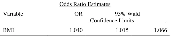
Multivariate Logistic Regression
Stepwise multivariate logistic regression was used to select which covariates had
a significant effect on HDP from all possible covariates. Only one variable, BMI (Body
Mass Index) remained. The goodness of fitness of the selected model was confirmed by
Hosmer-Lemeshow test: Chi-square test statistics is 3.3270 with d.f of 8, and p-value is
0.9122.
The estimated parameters for multivariate logistic regression and the estimated OR with
95% of confidence intervals are shown on the tables below. The results of the table 5
shows that increased BMI had significantly associated odds of HDP as indicated by odds
Table 1. BasicCharacteristics of the study population from NHANES 2001 to 2006
Variable Number (%)
Demographic Variables
Female 803
Age Group Mean
15-20 yrs
26.29 (SD=5.81) 118 (14.69)
21-35 yrs 440 (54.79)
36-44 245 (30.51)
Race/Ethnicity Non-Hispanic White 359 (44.71)
Non-Hispanic Black 135 (16.81)
Mixed/ Other 309 (38.48)
a
Annual Household Income
Less than $20,000 181(23.94)
$20,000.00 -$55,000 305 (40.34)
$55,000+ 270 (35.71)
b
Education Level Less than High School 179 (26.17)
High School Some College 332 (48.54)
College Grad 173 (25.29)
Dependent Variable
HDP Yes 197 (24.16)
No 609 (75.84)
Independent Variables Vitamin D Level/Status (ng/ml) Mean Low 22.50 (SD=8.97) 340 (42.34)
Not Low 463 (57.66)
Liver Disease Yes 13 (1.62)
No 790 (98.38)
c
Diabetic Status Yes 56 (7.04)
No 740 (92.96)
BMI Mean
Underweight
24.39 (SD=6.80) 237 (29.51)
Normal Weight 263 (32.75)
Overweight 170 (37.73)
Obese 133 (16.56)
Use of Supplements Yes 674 (83.94)
No 129 (16.06)
Vitamin D categories
Vitamin D Deficient 304 (37.85)
Vitamin D Insufficient 386 (48.07)
Vitamin D Sufficient 113 (14.07)
[image:51.612.107.415.98.678.2]